. 2008 Dec;22(12):2240-6.
doi: 10.1038/leu.2008.263. Epub 2008 Oct 2.

A role for IFN-lambda1 in multiple myeloma B cell growth

Affiliations

A role for IFN-lambda1 in multiple myeloma B cell growth

A J Novak et al. Leukemia. 2008 Dec.

Abstract

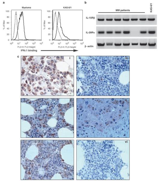
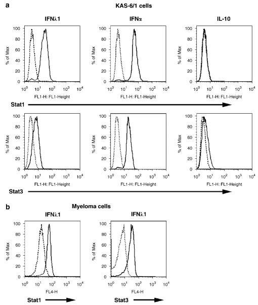
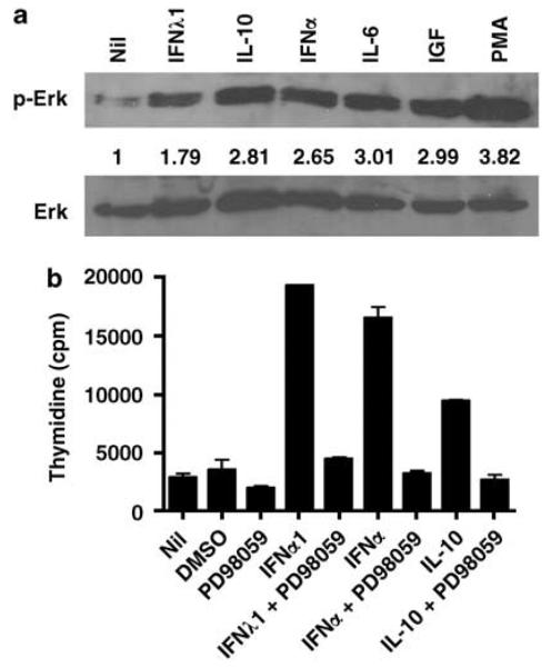
Multiple myeloma (MM) is a progressive disease that results from dysregulated proliferation of plasma cells. Although, causative factors such as genetic events and altered expression of anti-apoptotic factors have been described in a number of patients, the mechanistic details that drive myeloma development and continued growth of malignant cells remain largely undefined. Numerous growth factors, including interleukin (IL)-6, Insulin-like growth factor-1 and IL-10 have been shown to promote growth of MM cells suggesting a significant role for cytokines in this disease. Interferon (IFN)-lambda1 is a new member of the Class II cytokine family that, similar to IFN-alpha, has been shown to mediate viral immunity. In light of data supporting a role for cytokines in myeloma, we investigated the significance of IFN-lambda1 on myeloma cell biology. Our studies show for the first time that myeloma cells bind to soluble IFN-lambda1, and that IFN-lambda1 induces myeloma cell growth and protects against dexamethasone-induced cell death. Our data also show that IFN-lambda1 induces phosphorylation of STAT1, STAT3 and Erk. Taken together, our results suggest that IFN-lambda1 may regulate myeloma cell biology and could prove to be therapeutically important.


Figures

Figure 1
Figure 1
IFN-λ1 binding and expression in multiple myeloma. (a) KAS-6/1 cells or freshly isolated CD138+ MM cells were stained with biotin-conjugated IFN-λ1 for 30 min on ice, washed and incubated with PE-streptavidin (black line). Biotinylated mouse immunoglobulin served as a negative control (dashed line). (b) Expression of IL-10Rβ and IL-28Rα mRNA was determined by PCR. β-actin served as a loading control. (c) Multiple myeloma (i–iii), normal bone marrow (iv) and liver tissue sections were stained with an IFN-λ1-specific antibody as described in Materials and methods. Isotype controls were run for all specimens and a representive example is shown in (vi). IFN, Interferon; IL, interleukin; MM, multiple myeloma.
Figure 2
Figure 2
IFN-λ1 mediated cell growth of multiple myeloma cells. (a) KAS-6/1 cells were cultured in the presence of 0.01–1000 ng/ml IFN-λ1 for 3 days. Values represent the mean of triplicate values of a representative experiment (n = 3). (b) KAS-6/1 cells were cultured in the presence of 0.1 μg/ml IFN-λ1, 4000 U/ml IFN-α or 0.1 μg/ml BLyS, alone or in combination, for 3 days. Values represent the mean of triplicate values of a representative experiment (n = 5). (c) KAS-6/1 cells or freshly isolated CD138+ MM cells were cultured in RPMI + 10% FCS with the addition of 0.1 μg/ml IFN-λ1 and cell viability was determined after 3 days as described in Materials and methods. (d) KAS-6/1 cells were cultured in RPMI + 10% FCS with the addition of 0.1 μg/ml IFN-λ1, 1 ng/ml IL-6 or 10 nM dexamethasone alone or in combination, and cell viability was determined after 3 days. A representative experiment is shown (n = 3). IFN, Interferon; IL, interleukin; MM, multiple myeloma.
Figure 3
Figure 3
Activation of the STAT pathway by IFN-λ1. (a) KAS-6/1 or (b) CD138+ patient MM cells were stimulated with 0.1 mg/ml IFN-l1, 10 000 U/ml IFN-α or 0.1 mg/ml IL-10 for 10 min, and STAT1 and −3 phosphorylation was determined by FACS as described in Materials and methods. IFN, Interferon; IL, interleukin; MM, multiple myeloma.
Figure 4
Figure 4
IFN-λ1 mediated cell growth of multiple myeloma cells is Erk dependent. (a) Phosphorylated and total Erk levels were determined by western blot after stimulation of KAS-6/1 cells with 0.1 μg/ml IFN-λ1, 10 000 U/ml IFN-α, 0.1 μg/ml IL-10, 0.05 μg/ml IL-6, 0.1 μg/ml IGF-1 or 25 ng/ml PMA. The fold increase in phosphorylated Erk was calculated as described in Materials and methods and it is shown below the upper panel. (b) KAS-6/1 cells were cultured in the presence of 0.5 μg/ml IFN-λ1, 4000 U/ml IFN-α or 0.1 μg/ml IL-10 alone or with 50 μM PD98059 for 3 days. Values represent the mean of triplicate values of a representative experiment (n = 5). IGF-1, Insulin-like growth factor-1; IFN, Interferon; IL, interleukin.

References

    1. Kotenko SV, Gallagher G, Baurin VV, Lewis-Antes A, Shen M, Shah NK, et al. IFN-lambdas mediate antiviral protection through a distinct class II cytokine receptor complex. Nat Immunol. 2003;4:69–77. - PubMed
    1. Sheppard P, Kindsvogel W, Xu W, Henderson K, Schlutsmeyer S, Whitmore TE, et al. IL-28, IL-29 and their class II cytokine receptor IL-28R. Nat Immunol. 2003;4:63–68. - PubMed
    1. Dumoutier L, Tounsi A, Michiels T, Sommereyns C, Kotenko SV, Renauld JC. Role of the interleukin (IL)-28 receptor tyrosine residues for antiviral and antiproliferative activity of IL-29/interferon-lambda 1: similarities with type I interferon signaling. J Biol Chem. 2004;279:32269–32274. - PubMed
    1. Meager A, Visvalingam K, Dilger P, Bryan D, Wadhwa M. Biological activity of interleukins-28 and -29: comparison with type I interferons. Cytokine. 2005;31:109–118. - PubMed
    1. Pestka S, Krause CD, Walter MR. Interferons, interferon-like cytokines, and their receptors. Immunol Rev. 2004;202:8–32. - PubMed

Publication types

MeSH terms