. 2008 Oct 2;3(10):e3321.
doi: 10.1371/journal.pone.0003321.

Cerebrospinal fluid dendritic cells infiltrate the brain parenchyma and target the cervical lymph nodes under neuroinflammatory conditions

Affiliations

Cerebrospinal fluid dendritic cells infiltrate the brain parenchyma and target the cervical lymph nodes under neuroinflammatory conditions

Eric Hatterer et al. PLoS One. .

Abstract

Background: In many neuroinflammatory diseases, dendritic cells (DCs) accumulate in several compartments of the central nervous system (CNS), including the cerebrospinal fluid (CSF). Myeloid DCs invading the inflamed CNS are thus thought to play a major role in the initiation and perpetuation of CNS-targeted autoimmune responses. We previously reported that, in normal rats, DCs injected intra-CSF migrated outside the CNS and reached the B-cell zone of cervical lymph nodes. However, there is yet no information on the migratory behavior of CSF-circulating DCs under neuroinflammatory conditions.

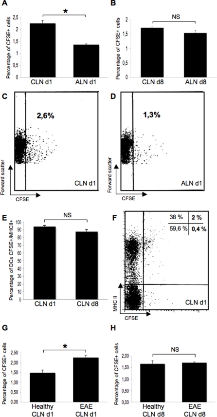
Methodology/principal findings: To address this issue, we performed in vivo transfer experiments in rats suffering from experimental autoimmune encephalomyelitis (EAE), a model of multiple sclerosis. EAE or control rats were injected intra-CSF with bone marrow-derived myeloid DCs labeled with the fluorescent marker carboxyfluorescein diacetate succinimidyl ester (CFSE). In parallel experiments, fluorescent microspheres were injected intra-CSF to EAE rats in order to track endogenous antigen-presenting cells (APCs). Animals were then sacrificed on day 1 or 8 post-injection and their brain and peripheral lymph nodes were assessed for the presence of microspheres(+) APCs or CFSE(+) DCs by immunohistology and/or FACS analysis. Data showed that in EAE rats, DCs injected intra-CSF substantially infiltrated several compartments of the inflamed CNS, including the periventricular demyelinating lesions. We also found that in EAE rats, as compared to controls, a larger number of intra-CSF injected DCs reached the cervical lymph nodes. This migratory behavior was accompanied by an accentuation of EAE clinical signs and an increased systemic antibody response against myelin oligodendrocyte glycoprotein, a major immunogenic myelin antigen.

Conclusions/significance: Altogether, these results indicate that CSF-circulating DCs are able to both survey the inflamed brain and to reach the cervical lymph nodes. In EAE and maybe multiple sclerosis, CSF-circulating DCs may thus support the immune responses that develop within and outside the inflamed CNS.


Conflict of interest statement

Competing Interests: The authors have declared that no competing interests exist.

Figures

Figure 1
Figure 1. CSF-circulating DCs infiltrate the periventricular parenchyma.
Fluorescent microspheres or DCs labeled with the cytoplasmic fluorescent marker CFSE were injected into the left lateral ventricle of EAE rats (n = 32) at the clinical peak of disease (day 12 post-immunization). EAE rats injected with fluorescent microspheres (n = 14) or CFSE-labeled DCs (n = 18) were then sacrificed on day 1 post-injection (n = 16) or 8 post-injection (n = 16). An immunohistological analysis of brains was then performed using antibodies directed against CD11b/CD11c (OX42) or MHC class II molecules. A–C: OX42+ cells (green) harboring intracytoplasmic fluorescent microspheres (red) are observed in the lumen of the injected lateral ventricle, on day 1 post-injection. D: Microspheres+/OX42+ cells localize in the periventricular parenchyma of the injected ventricle, on day 1 post-injection. Insert (solid square) shows a high magnification view of microspheres+/OX42+ cells infiltrating a periventricular inflammatory lesion (dashed square). E: On day 1 post-injection, MHC class II+ cells (green) harboring intracytoplasmic fluorescent microspheres (red) are detectable in the lumen of the third ventricle (dashed square) and in a large periventricular area infiltrated with MHC class II+ cells. Insert (solid square) shows a high magnification view of microspheres+/MHC class II+ cells that localize in the lumen of the third ventricle (dashed square). Arrows indicate microspheres+/MHC class II+ cells in the periventricular parenchyma. F–H: CFSE+ DCs (green) that express OX42 (red) are detected in the lumen of the injected lateral ventricle, on day 1 post-injection. I: A periventricular area bordering the injected lateral ventricle is infiltrated by OX42+ cells and contains OX42+/CFSE+ DCs, on day 1 post-injection. J: On day 8 post-injection, CFSE+ DCs are observed in a periventricular inflammatory lesion, adjacent to the injected lateral ventricle. LV: lateral ventricle, V3: third ventricle. Scale bars: 100 µm (B–J), 50 µm (A–C, E–I), 10 µm (inserts in panel D and E).
Figure 2
Figure 2. CSF-circulating DCs penetrate periventricular demyelinating lesion.
Fluorescent microspheres or DCs labeled with the cytoplasmic fluorescent marker CFSE were injected into the left lateral ventricle of EAE rats (n = 32) at the clinical peak of disease (day 12 post-immunization). EAE rats injected with fluorescent microspheres (n = 14) or CFSE-labeled DCs (n = 18) were then sacrificed on day 1 post-injection (n = 16) or 8 post-injection (n = 16). An immunohistological analysis of brains obtained from injected EAE rats or control healthy rats (n = 4) was then performed using antibodies directed against MHC class II molecules and/or myelin basic protein (MBP). A–C: In a normal rat (A), immunostaining of MBP (green) is homogenous and symmetric in the periventricular parenchyma adjacent to the third ventricle. In contrast, a large periventricular demyelinated area, adjacent to the third ventricle, is observed in an EAE rat injected intra-CSF with microspheres and sacrificed on day 8 post-injection (B). This demyelinating lesion is filled with microspheres+ cells (C). D–F: On day 1 post-injection, an area of periventricular infiltration adjacent to the injected lateral ventricle (D) is demyelinated (E) and contains CFSE+ DCs (F). G–I: On day 1 post-injection, periventricular parenchymal infiltrates (G) adjacent to the injected lateral ventricle are formed by MHC class II+ cells (H) and contain MHC class II+/CFSE+ DCs (I). LV: lateral ventricle, V3: third ventricle. Scale bars: 50 µm (A–I).
Figure 3
Figure 3. CFSE+ DCs specifically target the periventricular demyelinating lesions.
DCs labeled with the cytoplasmic fluorescent marker CFSE were injected into the left lateral ventricle of healthy control rats or EAE rats at the clinical peak of disease (day 12 post-immunization). Rats were then sacrificed on day 1 post-injection (EAE rats: n = 4, normal rats: n = 4) and an immunohistological analysis was performed on brain sections crossing the injected lateral ventricle. At least 3 to 5 sections per animal were examined. A: In brain sections from EAE rats, an immunostaining of the myelin basic protein (MBP) was performed and the number of CFSE+ cells/10−2 mm2 was counted in demyelinated vs normal-appearing periventricular white matter, as described in the Materials and Methods section. Data show that the mean number of CFSE+ cells was more than 4 times higher in periventricular demyelinating lesions than in normal-appearing periventricular white matter (9.4+/−2.5 cells/10−2 mm2 vs 1.6+/−0.5 cells/10−2 mm2 in demyelinated vs normal-apprearing periventricular white matter respectively, p = 0.02, Mann and Whitney test). B: Intraventricular vs periventricular CFSE+ cells were counted in brain sections obtained from injected EAE rats or injected control rats. Data are presented as percentages of intraventricular vs periventricular cells. Results show that in normal rats, 14.5+/−0.6% cells localized in the periventricular parenchyma, while, in EAE rats, more than 40% of the injected cells localized in the periventricular parenchyma (42+/−7%, p = 0.008, Mann and Whitney test). Conversely, 85.5+/−0.6% CFSE+ cells localized in the intraventricular lumen of normal rats while less than 60% of the injected cells localized in the intraventricular lumen of EAE rats (58+/−7%, p = 0.008, Mann and Whitney test). *: p<0.05, **: p<0.01.
Figure 4
Figure 4. CSF-circulating DCs migrate along the deep penetrating meninges.
Fluorescent microspheres or DCs labeled with the cytoplasmic fluorescent marker CFSE were injected into the left lateral ventricle of EAE rats (n = 32) at the clinical peak of disease (day 12 post-immunization). EAE rats injected with fluorescent microspheres (n = 14) or CFSE-labeled DCs (n = 18) were then sacrificed on day 1 post-injection (n = 16) and an immunohistological analysis of brains was performed using antibodies directed against CD11b/CD11c (OX42) or MHC class II molecules. Nuclei were counterstained with the fluorescent nuclear dye DAPI. A, B: Counterstaining of nuclei with DAPI coloration (A) shows that microspheres+ cells (visualized as white spots in B) localize in the deep penetrating meninges lining the inner parts of the brainstem (Bs) and cerebllum (Cb). C–E: Microspheres+ cells (white in D, red in E) expressing MHC class II molecules (green in C and E) are observed in the deep penetrating meninges covering the cerebellar convolutions. Insert in E shows a high magnification view of a ramified microsphere+/MHC class II+ cell observed in the penetrating pia matter. F, G: Counterstaining of nuclei with DAPI coloration (F) shows that CFSE+ cells (G) localize along the pia matter lining the inner parts of the brainstem (Bs) and cerebellum (Cb). H–J: An OX42+ infiltrate is observed in the deep penetrating meninges covering the inner parts of the brainstem (Bs) and cerebellum (Cb) (H). These infiltrating cells comprise CFSE+ cells (I) that express OX42 (J). Scale bars: 100 µm (F–G), 50 µm (A–E, H–J), 10 µm (insert in panel E).
Figure 5
Figure 5. CSF-circulating DCs infiltrate the brain parenchyma.
Fluorescent microspheres or DCs labeled with the cytoplasmic fluorescent marker CFSE were injected into the left lateral ventricle of EAE rats (n = 32) at the clinical peak of disease (day 12 post-immunization). EAE rats injected with fluorescent microspheres (n = 14) or CFSE-labeled DCs (n = 18) were then sacrificed on day 1 post-injection (n = 16) and their brains examined by immunohistology using antibodies directed against CD11b/CD11c (OX42) or MHC class II molecules. Nuclei were counterstained with the fluorescent nuclear dye DAPI. A–C: Visualization of nuclei with DAPI coloration (A) allows the localization of microspheres (white in B, red in C) to be determined (dashed squares in A and B). Microspheres are detected in the molecular layer of the cerebellum and localize in the cytoplasm of a MHC class II+ cell (C). Insert in C shows a high magnification view of this microspheres+/MHC class II+ cell. DAPI coloration (A) and MHC class II staining (C) shows that there is no detectable inflammatory infiltrate in this area of the cerebellum. D–F: In the brainstem, an intraparenchymal inflammatory infiltrate is formed by OX42+ cells (D) and contains a CFSE+ cell (E) that expresses OX42 (F). G–I: In the brainstem, an area of diffuse infiltration with OX42+ cells (G) contains a CFSE+ cell (H) that expresses OX42 (I). Inserts in G, H and I show high magnification views of this CFSE+/OX42+ cell. Scale bars: 100 µm (A, B), 50 µm (C–I), 10 µm (insert in panel C).
Figure 6
Figure 6. CSF-circulating DCs migrate toward intraparenchymal perivascular infiltrates.
Fluorescent microspheres or DCs labeled with the cytoplasmic fluorescent marker CFSE were injected into the left lateral ventricle of EAE rats (n = 32) at the clinical peak of disease (day 12 post-immunization). EAE rats injected with fluorescent microspheres (n = 14) or CFSE-labeled DCs (n = 18) were then sacrificed on day 1 post-injection (n = 16) or 8 post-injection (n = 16). Their brains were then examined by immunohistology using antibodies directed against MHC class II molecules. Nuclei were counterstained with the fluorescent nuclear dye DAPI. A, B: Two cuffed vessels (dashed square in A) are observed in the brain parenchyma, distant away from the injected lateral ventricle, on day 1 post-injection. These perivascular infiltrates contain microspheres+/MHC class II+ cells (arrow heads in B). One of the cuffed vessels presents a venule-like morphology (arrow). Inserts in B show high magnification views of these microspheres+/MHC class II+ cells. C, D: In the brainstem, a perivascular infiltrate is formed by MHC class II+ cells (C) and contains CFSE+/MHC class II+ cells (arrow heads in C and D) on day 1 post-injection. Insert in C shows a DAPI coloration of the brainstem area where this cuffed vessel localizes (arrow in dashed square). E, F: In the hippocampus, on day 8 post-injection, three cuffed vessels harboring a venule-like morphology (white stars in E) are surrounded by CFSE+ cells (F). Scale bars: 100 µm (A), 50 µm (B, E, F), 20 µm (C, D).
Figure 7
Figure 7. CSF-derived microspheres+ cells target the B-cell zone of CLNs.
Fluorescent microspheres were injected into the left lateral ventricle of EAE rats (n = 14) at the clinical peak of disease (day 12 post-immunization). EAE rats were then sacrificed on day 1 post-injection (n = 8) or 8 post-injection (n = 6) and their cervical lymph nodes and axillary lymph nodes were examined by histological methods. Nuclei were counterstained with the fluorescent nuclear dye DAPI. A–D: In the cervical lymph nodes, on day 1 (d1)(A, B) or 8 (d8)(C, D) post-injection, microspheres+ cells (visualized as white spots) localize preferentially in the cortical, B-cell rich zone. E, F: On day 8 post-injection (d8), there is no detectable microspheres+ cell in the axillary lymph nodes. G: On day 8 post-injection (d8), in the cervical lymph nodes, microspheres+ cells (arrow heads)(red spots) are observed in a B-cell follicle (arrow heads). H: On day 8 post-injection, analysis of cervical lymph nodes by transmission electron microscopy (TEM) shows, in the cortical zone, a phagocytic cell containing a latex bead (Lb, dashed square) and localizing in close contact with lymphocytes (Ly). A high magnification view of this engulfed latex bead (Lb) is shown in the insert (solid square). Scale bars: 100 µm (A–F), 50 µm (G), 1 µm (H).
Figure 8
Figure 8. CSF-circulating DCs target the CLNs.
In parallel experiments, DCs labeled with the cytoplasmic fluorescent marker CFSE were injected into the left lateral ventricle of control rats (n = 7) or EAE rats (n = 8) at the clinical peak of disease (day 12 post-immunization). Rats were then sacrificed on day 1 (d1) or 8 (d8) following injections. The cervical lymph nodes (CLNs) and axillary lymph nodes (ALN) were assessed by FACS analysis for the presence of CFSE+ cells. The level of autofluorescence was established on cells obtained from the CLNs or ALNs of non-injected EAE rats (n = 4) or control rats (n = 3). A–D: In injected EAE rats sacrificed on day 1 post-injection (n = 4), we found that 2.26+/−0.11% CFSE+ cells could be detected in the CLNs as compared to 1.37+/−0.03% in the axillary lymph nodes (p = 0.0202, Mann and Whitney test)(A). However, in injected EAE rats sacrificed on day 8 post-injection (n = 4), the percentage of CFSE+ cells was not statistically different between the CLNs and the ALNs (B). Pannels C and D show representative dot plots obtained from the analysis of injected EAE rats sacrificed on day 1 post-injection. E–F: In the CLNs of injected EAE rats, a great majority of CFSE+ cells express MHC class II molecules, on day 1 post-injection (94.25+/−3.3%) or 8 post-injection (87.65+/−5.2%). A representative dot plot is shown in F. G–H: When comparing injected EAE rats to injected control rats, data showed that on day 1 post-injection, a greater pourcentage of CSFE+ cells was detectable in the CLNs of injected EAE rats as compared to injected control rats (2.26+/−0.11% vs 1.49+/−0.13% in injected EAE and injected control rats respectively, p = 0.0339, Mann and Whitney test)(G). This difference did not reach significance on day 8 post-injection (1.71+/−0.03% vs 1.66+/−0.13% in injected EAE and injected control rats respectively) (H). *: p<0.05, NS: not significant.
Figure 9
Figure 9. DCs injected intra-CSF aggravates EAE clinical signs.
The clinical course of EAE was compared between control EAE rats (n = 11), EAE rats injected intra-CSF with microspheres (n = 6) and EAE rats injected intra-CSF with CFSE-labeled DCs (n = 10). Data show that in the time period between day 12 (intra-CSF injections) and day 20 (sacrifice), EAE rats injected with DCs presented higher clinical scores (cumulative clinical score: 22.25+/−1.4) than EAE control rats (cumulative clinical score: 17.45+/−7.09, p = 0.0409 as compared to EAE rats injected with DCs, Student's t test) or EAE rats injected with microspheres (cumulative clinical score: 14.83+/−1.6; p = 0.0020 as compared to EAE rats injected with DCs, Student's t test). In contrast, the cumulative clinical scores observed in EAE control rats and EAE rats injected with microspheres were not statistically different. *: p<0.05, **: p<0.01.
Figure 10
Figure 10. DCs injected intra-CSF stimulates the antibody response against myelin oligodendrocyte glycoprotein.
On day 20 post-immunization, blood samples were withdrawn from control EAE rats (n = 4) or EAE rats that had been injected intra-CSF with DCs on day 12 post-immunization (n = 4). Sera were then assessed by Western blot analysis or ELISA for the presence of antibodies directed against CNS antigens. A. The serum antibody repertoire against whole spinal cord homogenate (obtained from healthy rats) was profiled by Western blot analysis (left panel) followed by measures of optical densities (right panel). Data showed that, as compared to control EAE rats, sera from injected EAE rats contained higher concentrations of antibodies directed against a 28 Kda protein, distinct from myelin basic protein (MBP, a major immunogenic myelin antigen) (optic density: 5583.9+/−1161.5 vs 2343+/−677 in EAE+DC and EAE rats respectively; p = 0.0433, non parametric Mann and Whitney test). B. As myelin oligodendrocyte glycoprotein (MOG, another major immunogenic myelin antigen) is a 28 Kda protein, we performed ELISA experiments allowing serum antibodies against MOG peptide 35–55 to be measured. Data showed that higher concentrations of anti-MOG antibodies were detectable in injected EAE rats (EAE+DCs) as compared to control EAE rats (EAE) (84.23+/−22.8 ng/ml vs 20.53+/−4.1 ng/ml in EAE+DCs vs EAE rats respectively, p = 0.0273, non parametric Mann and Whitney test). C. Western blot experiments (left panel) followed by quantitative analysis of optical densities (right panel) showed that the serum antibody response against purified myelin basic protein (MBP) was not significantly different in EAE injected rats (EAE+DCs) as compared to control EAE rats (EAE) (optic density: 20572+/−1683.93 vs 18209+/−2932.34 in EAE+DCs vs EAE rats respectively, p: not significant). Membranes were stained with Red ponceau (lower left panel) to ensure that similar amounts of purified MBP had been loaded in the different lanes. *: p<0.05.

References

    1. Matsuno K, Ezaki T, Kudo S, Uehara Y. A life stage of particle-laden rat dendritic cells in vivo: their terminal division, active phagocytosis, and translocation from the liver to the draining lymph. J Exp Med. 1996;183:1865–1878. - PMC - PubMed
    1. Liu YJ. Dendritic cell subsets and lineages, and their functions in innate and adaptive immunity. Cell. 2001;106:259–262. - PubMed
    1. Pashenkov M, Huang YM, Kostulas V, Haglund M, Soderstrom M, et al. Two subsets of dendritic cells are present in human cerebrospinal fluid. Brain. 2001;124:480–492. - PubMed
    1. Pashenkov M, Teleshova N, Kouwenhoven M, Smirnova T, Jin YP, et al. Recruitment of dendritic cells to the cerebrospinal fluid in bacterial neuroinfections. J Neuroimmunol. 2002;122:106–116. - PubMed
    1. Serafini B, Rosicarelli B, Magliozzi R, Stigliano E, Capello E, et al. Dendritic cells in multiple sclerosis lesions: maturation stage, myelin uptake, and interaction with proliferating T cells. J Neuropathol Exp Neurol. 2006;65:124–141. - PubMed

Publication types

MeSH terms