

AA-amyloidosis can be transferred by peripheral blood monocytes
- PMID: 18830411
- PMCID: PMC2553266
- DOI: 10.1371/journal.pone.0003308
AA-amyloidosis can be transferred by peripheral blood monocytes
Abstract
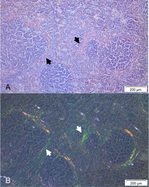
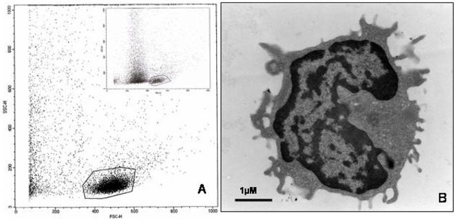
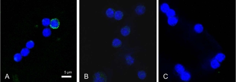
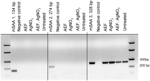
Spongiform encephalopathies have been reported to be transmitted by blood transfusion even prior to the clinical onset. Experimental AA-amyloidosis shows similarities with prion disease and amyloid-containing organ-extracts can prime a recipient for the disease. In this systemic form of amyloidosis N-terminal fragments of the acute-phase reactant apolipoprotein serum amyloid A are the main amyloid protein. Initial amyloid deposits appear in the perifollicular region of the spleen, followed by deposits in the liver. We used the established murine model and induced AA-amyloidosis in NMRI mice by intravenous injections of purified amyloid fibrils ('amyloid enhancing factor') combined with inflammatory challenge (silver nitrate subcutaneously). Blood plasma and peripheral blood monocytes were isolated, sonicated and re-injected into new recipients followed by an inflammatory challenge during a three week period. When the animals were sacrificed presence of amyloid was analyzed in spleen sections after Congo red staining. Our result shows that some of the peripheral blood monocytes, isolated from animals with detectable amyloid, contained amyloid-seed that primed for AA-amyloid. The seeding material seems to have been phagocytosed by the cells since the AA-precursor (SAA1) was found not be expressed by the monocytes. Plasma recovered from mice with AA amyloidosis lacked seeding capacity. Amyloid enhancing activity can reside in monocytes recovered from mice with AA-amyloidosis and in a prion-like way trigger amyloid formation in conjunction with an inflammatory disorder. Human AA-amyloidosis resembles the murine form and every individual is expected to be exposed to conditions that initiate production of the acute-phase reactant. The monocyte-transfer mechanism should be eligible for the human disease and we point out blood transfusion as a putative route for transfer of amyloidosis.
Conflict of interest statement
Figures
Similar articles
-
Cellular events associated with the initial phase of AA amyloidogenesis: insights from a human monocyte model.Amyloid. 2007 Mar;14(1):51-63. doi: 10.1080/13506120601116575. Amyloid. 2007. PMID: 17453625
-
Transmission of circulating cell-free AA amyloid oligomers in exosomes vectors via a prion-like mechanism.Biochem Biophys Res Commun. 2010 Oct 1;400(4):559-62. doi: 10.1016/j.bbrc.2010.08.101. Epub 2010 Aug 31. Biochem Biophys Res Commun. 2010. PMID: 20807507
-
Anti-interleukin 6 receptor antibody inhibits murine AA-amyloidosis.J Rheumatol. 2004 Jun;31(6):1132-8. J Rheumatol. 2004. PMID: 15170926
-
Review. Reflections on amyloidosis in Papua New Guinea.Philos Trans R Soc Lond B Biol Sci. 2008 Nov 27;363(1510):3701-5. doi: 10.1098/rstb.2008.0073. Philos Trans R Soc Lond B Biol Sci. 2008. PMID: 18849285 Free PMC article. Review.
-
Serum amyloid A and protein AA: molecular mechanisms of a transmissible amyloidosis.FEBS Lett. 2009 Aug 20;583(16):2685-90. doi: 10.1016/j.febslet.2009.04.026. Epub 2009 Apr 23. FEBS Lett. 2009. PMID: 19393650 Review.
Cited by
-
Peripheral administration of tau aggregates triggers intracerebral tauopathy in transgenic mice.Acta Neuropathol. 2014 Feb;127(2):299-301. doi: 10.1007/s00401-013-1231-5. Epub 2013 Dec 21. Acta Neuropathol. 2014. PMID: 24362441 Free PMC article. No abstract available.
-
The ASC inflammasome adapter governs SAA-derived protein aggregation in inflammatory amyloidosis.EMBO Mol Med. 2024 Sep;16(9):2024-2042. doi: 10.1038/s44321-024-00107-0. Epub 2024 Jul 30. EMBO Mol Med. 2024. PMID: 39080493 Free PMC article.
-
Prion-like disorders: blurring the divide between transmissibility and infectivity.J Cell Sci. 2010 Apr 15;123(Pt 8):1191-201. doi: 10.1242/jcs.051672. J Cell Sci. 2010. PMID: 20356930 Free PMC article.
-
Protein aggregate spreading in neurodegenerative diseases: problems and perspectives.Neurosci Res. 2011 Aug;70(4):339-48. doi: 10.1016/j.neures.2011.05.008. Epub 2011 May 20. Neurosci Res. 2011. PMID: 21624403 Free PMC article. Review.
-
A new era for understanding amyloid structures and disease.Nat Rev Mol Cell Biol. 2018 Dec;19(12):755-773. doi: 10.1038/s41580-018-0060-8. Nat Rev Mol Cell Biol. 2018. PMID: 30237470 Free PMC article. Review.
References
-
- Westermark P, Benson MD, Buxbaum JN, Cohen AS, Frangione B, et al. A primer of amyloid nomenclature. Amyloid. 2007;14:179–183. - PubMed
-
- Thorn CF, Lu ZY, Whitehead AS. Regulation of the human acute phase serum amyloid A genes by tumour necrosis factor-alpha, interleukin-6 and glucocorticoids in hepatic and epithelial cell lines. Scand J Immunol. 2004;59:152–158. - PubMed
-
- Steel DM, Whitehead AS. The major acute phase reactants: C-reactive protein, serum amyloid P component and serum amyloid A protein. Immunol Today. 1994;15:81–88. - PubMed
-
- Ein D, Kimura S, Glenner GG. An amyloid fibril protein of unknown origin: partial amino-acid sequence analysis. Biochem Biophys Res Commun. 1972;46:498–500. - PubMed
-
- Sletten K, Husby G. The complete amino-acid sequence of non-immunoglobulin amyloid fibril protein AS in rheumatoid arthritis. Eur J Biochem. 1974;41:117–125. - PubMed
Publication types
MeSH terms
Substances
LinkOut - more resources
Full Text Sources
Medical
Miscellaneous
