. 2008 Nov;118(11):3609-18.
doi: 10.1172/JCI36245. Epub 2008 Oct 1.

Deletion of GSK-3beta in mice leads to hypertrophic cardiomyopathy secondary to cardiomyoblast hyperproliferation

Affiliations

Deletion of GSK-3beta in mice leads to hypertrophic cardiomyopathy secondary to cardiomyoblast hyperproliferation

Risto Kerkela et al. J Clin Invest. 2008 Nov.

Abstract

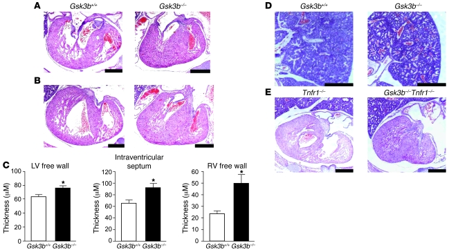
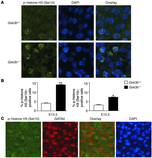
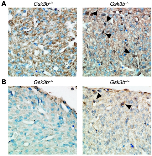
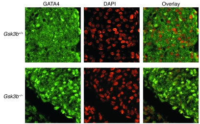
Based on extensive preclinical data, glycogen synthase kinase-3 (GSK-3) has been proposed to be a viable drug target for a wide variety of disease states, ranging from diabetes to bipolar disorder. Since these new drugs, which will be more powerful GSK-3 inhibitors than lithium, may potentially be given to women of childbearing potential, and since it has controversially been suggested that lithium therapy might be linked to congenital cardiac defects, we asked whether GSK-3 family members are required for normal heart development in mice. We report that terminal cardiomyocyte differentiation was substantially blunted in Gsk3b(-/-) embryoid bodies. While GSK-3alpha-deficient mice were born without a cardiac phenotype, no live-born Gsk3b(-/-) pups were recovered. The Gsk3b(-/-) embryos had a double outlet RV, ventricular septal defects, and hypertrophic myopathy, with near obliteration of the ventricular cavities. The hypertrophic myopathy was caused by cardiomyocyte hyperproliferation without hypertrophy and was associated with increased expression and nuclear localization of three regulators of proliferation - GATA4, cyclin D1, and c-Myc. These studies, which we believe are the first in mammals to examine the role of GSK-3alpha and GSK-3beta in the heart using loss-of-function approaches, implicate GSK-3beta as a central regulator of embryonic cardiomyocyte proliferation and differentiation, as well as of outflow tract development. Although controversy over the teratogenic effects of lithium remains, our studies suggest that caution should be exercised in the use of newer, more potent drugs targeting GSK-3 in women of childbearing age.


Figures

Figure 1
Figure 1. Histological analysis of heart development in GSK-3β–deficient mice.
Sequential rostral-to-caudal sections from an E15.5 Gsk3b–/– embryo from the pulmonary valve (top left) to the ventricles (bottom right) (original magnification, ×2.0; scale bar: 0.5 cm). The pulmonary valve and the pulmonary artery (P) are positioned over the RV (AC). Below the pulmonary valve, the aorta (A) descends toward the aortic valve (DF); the aortic valve is positioned over the RV (G). Caudal to the semilunar valves, the RV and the LV are connected by a VSD (HK). The location of the VSD is in the membranous portion of the interventricular septum.
Figure 2
Figure 2. Analysis of ventricular development.
H&E-stained sections of Gsk3b+/+ (left) and Gsk3b–/– (right) hearts (original magnification, ×2.5; scale bars: 0.1 mm) including the mitral valve leaflet (A) and the tricuspid valve leaflet (B) (original magnification, ×2.5; scale bars: 0.5 mm). The atrioventricular valve leaflets of Gsk3b–/– embryos showed cellularity, chordal attachment, and dimension comparable to those of control embryos. By comparison, the LV wall thickness was greatly increased in the Gsk3b–/– embryo hearts. (C) Quantification of thickness of the LV free wall, interventricular septum, and RV free wall in Gsk3b+/+ (n = 4) and Gsk3b–/– (n = 6) hearts. Error bars indicate mean ± SEM. *P < 0.05 versus WT. (D) The pulmonary vasculature and parenchyma showed congestion of blood in Gsk3b–/– (right) compared with Gsk3b+/+ (left) embryos (original magnification, ×1.6; scale bars: 0.5 mm). (E) TNFR1 deficiency does not rescue the cardiac phenotype of Gsk3b–/– embryo hearts. Sections of Tnfr1–/– (left) and Gsk3b–/–Tnfr1–/– (right) embryos, including the tricuspid valve leaflet (original magnification, ×1.6; scale bars: 0.5 mm).
Figure 3
Figure 3. Increased cardiomyocyte proliferation in GSK-3β–deficient hearts.
(A) Immunofluorescence staining for nuclear phospho–histone H3 (Ser10) (left panels) in GSK-3β–deficient (bottom) compared with WT (top) animals. Merging of the histone and the DAPI (middle) stains confirmed nuclear localization of the phospho–histone H3 (Ser10) staining (right panels). Original magnification, ×40. (B) Composite mean ± SEM percentage of phospho–histone H3 (Ser10)–staining cells at E13.5 and E15.5 in the myocardium of WT (white bars) and GSK-3β–deficient (back bars) animals.*P < 0.05, **P < 0.001 versus WT. (C) Coimmunostaining of heart sections with phospho–histone H3 and GATA4, a cardiomyocyte marker, demonstrating colocalization in the overlay. Thus, the phospho–histone H3 (Ser10)–positive cells in the myocardium are overwhelmingly GATA4 positive, consistent with cardiomyocytes. Also shown is nuclear staining with DAPI. Original magnification, ×40.
Figure 4
Figure 4. Altered transcription factor and cell cycle regulator expression and localization in GSK-3β–deficient hearts.
(A) Increased nuclear cyclin D1 in hearts of E15.5 GSK-3β–deficient embryos. Sections were stained with anti–cyclin D1 antibody (brown) and then counterstained with hematoxylin to identify nuclei (blue). Note the multiple dark brown nuclei (arrowheads) and the overall increase in cyclin D1 stain and corresponding decrease in intensity of the hematoxylin stain in the GSK-3β–deficient hearts. Original magnification, ×40. (B) c-Myc expression is increased in GSK-3β–deficient hearts. Sections from E15.5 embryos were stained with anti–c-Myc antibody (brown) and then counterstained with hematoxylin as described above. Note the enhanced brown staining of the nuclei (arrowheads) and the reduction in intensity of the hematoxylin stain, consistent with increases in nuclear c-Myc expression in the GSK-3β–deficient heart. Original magnification, ×40.
Figure 5
Figure 5. Increased nuclear GATA4 expression in GSK-3β–deficient hearts.
GATA4 staining (left) was located over nuclei in the hearts of E13.5 embryos stained by DAPI (center) in merged images (right). Original magnification, ×40.
Figure 6
Figure 6. GATA4 overexpression drives cardiomyocyte proliferation.
(A) GSK-3 inhibition increases neonatal rat ventricular myocyte (NRVM) proliferation. Shown is the mean ± SEM percentage of phospho–histone H3 (Ser10)–staining cells 20 hours following LiCl and 6-bromoindirubin-3ι-oxime (BIO) treatment at the indicated concentrations. (B) GATA4 overexpression induces NRVM proliferation. Immunoblot of lysates from NRVMs infected with adenovirus encoding LacZ (left 2 lanes) or GATA4 (right 2 lanes). NRVMs were treated with 100 viral particles per cell (first and third lanes) or 200 particles per cell (second and fourth lanes). NRVMs plated onto glass coverslips were stained for phospho–histone H3 (Ser10) 48 hours after infection of cells with adenoviruses (200 viral particles per cell) encoding GATA4 (Ad-GATA4) or LacZ (Ad-LacZ). Shown is the mean ± SEM percentage of phospho–histone H3 (Ser10)–staining cells following Ad-LacZ or Ad-GATA4 infection. *P < 0.05, **P < 0.01, ***P < 0.001 versus vehicle control (Ctrl); P < 0.001 versus Ad-LacZ.

References

    1. Gassmann M., et al. Aberrant neural and cardiac development in mice lacking the ErbB4 neuregulin receptor. Nature. 1995;378:390–394. doi: 10.1038/378390a0. - DOI - PubMed
    1. Lee K.F., et al. Requirement for neuregulin receptor erbB2 in neural and cardiac development. Nature. 1995;378:394–398. doi: 10.1038/378394a0. - DOI - PubMed
    1. Meyer D., Birchmeier C. Multiple essential functions of neuregulin in development. Nature. 1995;378:386–390. doi: 10.1038/378386a0. - DOI - PubMed
    1. Hertig C.M., Kubalak S.W., Wang Y., Chien K.R. Synergistic roles of neuregulin-1 and insulin-like growth factor-I in activation of the phosphatidylinositol 3-kinase pathway and cardiac chamber morphogenesis. J. Biol. Chem. 1999;274:37362–37369. doi: 10.1074/jbc.274.52.37362. - DOI - PubMed
    1. Lavine K.J., et al. Endocardial and epicardial derived FGF signals regulate myocardial proliferation and differentiation in vivo. Dev. Cell. 2005;8:85–95. doi: 10.1016/j.devcel.2004.12.002. - DOI - PubMed

Publication types

Substances