Preoperative cervical lymph node size evaluation in patients with malignant head/neck tumors: comparison between ultrasound and computer tomography
- PMID: 18830710
- PMCID: PMC12160172
- DOI: 10.1007/s00432-008-0487-y
Preoperative cervical lymph node size evaluation in patients with malignant head/neck tumors: comparison between ultrasound and computer tomography
Abstract
Purpose: The spread of malignant lymph nodes due to malignancies of the head and neck is systematically observed. However, sentinel lymph nodes in the cervical region, such as in the axillary or supraclavicular regions, are not described. Therefore, precise preoperative lymph node screening of all neck compartments is required.
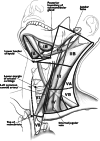
Materials and methods: Forty-five patients with a primary malignant tumor in the head and neck area underwent lymph node staging of the head by means of both CT and ultrasound as a preoperative evaluation. The lymph nodes were classified on the origin of the level system proposed by Som et al. (174:837-844, 2000), which is based on the recommendation of the American College of Radiology introduced in 1990. According to the manual measurement of World Health Organization and the Revised Response Evaluation Criteria in Solid Tumors, the longest transversal and longitudinal diameters were measured by ultrasound, while only the two longest transversal diameters were recorded by CT. The study was conducted by two independent observers. These results were compared with the histopathological results as references.
Results: Six hundred and twenty-four lymph nodes were detected, 64 of which were malignant. Most of the transformed lymph nodes were found in level IIa, II b and III. A more precise measurement was given using ultrasound. The correct positive rate of sonographically detected malignant lymph nodes was significantly higher compared to the CT reading.
Conclusion: Cervical lymph node staging can be performed safely by ultrasound. It is a cheap, easy-to-handle and cost-effective diagnostic method. However, only the uppermost regions of the neck are accessible with a linear transducer. Despite this restriction, ultrasound is a reliable and valuable tool for screening lymph nodes in the case of a head or neck malignancy.
Figures


Similar articles
-
Positron emission tomography (PET) and magnetic resonance imaging (MRI) for the assessment of axillary lymph node metastases in early breast cancer: systematic review and economic evaluation.Health Technol Assess. 2011 Jan;15(4):iii-iv, 1-134. doi: 10.3310/hta15040. Health Technol Assess. 2011. PMID: 21276372 Free PMC article.
-
Diagnostic accuracy of endoscopic ultrasonography (EUS) for the preoperative locoregional staging of primary gastric cancer.Cochrane Database Syst Rev. 2015 Feb 6;2015(2):CD009944. doi: 10.1002/14651858.CD009944.pub2. Cochrane Database Syst Rev. 2015. PMID: 25914908 Free PMC article.
-
Comparison of pre-operative ultrasonic depth of invasion (DOI) with histopathological depth of invasion (DOI) in gingivobuccal sulcus squamous cell carcinoma with neck nodal metastasis.Eur Arch Otorhinolaryngol. 2025 Jul;282(7):3715-3721. doi: 10.1007/s00405-025-09326-8. Epub 2025 Mar 22. Eur Arch Otorhinolaryngol. 2025. PMID: 40119905
-
Development of a preoperative nomogram to identify low-risk early-stage breast cancer patients eligible for SLNB omission.World J Surg Oncol. 2025 Jul 7;23(1):268. doi: 10.1186/s12957-025-03921-z. World J Surg Oncol. 2025. PMID: 40624650 Free PMC article.
-
Magnetic resonance imaging and ultrasound examination in preoperative pelvic staging of early-stage cervical cancer: post-hoc analysis of SENTIX study.Ultrasound Obstet Gynecol. 2025 Apr;65(4):495-502. doi: 10.1002/uog.29205. Epub 2025 Mar 25. Ultrasound Obstet Gynecol. 2025. PMID: 40130299 Free PMC article.
Cited by
-
Quantitative evaluation of vascularity within cervical lymph nodes using Doppler ultrasound in patients with oral cancer: relation to lymph node size.Dentomaxillofac Radiol. 2011 Oct;40(7):415-21. doi: 10.1259/dmfr/18694011. Dentomaxillofac Radiol. 2011. PMID: 21960398 Free PMC article.
-
Diagnostic accuracy of contrast-enhanced computed tomography in assessing cervical lymph node status in patients with oral squamous cell carcinoma.J Cancer Res Clin Oncol. 2023 Dec;149(19):17437-17450. doi: 10.1007/s00432-023-05470-y. Epub 2023 Oct 25. J Cancer Res Clin Oncol. 2023. PMID: 37875746 Free PMC article.
-
Evaluation of sentinel lymph node size and shape as a predictor of occult metastasis in patients with squamous cell carcinoma of the oral cavity.Eur Arch Otorhinolaryngol. 2013 Jan;270(1):249-54. doi: 10.1007/s00405-012-1959-x. Epub 2012 Feb 14. Eur Arch Otorhinolaryngol. 2013. PMID: 22331260
-
Clinical utility and prospective comparison of ultrasonography and computed tomography imaging in staging of neck metastases in head and neck squamous cell cancer in an Indian setup.Int J Clin Oncol. 2011 Dec;16(6):686-93. doi: 10.1007/s10147-011-0250-2. Epub 2011 Jun 16. Int J Clin Oncol. 2011. PMID: 21674359
-
Review of ultrasonography of malignant neck nodes: greyscale, Doppler, contrast enhancement and elastography.Cancer Imaging. 2014 Jan 6;13(4):658-69. doi: 10.1102/1470-7330.2013.0056. Cancer Imaging. 2014. PMID: 24434158 Free PMC article. Review.
References
-
- Ahn JE, Lee JH, Yi JS et al (2008) Diagnostic accuracy of CT and ultrasonography for evaluating metastatic cervical lymph nodes in patients with thyroid cancer. World J Surg 32:1552–1558. doi:10.1007/s00268-008-9588-7 - PubMed
-
- Akoglu E, Dutipek M, Bekis R et al (2005) Assessment of cervical lymph node metastasis with different imaging methods in patients with head and neck squamous cell carcinoma. J Otolaryngol 34:384–394. doi:10.2310/7070.2005.34605 - PubMed
-
- Bruneton JN, Balu-Maestro C, Marcy PY et al (1994) Very high frequency (13 MHz) ultrasonographic examination of the normal neck: detection of normal lymph nodes and thyroid nodules. J Ultrasound Med 13:87–90 - PubMed
-
- Close LG, Brown PM, Vuitch MF et al (1989) Microvascular invasion and survival in cancer of the oral cavity and oropharynx. Arch Otolaryngol Head Neck Surg 115:1304–1309 - PubMed
Publication types
MeSH terms
LinkOut - more resources
Full Text Sources
Medical

