. 2008 Oct 28;47(43):11273-84.
doi: 10.1021/bi800920j. Epub 2008 Oct 2.

Covalent binding of flavins to RnfG and RnfD in the Rnf complex from Vibrio cholerae

Affiliations

Covalent binding of flavins to RnfG and RnfD in the Rnf complex from Vibrio cholerae

Julianne Backiel et al. Biochemistry. .

Abstract

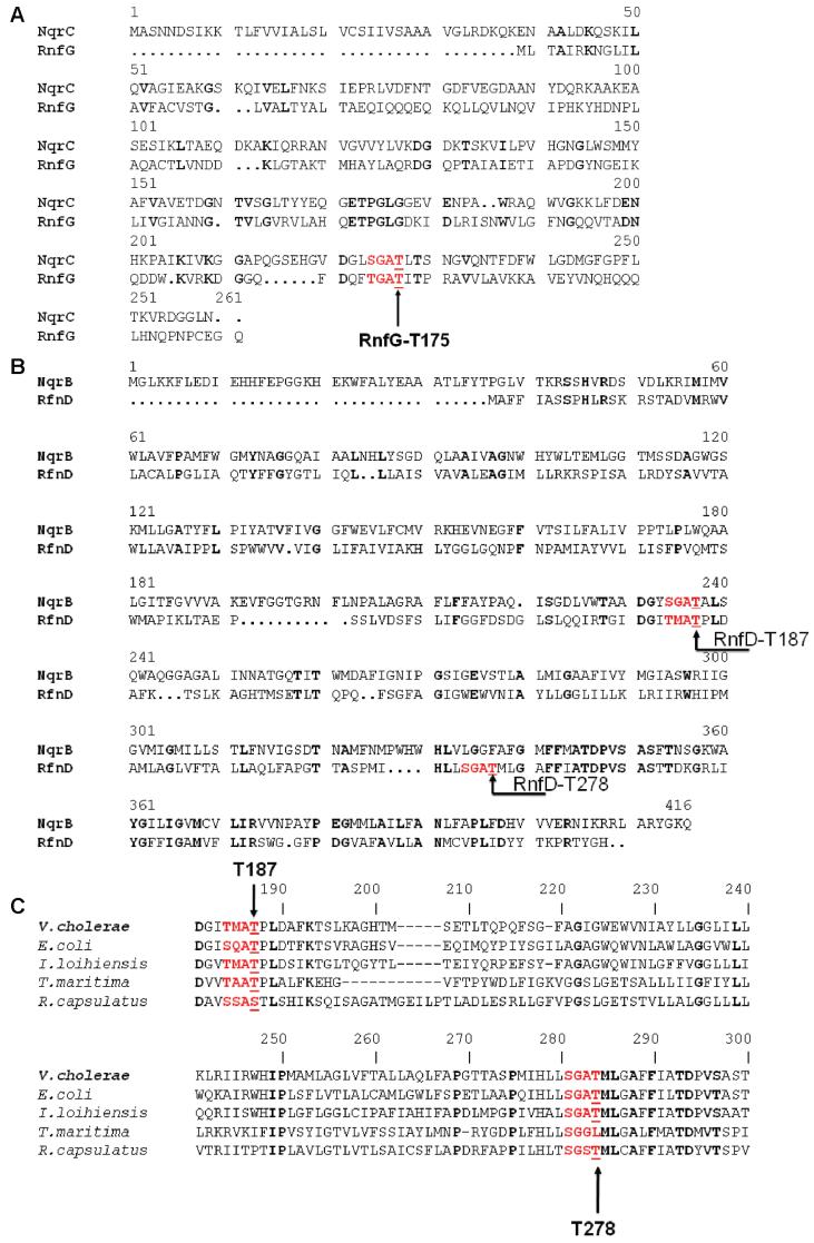
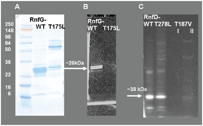
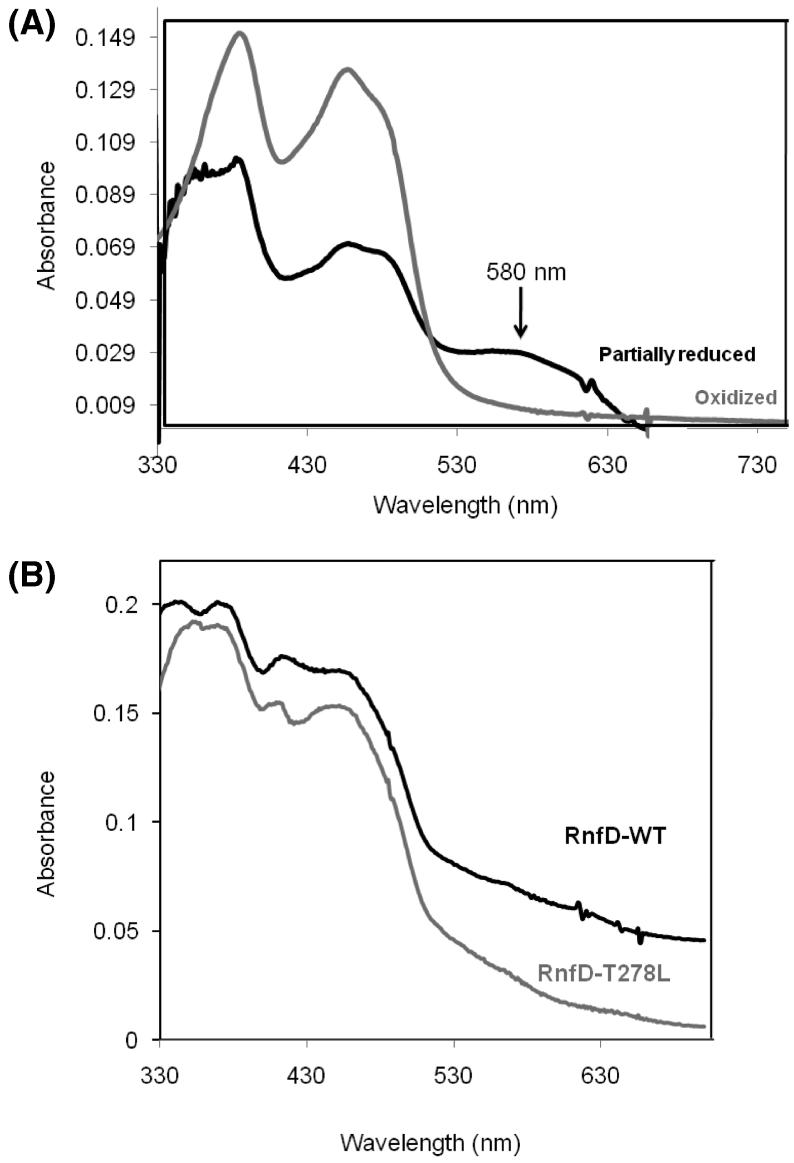
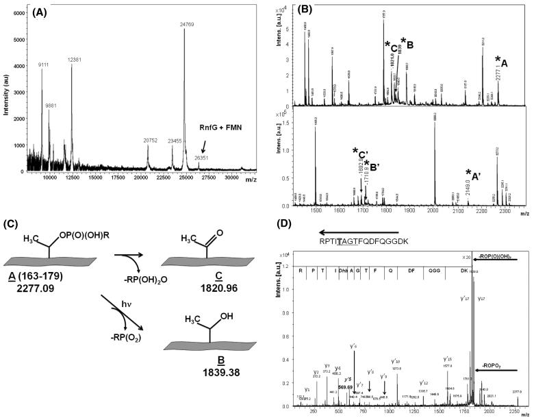
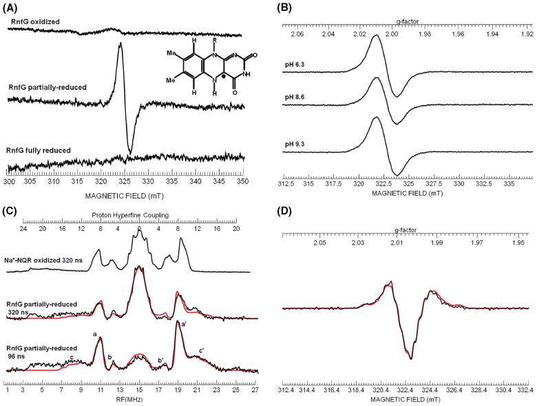
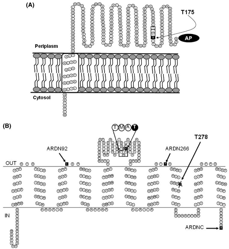
Enzymes of the Rnf family are believed to be bacterial redox-driven ion pumps, coupling an oxidoreduction process to the translocation of Na+ across the cell membrane. Here we show for the first time that Rnf is a flavoprotein, with FMN covalently bound to threonine-175 in RnfG and a second flavin bound to threonine-187 in RnfD. Rnf subunits D and G are homologous to subunits B and C of Na+-NQR, respectively. Each of these Na+-NQR subunits includes a conserved S(T)GAT motif, with FMN covalently bound to the final threonine. RnfD and RnfG both contain the same motif, suggesting that they bind flavins in a similar way. In order to investigate this, the genes for RnfD and RnfG from Vibrio cholerae were cloned and expressed individually in that organism. In both cases the produced protein fluoresced under UV illumination on an SDS gel, further indicating the presence of flavin. However, analysis of the mutants RnfG-T175L, RnfD-T278L, and RnfD-T187V showed that RnfG-T175 and RnfD-T187 are the likely flavin ligands. This indicates that, in the case of RnfD, the flavin is bound, not to the SGAT sequence but to the final residues of a TMAT sequence, a novel variant of the flavin binding motif. In the case of RnfG, flavin analysis, followed by MALDI-TOF-TOF mass spectrometry, showed that an FMN is covalently attached to threonine-175, the final threonine of the S(T)GAT sequence. Studies by visible, EPR, and ENDOR spectroscopy showed that, upon partial reduction, the isolated RnfG produces a neutral semiquinone intermediate. The semiquinone species disappeared upon full reduction and was not observed in the denatured protein. A topological analysis combining reporter protein fusion and computer predictions indicated that the flavins in RnfG and RnfD are localized in the periplasmic space. In contrast, in NqrC and NqrB the flavins are located in a cytoplasmic loop. This topological analysis suggests that there may be mechanistic differences between the Rnf and Na+-NQR complexes.


Figures

Figure 1
Figure 1
(A) Alignment between RnfG and NqrC from V. cholerae showing the conserved FMN binding motif, S(T)GAT. (B) Alignment between RnfD and NqrB from V. cholerae showing the two possible flavin binding sequences: the SGAT sequence containing Thr-278 and the TMAT sequence containing Thr-187. (C) Alignment between RnfD's from different sources showing that the two possible flavin binding sequences are both conserved.
Figure 2
Figure 2
SDS-PAGE comparing the wild-type RnfG to the RnfGT175L mutant and wild-type RnfD to the RnfD-T278L mutant. Panel A: RfnG and RnfG-T175L, gel stained with Coomassie. Panel B: The same gel under UV illumination prior to staining, showing flavin fluorescence. Panel C: RfnD, RnfD-T278L, and RnfD-T187V (I, after Ni-NTA; II, after gel filtration), gel under UV illumination prior to staining, showing flavin fluorescence. Approximately 20 μg of protein was loaded in each lane.
Figure 3
Figure 3
(A) Visible spectra of oxidized and partially reduced RnfG. Approximately 30 μg of protein was used. Gray spectrum: oxidized (as isolated) RnfG. Black spectrum: partially reduced RnfG. The partially reduced spectrum was acquired under anaerobic conditions using dithionite as reductant. (B) Visible spectrum of oxidized RnfD wild type (black line) and the RnfD-T278L mutant (gray line). Approximately 30 μg of protein was used.
Figure 4
Figure 4
(A) MALDI mass spectrum of RnfG. The peak corresponding to RnfG with the covalently attached FMN is shown. (B) MALDI mass spectra of two fluorescent FMN-containing peptide fractions in m/z range 1200-2400. Peaks corresponding to modified threonine are marked as A, A′, B, B′, C, and C′. (C) Scheme illustrating the two mechanisms of phosphoester bond breaking leading to the results shown in (B). (D) MS/MS (postsource decay) mass spectra of the MS peak at m/z 2277, showing the protein sequence in the vicinity of T175 including the TGAT flavin binding motif. The dephosphorylated state of Thr-175 is evident.
Figure 5
Figure 5
(A) X-band EPR spectra of oxidized, partially reduced, and fully reduced RnfG. Inset: Structure of the neutral flavin radical showing the location of the unpaired electron spin on the isoalloxazine ring. (B) X-band EPR spectra of partially reduced RnfG samples at different pH values. (C) ENDOR spectra of the partially reduced RnfG at π pulse lengths of 320 and 96 ns. The spectrum of the neutral flavin semiquinone radical of Na+-NQR for a π pulse length of 320 ns is shown for comparison. The peaks marked a and a′ are due to the C(8α) methyl protons; peaks marked b and b′ are due to the C(6) proton, and peaks marked c and c′ are due to the C(1′) β-methylene protons. The simulation of the partially reduced RnfG is presented in red overlaying the actual spectrum. (D) Second derivative X-band EPR spectrum of partially reduced RnfG. Simulated spectrum is shown in red.
Figure 6
Figure 6
(A) Schematic showing membrane topology of RnfG. The figure highlights the FMN binding motif, TGAT, where last threonine of the motif, Thr-175, is the ligand of the flavin. (B) Schematic showing the most probable topology of RnfD. The figure highlights the new motif for binding of flavin, TMAT, where Thr-187 is the ligand. The alkaline phosphatase fusion sites are labeled as follows: ARDN92, alkaline phosphatase fused to the C-terminus of a subset of RnfD from the N-terminus to residue 92; ARDN266, alkaline phosphatase fused to the C-terminus of a subset of RnfD from the N-terminus to residue 266; ARDNC, alkaline phosphatase fused to the C-terminus of the complete RnfD. Thr-278 is part of the conserved SGAT motif labeled in the figure.

References

    1. Schmehl M, Jahn A, Meyer zu Vilsendorf A, Hennecke S, Masepohl B, Schuppler M, Marxer M, Oelze J, Klipp W. Identification of a new class of nitrogen fixation genes in Rhodobacter capsulatus: a putative membrane complex involved in electron transport to nitrogenase. Mol. Gen. Genet. 1993;241:602–615. - PubMed
    1. Jouanneau Y, Jeong HS, Hugo N, Meyer C, Willison JC. Overexpression in Escherichia coli of the rnf genes from Rhodobacter capsulatus-characterization of two membrane-bound iron-sulfur proteins. Eur. J. Biochem. 1998;251:54–64. - PubMed
    1. Kumagai H, Fujiwara T, Matsubara H, Saeki K. Membrane localization, topology, and mutual stabilization of the rnfABC gene products in Rhodobacter capsulatus and implications for a new family of energy-coupling NADH oxidoreductases. Biochemistry. 1997;36:5509–5521. - PubMed
    1. Jeong HS, Jouanneau Y. Enhanced nitrogenase activity in strains of Rhodobacter capsulatus that overexpress the rnf genes. J. Bacteriol. 2000;182:1208–1214. - PMC - PubMed
    1. Duran-Pinedo AENK, Duncan MJ. The RprY response regulator of Porphyromonas gingivalis. Mol. Microbiol. 2007;64:1061–1074. - PubMed

Publication types

MeSH terms

LinkOut - more resources