. 2008 Oct 29;130(43):14207-16.
doi: 10.1021/ja8035916. Epub 2008 Oct 3.

Role of the Zn1 and Zn2 sites in metallo-beta-lactamase L1

Affiliations

Role of the Zn1 and Zn2 sites in metallo-beta-lactamase L1

Zhenxin Hu et al. J Am Chem Soc. .

Abstract

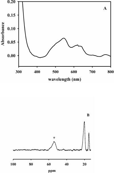
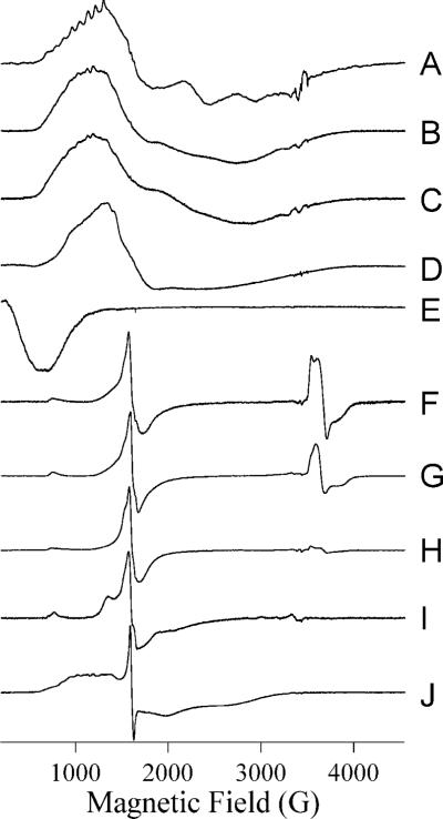
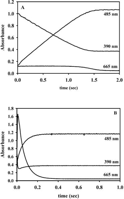
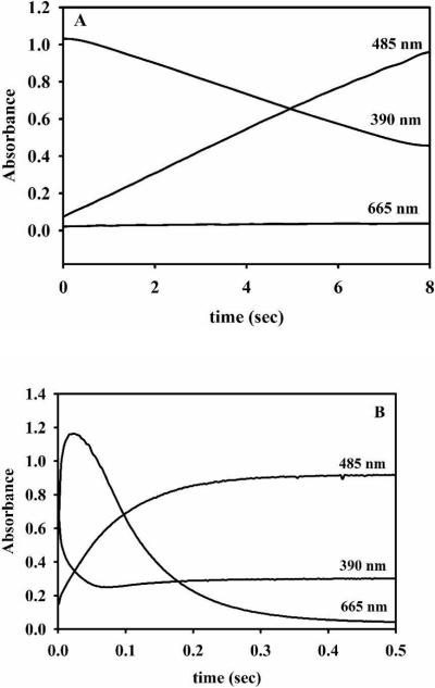
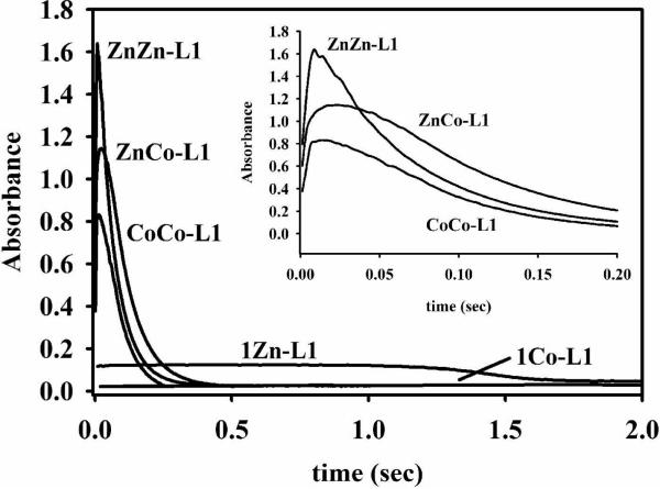
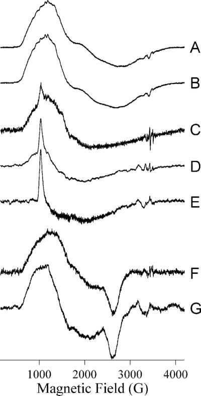
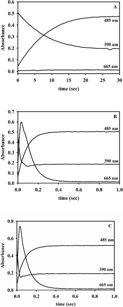
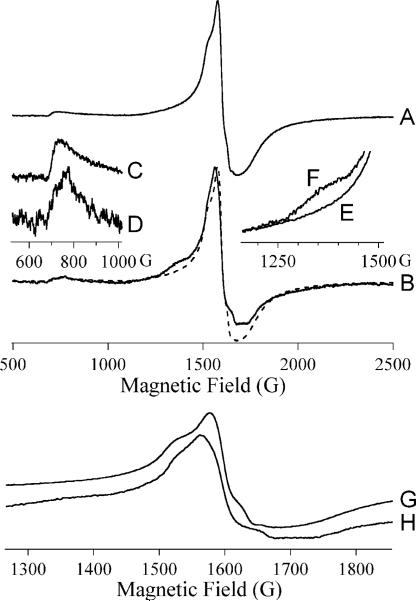
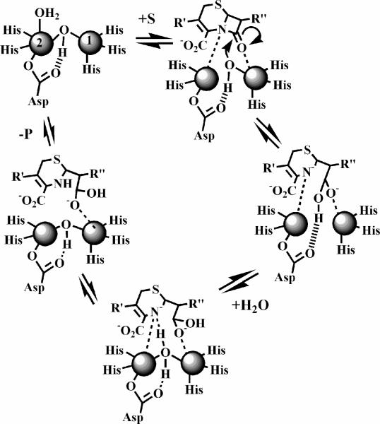
In an effort to probe the role of the Zn(II) sites in metallo-beta-lactamase L1, mononuclear metal ion containing and heterobimetallic analogues of the enzyme were generated and characterized using kinetic and spectroscopic studies. Mononuclear Zn(II)-containing L1, which binds Zn(II) in the consensus Zn1 site, was shown to be slightly active; however, this enzyme did not stabilize a nitrocefin-derived reaction intermediate that had been previously detected. Mononuclear Co(II)- and Fe(III)-containing L1 were essentially inactive, and NMR and EPR studies suggest that these metal ions bind to the consensus Zn2 site in L1. Heterobimetallic analogues (ZnCo and ZnFe) analogues of L1 were generated, and stopped-flow kinetic studies revealed that these enzymes rapidly hydrolyze nitrocefin and that there are large amounts of the reaction intermediate formed during the reaction. The heterobimetallic analogues were reacted with nitrocefin, and the reactions were rapidly freeze quenched. EPR studies on these samples demonstrate that Co(II) is 5-coordinate in the resting state, proceeds through a 4-coordinate species during the reaction, and is 5-coordinate in the enzyme-product complex. These studies demonstrate that the metal ion in the Zn1 site is essential for catalysis in L1 and that the metal ion in the Zn2 site is crucial for stabilization of the nitrocefin-derived reaction intermediate.


Figures

Figure 1
Figure 1. UV-Vis and NMR spectra of 1Co-L1
A: UV-Vis difference spectrum of 1Co-L1 prepared using biological incorporation method. The enzyme concentration was 550 μM, and the buffer was 50 mM Hepes, pH 7.0. B: 1H NMR spectrum of 550 μM 1Co-L1. The asterisk signifies the peak that is solvent-exchangeable.
Figure 2
Figure 2. EPR spectra from metal-containing species of L1
Spectra are of the following species of L1: (A) L1 containing 0.8 eq Co(II) at 12 K, 25 mW; (B) 1Co-L1 at 12 K, 2 mW; (C) ZnCo-L1 at 10 K, 2 mW; (D) CoCo-L1 at 12 K, 10 mW; (E) CoCo-L1 at 7 K, 20 mW, B0B1; (F) 1Fe-L1 at 10 K, 2 mW; (G) ZnFe-L1 at 12 K, 10 mW; (H) FeFe-L1 at 10 K, 2 mW; (I) FeNi-L1 at 7 K, 50 mW; (J) FeCo-L1 at 10 K, 2 mW. Spectra are shown with arbitrary intensities.
Figure 3
Figure 3. Stopped-flow traces of reaction of Zn(II)-containing L1 analogs and nitrocefin
Stopped-flow traces of 50 μM 1Zn- (A) and ZnZn-L1 (B) analogs when reacted with 50 μM nitrocefin at 4 °C. The absorbance at 485 nm is due to the product, the absorbance at 390 nm is due to the substrate, and the absorbance at 665 nm is due to the intermediate .
Figure 4
Figure 4. Stopped-flow traces of the reaction of Co(II)-containing L1 analogs with nitrocefin
50 μM 1Co- (A) and ZnCo-L1 (B) analogs were reacted with 50 μM nitrocefin at 4 °C. The absorbance at 485 nm is due to the product, the absorbance at 390 nm is due to the substrate, and the absorbance at 665 nm is due to the intermediate.
Figure 5
Figure 5. Intermediate formation by L1 analogs
The absorbance at 665 nm arises from the presence of intermediate. Each reaction contained 50 μM L1 analog and 50 μM nitrocefin at 4 °C in 50 mM cacodylate, pH 7.0. Inset: Intermediate formation for ZnZn-, ZnCo-, and CoCo-L1 analogs over 200 ms.
Figure 6
Figure 6. RFQ-EPR of ZnCo-L1 with nitrocefin
Spectra (A) and (B) are from resting ZnCo-L1. Spectra (C – E) are from ZnCo-L1 after reaction with nitrocefin for 10 ms at 3 °C. Spectra (F) and (G) are from ZnCo-L1 after incubation with nitrocefin for 2 min (at which time all of the added nitrocefin has been hydrolyzed). Spectra (A), (C) and (F) were recorded at 10 K, 2 mW, spectra (B), (D) and (G) at 7 K, 80 mW, and spectrum (E) at 5 K, 126 mW. Spectra are shown with arbitrary intensities.
Figure 7
Figure 7. Stopped-flow traces of Fe-containing L1 analogs reacted with nitrocefin
50 μM 1Fe- (A), ZnFe-L1 (made by adding Fe(II) to 1Zn-L1) (B), and ZnFe-L1 (made by adding Zn(II) to 1Fe-L1) (C) were reacted with 50 μM nitrocefin at 4 °C. The absorbance at 485 nm is due to the product, the absorbance at 390 nm is due to the substrate, and the absorbance at 665 nm is due to the intermediate.
Figure 8
Figure 8. RFQ-EPR of ZnFe-L1 with nitrocefin
Trace A shows the 500 − 2500 G region of the spectrum of resting ZnFe-L1. Trace B shows the 500 − 2500 G region of the spectrum of ZnFe-L1 upon reaction with nitrocefin for 10 ms at 3 °C (solid) overlaid with that of resting ZnFe-L1 (dashed). The inserts show more detailed comparisons between the spectra over particular field ranges; traces (C), (F) and (G) are from resting ZnFe-L1 and traces (D), (E) and (H) are from ZnFe-L1 after reaction with nitrocefin for 10 ms at 4 °C.
Figure 9
Figure 9
Proposed reaction mechanism of L1 for the hydrolysis of nitrocefin.

References

    1. Fisher JF, Meroueh SO, Mobashery S. Chem. Rev. 2005;105:395–424. - PubMed
    1. Perez F, Endimiani A, Hujer KM, Bonomo RA. Curr. Opin. Pharmacol. 2007;7(5):459–469. - PMC - PubMed
    1. Walsh TR, Toleman MA, Poirel L, Nordmann P. Clin. Microbiol. Rev. 2005;18(2):306–325. - PMC - PubMed
    1. Crowder MW, Spencer J, Vila AJ. Acc. Chem. Res. 2006;39(10):721–728. - PubMed
    1. Moran-Barrio J, Gonzalez JM, Lisa MN, Costello AL, Dal Peraro M, Carloni P, Bennett B, Tierney DL, Limansky AS, Viale AM, Vila AJ. J. Biol. Chem. 2007;282:18286–18293. - PubMed

Publication types