Review
. 2009 Feb;11(2):251-66.
doi: 10.1089/ars.2008.2117.

Gap junctions in the control of vascular function

Affiliations
Review

Gap junctions in the control of vascular function

Xavier F Figueroa et al. Antioxid Redox Signal. 2009 Feb.

Abstract

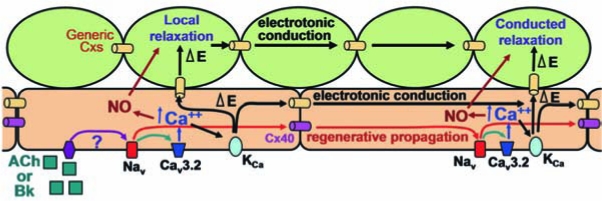
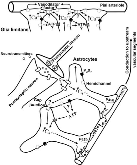
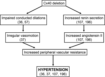
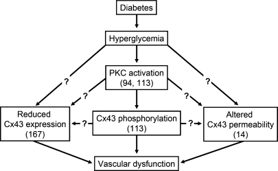
Direct intercellular communication via gap junctions is critical in the control and coordination of vascular function. In the cardiovascular system, gap junctions are made up of one or more of four connexin proteins: Cx37, Cx40, Cx43, and Cx45. The expression of more than one gap-junction protein in the vasculature is not redundant. Rather, vascular connexins work in concert, first during the development of the cardiovascular system, and then in integrating smooth muscle and endothelial cell function, and in coordinating cell function along the length of the vessel wall. In addition, connexin-based channels have emerged as an important signaling pathway in the astrocyte-mediated neurovascular coupling. Direct electrical communication between endothelial cells and vascular smooth muscle cells via gap junctions is thought to play a relevant role in the control of vasomotor tone, providing the signaling pathway known as endothelium-derived hyperpolarizing factor (EDHF). Consistent with the importance of gap junctions in the regulation of vasomotor tone and arterial blood pressure, the expression of connexins is altered in diseases associated with vascular complications. In this review, we discuss the participation of connexin-based channels in the control of vascular function in physiologic and pathologic conditions, with a special emphasis on hypertension and diabetes.


Figures

FIG. 1.
FIG. 1.
Gap-junction channels in the plasma membrane. The family of proteins known as connexins constitutes the structural and functional unit of gap-junctional intercellular communication. Oligomerization of six connexin proteins forms a connexon or hemichannel (49, 162). In response to stimuli such as phosphorylation or Ca2+, the associated six connexin subunits may coordinately change configuration to open the hemichannel and allow movement of paracrine signaling molecules such as ATP (70, 163). Hemichannels may diffuse laterally to the junctional membrane where they are in a position to dock with another hemichannel on the apposed membrane of the adjacent cell to complete a gap-junction channel (49, 162).
FIG. 2.
FIG. 2.
Control of vasomotor tone by the endothelium-derived hyperpolarizing factor. Extensions derived from either the smooth muscle cells or endothelial cells may penetrate the internal elastic lamina (IEL) to make close contact with the other cell type. These points of contact, known as myoendothelial junctions (MEJs), provide the structural organization to achieve direct heterocellular coupling between the two cell types via gap junctions (40, 55, 166), and thus, one basis for endothelium-mediated smooth muscle hyper-polarization (often referred to as endothelium-derived hyperpolarizing factor, EDHF). Endothelium-dependent vasodilators such as acetylcholine (ACh) induce an increase in endothelial cell intracellular Ca2+ concentration, which, in turn, activates the Ca2+-activated K+ channels (KCa) of small (SKCa) and intermediate conductance (IKCa). The endothelial cell hyperpolarization is transmitted electrotonically to the underlying smooth muscle cells via gap junctions located at the MEJ, contributing to the endothelium-dependent vasodilation (18, 44, 51). The question mark highlights that the participation of a diffusible EDHF has not been definitely discarded.
FIG. 3.
FIG. 3.
Possible sex differences in the regulation of the endothelium-derived hyperpolarizing factor (EDHF). Nitric oxide (NO) and EDHF are the major endothelium-derived vasodilator signals in resistance vessels. The involvement of these two vasodilator signals differs between male and female animals. NO prevails over EDHF in males, and the contrary is observed in females (130, 169). This gender difference may be explained by the estrogen modulation of the myoendothelial gap junction–mediated smooth muscle hyperpolarization. In female animals, estrogen upregulates the expression of caveolin-1 (Cav-1) (128, 143), a structural protein of caveolae that, in turn, inhibits the activity of eNOS (52, 65, 100). In addition, this hormone enhances the expression of Cx43 and Cx40 (128, 143), two gap-junction proteins found at the myoendothelial junctions (35, 97). As a result, the NO-dependent vasodilation is reduced, and the gap junction–mediated EDHF signaling is increased. The involvement of the estrogen receptor α (ERα) or β (ERβ) remains to be determined. In contrast, in male animals, the activation of ERβ reduces the gap junction–dependent smooth muscle hyperpolarization (130).
FIG. 4.
FIG. 4.
Conduction of vasodilator responses induced by the stimulation of cremasteric arterioles with a pulse of ACh. The microcirculation of the cremaster muscle was prepared as described previously (54, 57), and arterioles were stimulated focally with the ejection of ACh (10 μM) by a pressure-pulse from a micropipette (inner diameter, 3–4 μm). The changes in diameter were measured first at the stimulation site (local), and then, at locations 500, 1,000, and 2,000 μm upstream in four separate stimuli. Variations in diameter were expressed as percentage of the maximal dilation possible (% maximum). The duration of the pulse (300–700 ms) and the ejection pressure (10–20 psi) were set to induce a local vasodilation of ~100%, ~50%, or ~25%. Maximal diameter was estimated during superfusion of 1 mM adenosine. Note that the magnitude of the response decays only from the local site to the 500-μm conducted site and does not show a further reduction thereafter.
FIG. 5.
FIG. 5.
Representative tracings of the conduction of the vasomotor responses induced by focal electrical stimulation of cremasteric arterioles. The microcirculation of the cremaster muscle was prepared as described previously (54, 57). An Ag/AgCl reference electrode immersed in the superfusate was positioned symmetrically around the cremaster, and the arteriole was stimulated with a depolarizing train of pulses (30 Hz, 2 ms, 30 V) for 10 s by using a beveled micropipette (inner diameter, 3–4 μm) filled with 1 M NaCl. The stimulating pipette was inserted under the cremasteric mesothelium and positioned directly above the arteriole at a distance selected to evoke a local constriction of ~50% (54, 57). In separate stimuli, the changes in diameter were observed at the stimulation site (local), and at locations 500, 2,000, and 4,000 μm upstream. Variations in diameter were expressed as percentage of the maximal constriction or dilation possible (% maximum). Maximal diameter was estimated during superfusion of 1 mM adenosine. Focal electrical stimulation of the arteriole evoked a vasoconstriction that was restricted to a short vessel segment (~70–100 μm) at the stimulation site and, in addition, activated a rapid conducted vasodilation that spread along the length of the entire vessel without decay. Horizontal bars indicate the stimulation period.
FIG. 6.
FIG. 6.
Hypothetical model of conducted vasodilator responses activated by endothelium-dependent vasodilators [based on data from (36, 54, 57, 206)]. Stimulation of the endothelial cells with ACh or bradykinin triggers a regenerative, conducted vasodilator signal that is mediated by the activation of voltage-dependent Na+ channels (Nav) and rapidly propagated along the endothelium selectively via Cx40 gap junctions (red lines). The Nav-mediated conducted electrical signal is transduced into vasodilation by activation of the T-type, voltage-dependent Ca2+ channels Cav3.2 and the subsequent initiation of the production of Ca2+-sensitive vasodilator signals such as NO and Ca2+-activated K+ channel (KCa)-mediated smooth muscle hyperpolarization (black lines). The KCa-dependent hyperpolarization may be conducted electrotonically along the longitudinal axis of the vessel by either the smooth muscle cells or the endothelial cells via Cx40 or other connexins present in the vascular wall, which, in this schematic, we designated “generic” Cxs. This hypothetical model is compatible with the evidence showing that deletion of Cx40 completely eliminates the regenerative component of the conducted vasodilator response (36, 57, 206). The decremental conducted vasodilation that remains in the absence of Cx40 may correspond to the electrotonic conduction of the KCa-dependent hyperpolarizing signal. (For interpretation of the references to color in this figure legend, the reader is referred to the web version of this article at www.lieberonline.com/ars)
FIG. 7.
FIG. 7.
Connexin-based channels likely to be involved in neurovascular coupling. Neurotransmitters released on an increase in neuronal activity may exit the synaptic cleft and activate receptors on astrocytes (2, 218). These receptors cause an increase in astrocyte intracellular Ca2+ concentration that leads to the activation of large conductance Ca2+-activated K+ channels (BKCa) and/or cytochrome P450 epoxygenase (P450) at the astrocytic endfeet. The efflux of K+ through BKCa and/or production of epoxyeicosatrienoic acids (EETs) by P450 results in vasodilation of the parenchymal arterioles (2, 58, 59, 82, 187). The astrocyte-mediated vasodilator signal may be coordinated by the propagation of an interastrocyte Ca2+ signal via ATP release or directly by gap-junction communication. ATP may be released by either P2X7 receptors or unpaired hemichannels (70, 188). The potential role of interastrocyte Ca2+ waves in the coordination of the neurovascular coupling remains to be clearly defined. Local vasodilation of parenchymal cerebral arterioles must be communicated to upstream vascular segments to produce a functional increase of blood supply, and astrocytes may also communicate the vasodilator response to upstream vessels, such as the pial arterioles. These arterioles overlie a thick layer of astrocytic processes, called the glia limitans. The vasodilator signal triggered by neuronal activity reaches the glia limitans and induces a Cx43-based channel-dependent vasodilation (210). The mechanism by which Cx43 controls the astrocyte-mediated vasodilation has not been established, but coordination of the response between astrocytes, via gap-junction communication, or ATP release by hemichannels, is an interesting hypothesis that must be explored. An alternative hypothesis may be that a vasodilator factor is released at the endfeet via Cx43-based hemichannels.
FIG. 8.
FIG. 8.
Mechanisms of the control of arterial blood pressure by Cx40. Cx40 seems to play a central role in the control of arterial blood pressure by the parallel coordination of the vasomotor tone of resistance vessels and by renin secretion in the juxtaglomerular apparatus (JGA). Deletion of Cx40 affects the endothelial cell–dependent longitudinal synchronization of the vessel wall function, which results in an impaired conduction of vasodilator responses (36, 57), an irregular vasomotion and segmental constrictions (37). At the same time, the absence of Cx40 in the renin-secreting cells at the JGA disrupts the tonic inhibition of renin system, leading to an increase in renin secretion with the consequent increase in angiotensin II levels (107, 196). The dysregulation in the longitudinal communication of microvessels and renin secretion converges to increase the peripheral vascular resistance and produce the hypertension observed in Cx40-knockout animals (36, 37, 107, 196).
FIG. 9.
FIG. 9.
Diabetes affects the Cx43-mediated communication in the vascular wall. The hyperglycemia associated with diabetes leads to a reduction in the gap-junction intercellular communication in the endothelial cells and smooth muscle cells through the activation of PKC (94, 113). The reduction in gap-junction communication induced by hyperglycemia is associated with a decrease in Cx43 expression (167), Cx43 phosphorylation (113), and changes in the permeability properties of the Cx43-based channels (14). Although the phosphorylation of Cx43 is mediated by PKC, the participation of this protein kinase in the changes of Cx43 expression and permeability remains to be established. Cx43 is an important gap-junction protein in the vasculature, and the disruption of Cx43-mediated gap-junction communication induced by hyperglycemia may contribute to the vascular dysfunction typically observed in diabetes.

References

    1. Ahluwalia A. Hobbs AJ. Endothelium-derived C-type natriuretic peptide: more than just a hyperpolarizing factor. Trends Pharmacol Sci. 2005;26:162–167. - PubMed
    1. Anderson CM. Nedergaard M. Astrocyte-mediated control of cerebral microcirculation. Trends Neurosci. 2003;26:340–344. - PubMed
    1. Andresen J. Shafi NI. Bryan RM., Jr Endothelial influences on cerebrovascular tone. J Appl Physiol. 2006;100:318–327. - PubMed
    1. Archer SL. Gragasin FS. Wu X. Wang S. McMurtry S. Kim DH. Platonov M. Koshal A. Hashimoto K. Campbell WB. Falck JR. Michelakis ED. Endothelium-derived hyperpolarizing factor in human internal mammary artery is 11,12-epoxyeicosatrienoic acid and causes relaxation by activating smooth muscle BK(Ca) channels. Circulation. 2003;107:769–776. - PubMed
    1. Arensbak B. Mikkelsen HB. Gustafsson F. Christensen T. Holstein-Rathlou NH. Expression of connexin 37, 40, and 43 mRNA and protein in renal preglomerular arterioles. Histochem Cell Biol. 2001;115:479–487. - PubMed

Publication types

LinkOut - more resources