. 2008 Oct 2:2:323.
doi: 10.1186/1752-1947-2-323.

Bezoar in gastro-jejunostomy presenting with symptoms of gastric outlet obstruction: a case report and review of the literature

Affiliations

Bezoar in gastro-jejunostomy presenting with symptoms of gastric outlet obstruction: a case report and review of the literature

Edmund Leung et al. J Med Case Rep. .

Abstract

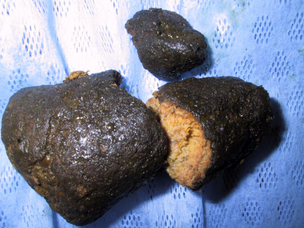
Introduction: Gastric outlet obstruction usually presents with non-bilious vomiting, colicky epigastric pain, loss of appetite and occasionally, upper gastrointestinal bleeding. Causes can be classified as benign or malignant, or as extra- or intraluminal. Gastrojejunostomy is a well-recognised surgical procedure performed to bypass gastric outlet obstruction. A bezoar occurs most commonly in patients with impaired gastrointestinal motility or with a history of gastric surgery. It is an intestinal concretion, which fails to pass along the alimentary canal.

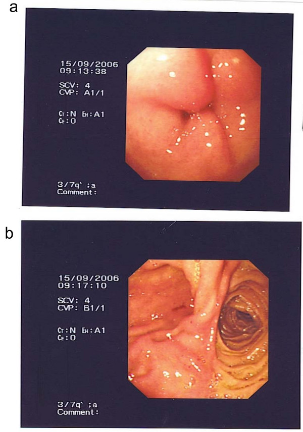
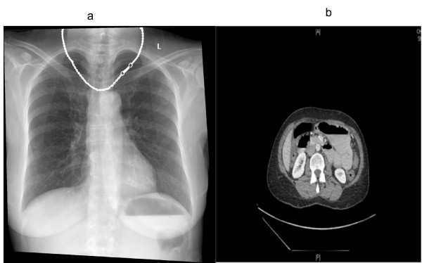
Case presentation: A 62-year-old Asian woman with a history of gastrojejunostomy for peptic ulcer disease was admitted to hospital with epigastric pain, vomiting and dehydration. All investigations concluded gastric outlet obstruction secondary to a "stricture" at the site of gastrojejunostomy. Subsequent laparotomy revealed that the cause of the obstruction was a bezoar.

Conclusion: Many bezoars can be removed endoscopically, but some will require operative intervention. Once removed, emphasis must be placed upon prevention of recurrence. Surgeons must learn to recognise and classify bezoars in order to provide the most effective therapy.


Figures

Figure 1
Figure 1
Image taken during upper endoscopy. a) Oedema present at the anastomotic site of the gastrojejunostomy. b) No evidence of obstruction beyond the anastomosis.
Figure 2
Figure 2
Imaging. a) Erect chest radiograph showing an air-fluid level within a dilated stomach. Lung fields were clear. There is no air under the diaphragm. b) Contrasted abdominal computed tomography showed possible stricture at the site of the gastrojejunostomy.
Figure 3
Figure 3
A 10 cm conical phytobezoar was found 20 cm distal to the gastrojejunostomy. It was removed by an enterotomy.

References

    1. Gibson JB, Behrman SW, Fabian TC. Gastric outlet obstruction resulting from peptic ulcer disease requiring surgical intervention is infrequently associated with Helicobacter pylori infection. J Am Coll Surg. 2000;191:32–37. doi: 10.1016/S1072-7515(00)00298-2. - DOI - PubMed
    1. Andersson A, Bergdahl L. Carcinoid tumours of the appendix in children. A report of 25 cases. Acta Chir Scand. 1977;143:173–175. - PubMed
    1. Doberneck RC, Berndt GA. Delayed gastric emptying after palliative gastrojejunostomy for carcinoma of the pancreas. Arch Surg. 1987;122:827–829. - PubMed
    1. Yau KK, Siu WT, Law BK, Cheung HY, Ha JP, Li MK. Laparoscopic approach compared with conventional open approach for bezoar-induced small-bowel obstruction. Arch Surg. 2005;140:972–975. doi: 10.1001/archsurg.140.10.972. - DOI - PubMed
    1. Yol S, Bostanci B, Akoglu M. Laparoscopic treatment of small bowel phytobezoar obstruction. J Laparoendosc Adv Surg Tech A. 2003;13:325–326. doi: 10.1089/109264203769681727. - DOI - PubMed

LinkOut - more resources