. 2008 Oct 2:8:167.
doi: 10.1186/1471-2180-8-167.

The Mycoplasma pneumoniae MPN229 gene encodes a protein that selectively binds single-stranded DNA and stimulates Recombinase A-mediated DNA strand exchange

Affiliations

The Mycoplasma pneumoniae MPN229 gene encodes a protein that selectively binds single-stranded DNA and stimulates Recombinase A-mediated DNA strand exchange

Marcel Sluijter et al. BMC Microbiol. .

Abstract

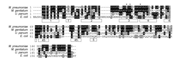
Background: Mycoplasma pneumoniae has previously been characterized as a micro-organism that is genetically highly stable. In spite of this genetic stability, homologous DNA recombination has been hypothesized to lie at the basis of antigenic variation of the major surface protein, P1, of M. pneumoniae. In order to identify the proteins that may be involved in homologous DNA recombination in M. pneumoniae, we set out to characterize the MPN229 open reading frame (ORF), which bears sequence similarity to the gene encoding the single-stranded DNA-binding (SSB) protein of other micro-organisms.

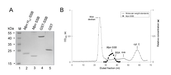
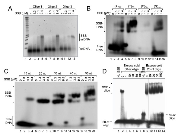
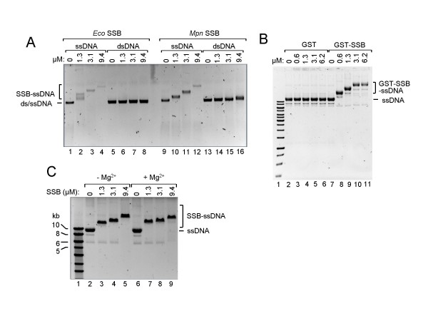
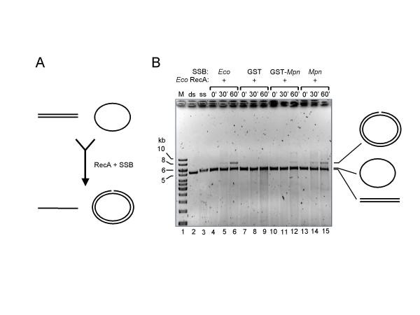
Results: The MPN229 ORF has the capacity to encode a 166-amino acid protein with a calculated molecular mass of 18.4 kDa. The amino acid sequence of this protein (Mpn SSB) is most closely related to that of the protein predicted to be encoded by the MG091 gene from Mycoplasma genitalium (61% identity). The MPN229 ORF was cloned, and different versions of Mpn SSB were expressed in E. coli and purified to > 95% homogeneity. The purified protein was found to exist primarily as a homo-tetramer in solution, and to strongly and selectively bind single-stranded DNA (ssDNA) in a divalent cation- and DNA substrate sequence-independent manner. Mpn SSB was found to bind with a higher affinity to ssDNA substrates larger than 20 nucleotides than to smaller substrates. In addition, the protein strongly stimulated E. coli Recombinase A (RecA)-promoted DNA strand exchange, which indicated that Mpn SSB may play an important role in DNA recombination processes in M. pneumoniae.

Conclusion: The M. pneumoniae MPN229 gene encodes a protein, Mpn SSB, which selectively and efficiently binds ssDNA, and stimulates E. coli RecA-promoted homologous DNA recombination. Consequently, the Mpn SSB protein may play a crucial role in DNA recombinatorial pathways in M. pneumoniae. The results from this study will pave the way for unraveling these pathways and assess their role in antigenic variation of M. pneumoniae.


Figures

Figure 1
Figure 1
Multiple alignment of the amino acid sequences predicted to be encoded by the M. pneumoniae MPN229 [3], M. genitalium MG091[17], U. parvum ssb [39], and E. coli ssb [40] genes. The amino acid motifs that are characteristic for the OB DNA-binding fold are indicated by the open boxes below the aligned sequences [18]. The asterisk above Y44 of the M. pneumoniae sequence indicates the position of a Trp residue (W55) in E. coli SSB that was shown to play a role in DNA-binding. The other two asterisks indicate conserved arginine (R) and lysine (K) residues (R46 and K52, respectively, in the M. pneumoniae sequence); for E. coli SSB these residues were shown to be oriented toward the DNA binding cleft [32]. The multiple aligment was performed using Clustal W . The program BOXSHADE 3.21 was used to generate white letters on black boxes (for residues that are identical in at least two out of four sequences) and white letters on grey boxes (for residues that are similar in at least two out of four sequences).
Figure 2
Figure 2
Purification of recombinant Mpn SSB proteins. (A) Samples of purified recombinant Mpn SSB proteins, i.e. Mpn H10-SSB (lane 2), Mpn SSB (lane 3), GST-SSB (lane 4), and purified GST (lane 5), were analyzed by SDS-PAGE (12%) and Coomassie brilliant blue (CBB)-staining. The sizes of protein markers (lane 1; PageRuler™ Prestained Protein Ladder [Fermentas]) are shown on the left-hand side of the gel. (B) Gel filtration analysis of Mpn SSB. Gel filtration chromatography was performed by applying Mpn SSB to a Sephadex G-150 column. The column was calibrated with blue dextran (2,000 kDa), bovine serum albumin (BSA, 66.4 kDa), ovalbumin (ova, 42.9 kDa), and cytochrome C (cyt. C, 12.3 kDa). Fractions of 1.0 ml were collected and monitored by measuring the optical density at 280 nm (OD280, Y-axis at the left-hand side of the graph). The fractions eluted from the subsequent run containing Mpn SSB were precipitated with trichloroacetic acid, and separated on 14% SDS-PAGE gels. Gels were stained with CBB and recorded using the GelDoc XR system (Bio-Rad). In fractions that contained Mpn SSB, the amount of protein was determined semi-quantitatively using BioNumerics Version 3.0 software (Applied Maths). The relative concentration of Mpn SSB (Y-axis on the right-hand side, in arbitrary units) in these fractions is plotted. In all other fractions, Mpn SSB was not detected by SDS-PAGE analysis and Coomassie brilliant blue-staining.
Figure 3
Figure 3
The Mpn SSB protein binds oligonucleotide substrates in a DNA sequence-independent fashion. (A) Binding of Mpn H10-SSB to three different oligonucleotide substrates. Reactions were performed in volumes of 20 μl and contained either 0 μM (lanes 2, 6 and 10), 1.3 μM (lanes 3, 7 and 11), 3.1 μM (lanes 4, 8 and 12) or 9.4 μM (lanes 5, 9 and 13) of Mpn H10-SSB, and 5 μM of either of three different single-stranded oligonucleotides (Oligo 1, 2 or 3; Table 1). After incubation for 15 min at 37°C, the samples were electrophoresed on a 1.0% agarose gel in 0.5 × TBE buffer. A black/white inverted image of a typical ethidium bromide-stained gel is shown. (B) Binding of Mpn H10-SSB (at 0, 1.3, 3.1 or 9.4 μM in lanes 1–4, 5–8, 9–12 and 13–16, respectively) to 5'-32P-labeled homooligomeric DNA substrates (at 1 μM; Table 1). The samples were separated on 5% polyacrylamide gels in 0.5 × TBE buffer. An autoradiograph is shown. (C) Binding of Mpn H10-SSB (at 0, 1.3, 3.1 or 9.4 μM) to a series of 15- to 50-mer 5'-32P-labeled oligonucleotides (at 1 μM), each containing the same 15-nucleotide core sequence (Table 1). The samples were processed as described above in (B). (D) A DNA-binding competition experiment in which Mpn H10-SSB (at 3.1 μM) was incubated with a constant amount (1 μM) of either the 5'-32P-labeled '20 nt' oligonucleotide (lanes 1–7) or '50-nt' oligonucleotide (lanes 8–13) (Table 1), and increasing amounts of the other, unlabeled ('cold') oligonucleotide. A molar excess of 5 to 100 times unlabeled oligonucleotide over labeled oligonucleotide was tested, as indicated above the lanes. From the samples loaded in lanes 1 and 8, Mpn H10-SSB was omitted; the gel was processed similarly as in (B).
Figure 4
Figure 4
Mpn SSB binding to long DNA substrates and Mg2+-dependence of DNA-binding. (A) Binding of E. coli SSB (Eco SSB, lanes 1–8) and Mpn SSB (lanes 9–16) to either 2 nM of ss φX174 DNA (ssDNA, lanes 1–4 and lanes 9–12) or 1 nM of ds φX174 DNA (dsDNA, lanes 5–8 and lanes 13–16). Reactions were performed in volumes of 20 μl and contained various concentrations of protein, as indicated above the lanes. The samples were separated on a 0.6% agarose gel in 0.5 × TBE buffer. A black/white inverted image of a typical ethidium bromide-stained gel is shown. (B) Binding of GST-SSB to circular, single-stranded M13mp18 DNA (ssDNA). Binding reactions were carried out with various concentrations of either GST-SSB (lanes 8–11) or GST (lanes 3–6), as indicated above the lanes, and 2 nM of DNA. The samples were separated on 0.6% agarose gels in 0.5 × TBE buffer and processed as described above. (C) DNA-binding by Mpn SSB is Mg2+-independent. Reactions with Mpn H10-SSB were executed similarly as described in (B), except for the omission of Mg(OAc)2 in the reaction mixtures of the samples loaded in lanes 2–5. Complexes of Mpn SSB bound to ssDNA (SSB-ssDNA), and the position of unbound DNA (ssDNA and dsDNA), are indicated alongside the gel. The marker DNA loaded in lane 1 of both (B) and (C) is the SmartLadder (Eurogentec).
Figure 5
Figure 5
Mpn SSB promotes E. coli RecA-catalyzed three-strand DNA transfer. (A) Schematic representation of the three-strand transfer reaction. E. coli RecA catalyzes the transfer of a single strand from a linear, double-stranded DNA donor molecule (at the top left) to a complementary, single-stranded, circular acceptor molecule (at the top right), resulting in a linear, single-stranded product (bottom, left) and a (nicked) circular, double-stranded product (bottom, right). This reaction is strongly promoted by the E. coli SSB protein. (B) E. coli RecA-promoted DNA strand transfer reactions using φX174 DNA. Reactions were performed at 37°C in the presence of either E. coli SSB (Eco SSB, lanes 4–6), GST (lanes 7–9), GST-SSB (GST-Mpn, lanes 10–12) or Mpn SSB (lanes 13–15). DNA concentrations used were 1 nM and 2 nM for the ssDNA and dsDNA, respectively. Reactions were terminated at either 0, 30 or 60 min of incubation (0', 30' and 60', respectively, above the lanes). The samples were separated on 0.6% agarose gels in 0.5 × TBE buffer. A black/white inverted image of an ethidium bromide-stained gel is shown. A schematic representation of the major DNA products is indicated at the right-hand side of the gel. The DNA marker (M) is the SmartLadder (Eurogentec). ds (lane 2), linear, dsDNA donor; ss (lane 3), circular, ssDNA acceptor.

Similar articles

Cited by

References

    1. Waites KB, Talkington DF. Mycoplasma pneumoniae and its role as a human pathogen. Clin Microbiol Rev. 2004;17:697–728. doi: 10.1128/CMR.17.4.697-728.2004. - DOI - PMC - PubMed
    1. Wilson MH, Collier AM. Ultrastructural study of Mycoplasma pneumoniae in organ culture. J Bacteriol. 1976;125:332–339. - PMC - PubMed
    1. Himmelreich R, Hilbert H, Plagens H, Pirkl E, Li BC, Herrmann R. Complete sequence analysis of the genome of the bacterium Mycoplasma pneumoniae. Nucleic Acids Res. 1996;24:4420–4449. doi: 10.1093/nar/24.22.4420. - DOI - PMC - PubMed
    1. Dandekar T, Huynen M, Regula JT, Ueberle B, Zimmermann CU, Andrade MA, Doerks T, Sanchez-Pulido L, Snel B, Suyama M, et al. Re-annotating the Mycoplasma pneumoniae genome sequence: adding value, function and reading frames. Nucleic Acids Res. 2000;28:3278–3288. doi: 10.1093/nar/28.17.3278. - DOI - PMC - PubMed
    1. Dandekar T, Sauerborn R. Comparative genome analysis and pathway reconstruction. Pharmacogenomics. 2002;3:245–256. doi: 10.1517/14622416.3.2.245. - DOI - PubMed

MeSH terms

LinkOut - more resources