. 2008 Nov 25;26(50):6329-37.
doi: 10.1016/j.vaccine.2008.09.031. Epub 2008 Oct 1.

Single-dose, virus-vectored vaccine protection against Yersinia pestis challenge: CD4+ cells are required at the time of challenge for optimal protection

Affiliations

Single-dose, virus-vectored vaccine protection against Yersinia pestis challenge: CD4+ cells are required at the time of challenge for optimal protection

Anasuya Chattopadhyay et al. Vaccine. .

Abstract

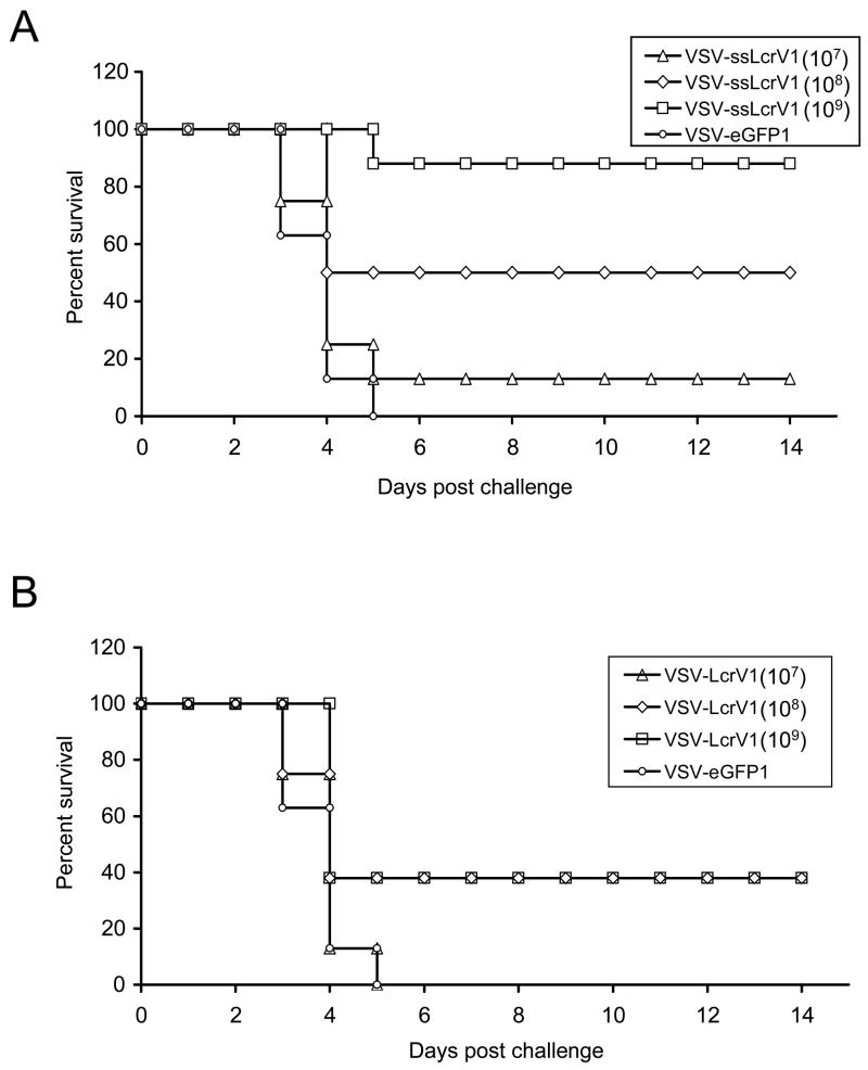
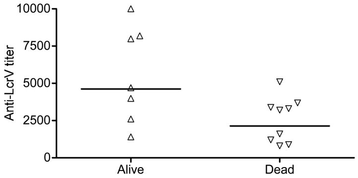
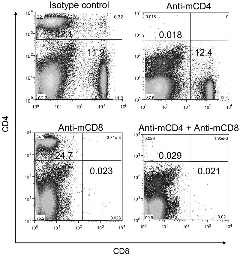
We have developed an experimental recombinant vesicular stomatitis virus (VSV) vectored plague vaccine expressing a secreted form of Yersinia pestis low calcium response protein V (LcrV) from the first position of the VSV genome. This vector, given intramuscularly in a single dose, induced high-level antibody titers to LcrV and gave 90-100% protection against pneumonic plague challenge in mice. This single-dose protection was significantly better than that generated by VSV expressing the non-secreted LcrV protein. Increased protection correlated with increased anti-LcrV antibody and a bias toward IgG2a and away from IgG1 isotypes. We also found that the depletion of CD4+ cells, but not CD8+ cells, at the time of challenge resulted in reduced vaccine protection, indicating a role for cellular immunity in protection.


Figures

Figure 1
Figure 1
Recombinant VSV expressing the secreted form of Yernisia pestis lcrV gene (A) Schematic representation of the recombinant VSV with ssLcrV inserted upstream of the N gene (VSV-ssLcrV1) used in this study, showing the gene order in 3′ to 5′ direction on the negative-strand genome. (B) Indirect immunofluorescence microscopy of BHK-21 cells infected with the indicated viruses. Cells were fixed at 3 hrs post infection, permeabilized and stained using anti-LcrV monoclonal antibodies (MAbs), followed by an AlexaFlour-594 secondary antibody. The differential interference contrast (DIC) images are shown in the left column, the fluorescence images are shown in the middle column and the merged images are shown in the right column.
Figure 2
Figure 2
Metabolic labeling of proteins expressed from recombinant VSV vectors. BHK-21 cells, were infected with the indicated viruses or mock infected and then metabolically labeled with [35S]-methionine. Cell lysates and cell supernatants were fractionated by 10% SDS-PAGE and the gel image collected on a phosphorimager. Positions of VSV encoded proteins are indicated on the left side of the gel image.
Figure 3
Figure 3
Dose-response of anti-LcrV serum titers in VSV-ssLcrV1 immunized mice. Mice in five different groups were immunized intramuscularly (IM) on day 0 with 105, 106, 107, 108 and 109 pfu of VSV-ssLcrV1. Blood was collected, by retro-orbital bleeding, on days 28 and 53 post inoculation. Serum samples were pooled and total IgG titers, specific to LcrV, were determined at the indicated days using ELISA.
Figure 4
Figure 4
Comparison of humoral immune responses of mice to VSV-ssLcrV1 and VSV-LcrV1 vaccinations. (A) Timeline for dose response vaccination-challenge study. Mice were vaccinated IM with 107, 108 and 109 pfu doses of rVSVs encoding either ssLcrV or LcrV and challenged IN with Y. pestis (CO92 strain). VSV-eGFP1 immunization (107 pfu) was used as control. (B) Anti-LcrV titer profile at 56 and 82 days post immunization, in mice immunized with 107, 108 and 109 pfu doses of either VSV-ssLcrV1 (left panel) or VSV-LcrV1 (right panel). (C) Amount of anti-LcrV specific IgG1 and IgG2a isotypes in VSV-ssLcrV1 (left) of VSV-LcrV1 (right) vaccinated mice in the 109 pfu dose group. Serum antibodies that bound LcrV were quantitated using secondary IgG1- and IgG2-specific antibodies. Concentrations of mouse IgG1 or IgG2a were then calculated from standard curves run in parallel using a range of concentrations of purified mouse IgG1 or IgG2a.
Figure 5
Figure 5
Protective efficacy of single-dose VSV-ssLcrV or VSV-LcrV vaccine in a plague challenge assay. (A) Survival profile of VSV-ssLcrV1 vaccinated mice following lethal plague challenge. Mice were immunized (IM) with VSV-ssLcrV1 at three different doses - 107 pfu (open triangle), 108 pfu (open diamond) or 109 pfu (open square). The control mice received VSV-eGFP1 (open circle). All animals were challenged at 3 months post vaccination with 10LD50 Y. pestis (CO92) and observed for 14 days. (B) Similar to A, except that animals were vaccinated with three different doses of VSV-LcrV1 - 107 pfu (open triangle), 108 pfu (open diamond) or 109 pfu (open square).
Figure 6
Figure 6
Correlation of pre-challenge anti-LcrV titers and mouse survival. Mice were vaccinated with 108 pfu of either VSV-ssLcrV1 or VSV-LcrV1 and challenged at day 84 post vaccination with 10 LD50 (10,000 cfu) of Y. pestis (CO92 strain). Anti-LcrV serum titers in individual mice at day 82 post vaccination are plotted for those mice that survived challenge (alive, filled triangle) or succumbed to challenge (dead, open inverted triangle). The horizontal bars represent the geometric means for the two groups.
Figure 7
Figure 7
Depletion of CD4+ and CD8+ T-cells in mice using mouse specific MAbs. Representative FACS plots are shown for splenocytes obtained from mice injected intraperitonealy (IP) with anti-mouse isotype control (top left), anti-mCD4 (top right), anti-mCD8 (bottom left) or combined anti-mCD4 + anti-mCD8 (bottom right) MAbs and stained with approrpiately conjugated rat anti-mouse CD4 and CD8 antibodies on day 3 post injection.
Figure 8
Figure 8
CD4+ cells are required at time of challenge for complete protection against plague challenge. (A) Timeline for the depletion-challenge study. Mice in four groups (A-D) were immunized IM with 109 pfu of VSV-ssLcrV1. Serum sampling was done on day 28, 56 and 82 post immunization. Mice were injected with CD4+/CD8+ depleting MAbs (IP) on days 90 and 91 post immunization followed by challenge on day 91 with 10 LD50 (10,000 cfu) of Y. pestis (CO92 strain). VSV-eGFP1 vaccinated mice were used as the control group. Rat IgG2b isotype MAb was used as a control. (B) Survival profile for mice in the depletion-challenge experiment. VSV-ssLcrV1 vaccinated mice were injected with isotype-matched control (filled square), anti-mCD4 (filled triangle), anti-mCD8 (open diamond) or mCD4+mCD8 (open circle) MAbs. Control animals receiving VSV-eGFP1 (open triangle) were not treated with MAbs.

Similar articles

Cited by

References

    1. Perry RD, Fetherston JD. Yersinia pestis--etiologic agent of plague. Clin Microbiol Rev. 1997;10(1):35–66. - PMC - PubMed
    1. Meyer KF. Pneumonic plague. Bacteriol Rev. 1961;25:249–61. - PMC - PubMed
    1. Galimand M, Guiyoule A, Gerbaud G, Rasoamanana B, Chanteau S, Carniel E, et al. Multidrug resistance in Yersinia pestis mediated by a transferable plasmid. N Engl J Med. 1997;337(10):677–80. - PubMed
    1. Hinnebusch BJ, Rosso ML, Schwan TG, Carniel E. High-frequency conjugative transfer of antibiotic resistance genes to Yersinia pestis in the flea midgut. Mol Microbiol. 2002;46(2):349–54. - PubMed
    1. Inglesby TV, Dennis DT, Henderson DA, Bartlett JG, Ascher MS, Eitzen E, et al. Plague as a biological weapon: medical and public health management. Working Group on Civilian Biodefense Jama. 2000;283(17):2281–90. - PubMed

Publication types

MeSH terms