. 2008 Nov 11;157(1):70-9.
doi: 10.1016/j.neuroscience.2008.08.064. Epub 2008 Sep 9.

Time course of morphine's effects on adult hippocampal subgranular zone reveals preferential inhibition of cells in S phase of the cell cycle and a subpopulation of immature neurons

Affiliations

Time course of morphine's effects on adult hippocampal subgranular zone reveals preferential inhibition of cells in S phase of the cell cycle and a subpopulation of immature neurons

A A Arguello et al. Neuroscience. .

Abstract

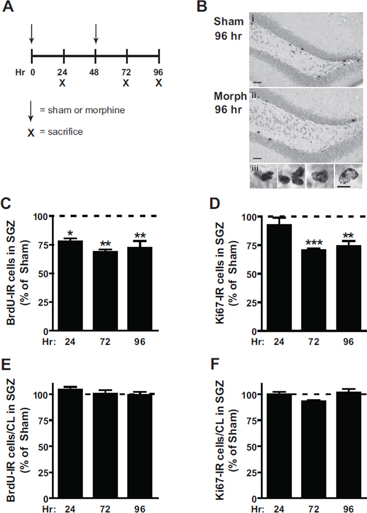
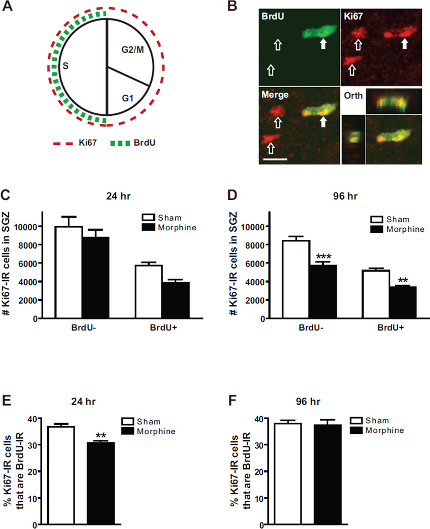
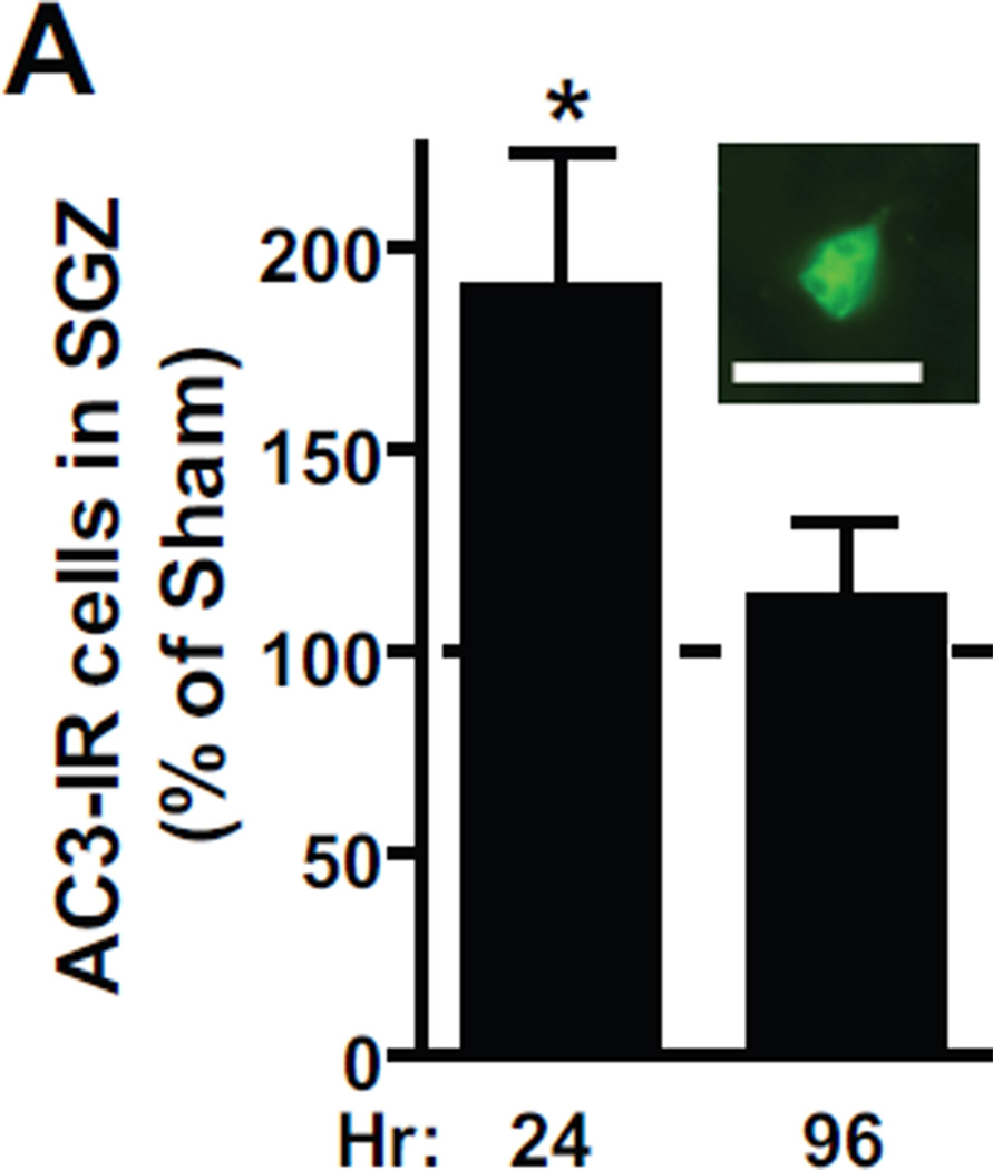
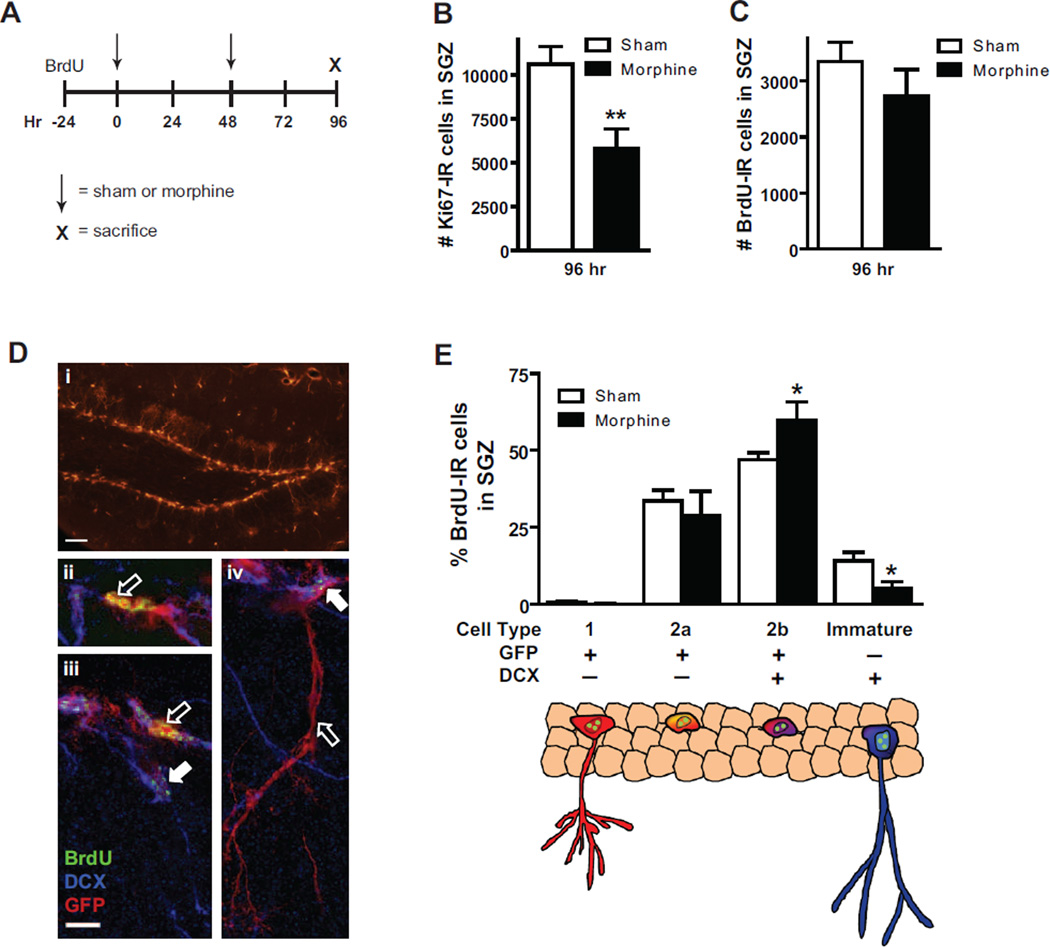
Opiates, such as morphine, decrease neurogenesis in the adult hippocampal subgranular zone (SGZ), raising the possibility that decreased neurogenesis contributes to opiate-induced cognitive deficits. However, there is an incomplete understanding of how alterations in cell cycle progression and progenitor maturation contribute to this decrease. The present study examined how morphine regulates progenitor cell cycle, cell death and immature SGZ neurons (experiment 1) as well as the progression of SGZ progenitors through key stages of maturation (experiment 2). In experiment 1, mice received sham or morphine pellets (s.c., 0 and 48 h) and bromodeoxyuridine (BrdU) 2 h prior to sacrifice (24, 72 or 96 h). Morphine decreased both the number of S phase and total cycling cells, as there were fewer cells immunoreactive (IR) for the S phase marker BrdU and the cell cycle marker Ki67. The percentage of Ki67-IR cells that were BrdU-IR was decreased after 24 but not 96 h of morphine, suggesting a disproportionate effect on S phase cells relative to all cycling cells at this time point. Cell death (activated caspase-3 counts) was increased after 24 but not 96 h. In experiment 2, nestin-green fluorescent protein (GFP) mice given BrdU 1 day prior to morphine or sham surgery (0 and 48 h, sacrifice 96 h) had fewer Ki67-IR cells, but no change in BrdU-IR cell number, suggesting that this population of BrdU-IR cells was less sensitive to morphine. Interestingly, examination of key stages of progenitor cell maturation revealed that morphine increased the percent of BrdU-IR cells that were type 2b and decreased the percent that were immature neurons. These data suggest that chronic morphine decreases SGZ neurogenesis by inhibiting dividing cells, particularly those in S phase, and progenitor cell progression to a more mature neuronal stage.


Figures

Figure 1
Figure 1. Chronic morphine decreases SGZ proliferation
For Experiment 1, morphine or sham pellets were implanted subcutaneously at 0 and 48 hrs. Two hrs prior to sacrifice (24, 72 and 96 hrs) mice were given an injection of 150 mg/kg BrdU. (B) Qualitative IHC shows decreased numbers of BrdU-IR cells in the SGZ of (ii) morphine exposed mice compared to (i) sham exposed mice. Scale bar = 50 um. (iii) BrdU-IR cells, from a representative sham exposed mouse, divide in clusters and have dark or punctate heterochromatin. Scale bar = 10 um. (C) The number of BrdU-IR cells expressed as percent of sham is decreased at all time points. (D) The number of Ki67-IR cells expressed as percent of sham is decreased at 72 and 96 hr. (E–F) The numbers of BrdU-IR or Ki67-IR cells in a cluster are not changed after 24, 72, or 96 hrs of exposure to morphine. At all time points sham: n=6, morphine: n=6. *p<0.05, **p<0.01, ***p<0.001.
Figure 2
Figure 2. Chronic morphine alters cell cycle dynamics
(A) Schematic depicting putative expression of Ki67 and BrdU within the cell cycle. Cycling cells in non-S phases do not co-label with Ki67 and BrdU (Ki67+/BrdU−; dashed red line only), whereas S phase cycling cells co-label with Ki67 and BrdU (Ki67+/BrdU+; dashed red and green lines). (B) Confocal image of cells that are Ki67+/BrdU− (open arrows) and Ki67+/BrdU+ (closed arrows). Orthogonal view shows co-localization of Ki67 (red) and BrdU (green). Scale bar=10 um. (C–D) Quantification of the number of Ki67-IR cells that are BrdU– or BrdU+ reveals significant main effect for Ki67 cell type at 24 hrs. In contrast there was a significant main effect for Ki67 cell type and drug treatment at 96 hrs with no significant interaction. Bonferroni post hoc tests revealed significant differences between sham and morphine groups within Ki67 (BrdU− and BrdU+) cell type. (E) The percentage of Ki67-IR cells that were BrdU-IR was decreased after 24 hrs exposure to morphine (F) but not after 96 hrs. (C, E 24 hr) sham: n=4, morphine: n=4. (D, E 96 hr) sham: n=6, morphine: n=7. **p<0.01, ***p<0.001.
Figure 3
Figure 3. Chronic morphine transiently increases cell death
The number of AC3-IR cells, expressed as percent of sham, was increased after 24 hrs but not after 96 hrs morphine exposure. (Inset) AC3-IR cells were identified by their distinct cell morphology. For both time points sham: n=6, morphine: n=6. Scale bar = 10 um. *p<0.05.
Figure 4
Figure 4. Chronic morphine alters specific stages of progenitor cell maturation
(A) A single 150 mg/kg BrdU injection was given to nestin-GFP mice 24 hrs before sham surgery or implantation of morphine pellet at 0 and 48. Mice were killed after 96 hrs of exposure. (B) The number of Ki67-IR proliferating cells was decreased as previously shown (Figure 1D). (C) The number of BrdU-IR cells was not significantly decreased. (D) Phenotypic analysis of BrdU-IR cells was assessed by cell morphology and co-localization with GFP and DCX with confocal microscopy. (i) GFP was robustly expressed in the SGZ. Scale bar = 50 um. (ii–iv) BrdU-IR cells were classified as Type 1 (iv, open arrow; GFP+/DCX–, triangular cell body, dendrite extending into molecular layer), Type 2a (ii, iii, open arrows; GFP+/DCX–, ovoid cell body, no dendrites), Type 2b (iv, closed arrow; GFP+/DCX+, ovoid cell body, no dendrites;) or immature neuron (iii, closed arrow; GFP−/DCX+, mature morphology). Scale bar= 10 um in iii. (E) The percent of BrdU-IR cells that were Type 2b increased whereas those that were immature neurons decreased. Schematic diagram depicts cell morphology and marker expression of cell type affected by morphine. Green dots represent BrdU (as BrdU-IR cells were phenotyped). (B,C) sham: n=8, morphine: n=7. (E) sham: n=7, morphine: n=6. *p≤0.05, **p<0.01.

Similar articles

Cited by

References

    1. Altman J, Das GD. Autoradiographic and histological evidence of postnatal hippocampal neurogenesis in rats. J Comp Neurol. 1965;124(3):319–335. - PubMed
    1. Bauer S, Patterson PH. The cell cycle-apoptosis connection revisited in the adult brain. J Cell Biol. 2005;171(4):641–650. - PMC - PubMed
    1. Cameron HA, McKay RD. Adult neurogenesis produces a large pool of new granule cells in the dentate gyrus. J Comp Neurol. 2001;435(4):406–417. - PubMed
    1. Canales JJ. Adult neurogenesis and the memories of drug addiction. Eur Arch Psychiatry Clin Neurosci. 2007;257(5):261–270. - PubMed
    1. Chumley MJ, Catchpole T, Silvany RE, Kernie SG, Henkemeyer M. EphB receptors regulate stem/progenitor cell proliferation, migration, and polarity during hippocampal neurogenesis. J Neurosci. 2007;27(49):13481–13490. - PMC - PubMed

Publication types

MeSH terms