. 2008 Oct 1;22(19):2707-20.
doi: 10.1101/gad.486808.

Sequestration of Polo kinase to microtubules by phosphopriming-independent binding to Map205 is relieved by phosphorylation at a CDK site in mitosis

Affiliations

Sequestration of Polo kinase to microtubules by phosphopriming-independent binding to Map205 is relieved by phosphorylation at a CDK site in mitosis

Vincent Archambault et al. Genes Dev. .

Abstract

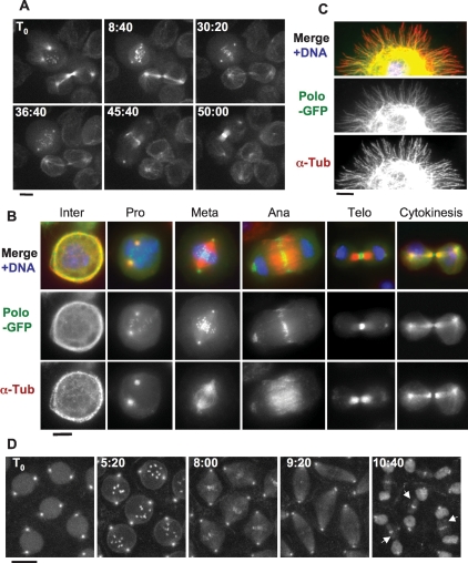
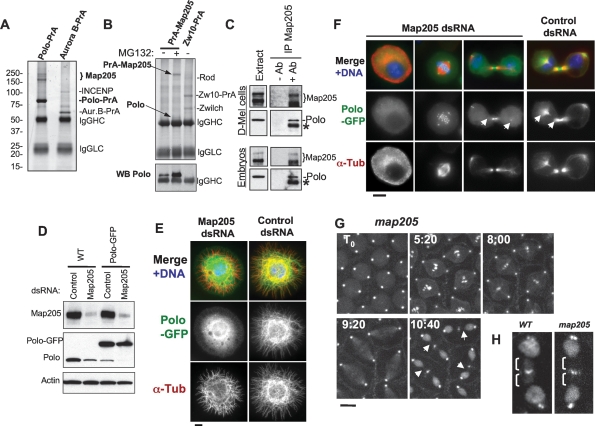
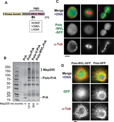
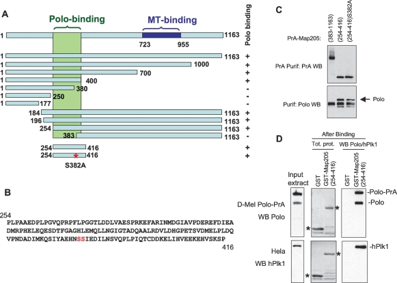
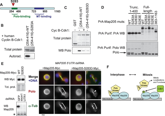
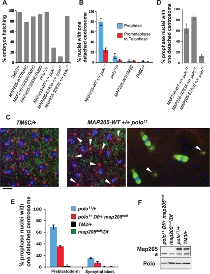
The conserved Polo kinase controls multiple events in mitosis and cytokinesis. Although Polo-like kinases are regulated by phosphorylation and proteolysis, control of subcellular localization plays a major role in coordinating their mitotic functions. This is achieved largely by the Polo-Box Domain, which binds prephosphorylated targets. However, it remains unclear whether and how Polo might interact with partner proteins when priming mitotic kinases are inactive. Here we show that Polo associates with microtubules in interphase and cytokinesis, through a strong interaction with the microtubule-associated protein Map205. Surprisingly, this interaction does not require priming phosphorylation of Map205, and the Polo-Box Domain of Polo is required but not sufficient for this interaction. Moreover, phosphorylation of Map205 at a CDK site relieves this interaction. Map205 can stabilize Polo and inhibit its cellular activity in vivo. In syncytial embryos, the centrosome defects observed in polo hypomorphs are enhanced by overexpression of Map205 and suppressed by its deletion. We propose that Map205-dependent targeting of Polo to microtubules provides a stable reservoir of Polo that can be rapidly mobilized by the activity of Cdk1 at mitotic entry.


Figures

Figure 1.
Figure 1.
Polo localizes to MTs in a cell cycle-dependent manner. In addition to centrosomes, kinetochores, and the midbody, Polo-GFP localizes to MTs in cytokinesis and interphase. (A) Polo-GFP dynamics in D-Mel cells by time lapse (time is shown in minutes:seconds). Bar, 5 μm. See Supplemental Movie 1 and the text for details. (B) Polo-GFP colocalizes with MTs during interphase and cytokinesis. Cells were fixed with formaldehyde. Polo-GFP appears in green, α-Tubulin is stained in red, and DNA is DAPI-stained in blue. Bar, 5 μm. (C) Polo-GFP colocalizes with MTs in a cell spreading on a concanavalin-A-coated surface and fixed with methanol/formaldehyde (only a portion of the cell is shown for clarity). Stainings are anti-GFP (green), α-Tubulin (red), and DNA (blue). Bar, 5 μm. (D) GFP-Polo dynamics in a transgenic syncytial embryo (cycle 13) by time lapse (time is shown in minutes:seconds). Note that GFP-Polo localizes to the MTs of the central spindle during karyokinesis (arrows). Bar, 10 μm.
Figure 2.
Figure 2.
Polo interacts with Map205, and this is required for its localization to MTs. (A) Polo specifically interacts with Map205. Polo-PrA and Aurora B-PrA (control) were purified from stably expressing cell lines, and purified proteins were resolved on SDS-PAGE and stained with Coomassie Blue. The indicated proteins were identified by mass spectrometry. (IgGHC) IgG heavy chain; (IgHLC) IgG light chain. (Left) Molecular mass markers are in kilodaltons (kDa). (B) Reciprocal purification. (Top) PrA-Map205 and Zw10-PrA (control) were purified from stably expressing cell lines, and products were analyzed as in A. In one sample, cells were treated overnight with 25 μM MG132 to inhibit the proteasome, which results in a stabilization of Map205 (but does not lead to an arrest of the majority of the cells in mitosis). (Bottom) The same purification products were probed for Polo by Western blot. (C) Endogenous Polo and Map205 interact in D-Mel cells and embryos. Polo was coimmunoprecipitated with Map205 using anti-Map205 antibodies from D-Mel cells and from syncytial embryos. As a control, antibodies were not added. (*) Antibodies from the IP cross-reacting in the Polo Western blot. (D) Depletion of Map205 by RNAi. D-Mel cells, wild-type or stably expressing Polo-GFP, were treated with dsRNA against Map205 or the bacterial kanamycin-resistance gene (control). Four days later, cells were analyzed by Western blotting against Map205, Polo, and actin (loading control). Note that in addition to the depletion of Map205, Polo levels are also reduced. (E,F) Polo requires Map205 for its localization to MTs. (E) Polo-GFP cells were treated with Map205 dsRNA or control dsRNA (as in D), spread on concanavalin-A, fixed with methanol/formaldehyde, and stained for α-tubulin (red) GFP (anti-GFP; green), and DNA (blue). Bar, 5 μm. (F) Polo-GFP cells were treated with Map205 or control dsRNA (as in D and E), fixed with formaldehyde, and stained for α-tubulin (red) and DNA (blue). (Right) Note that unlike in the control, Polo-GFP fails to colocalize with MTs in cytokinesis after Map205 RNAi (arrows). Also compare with cells in Figure 1B. Bar, 5 μm. (G) GFP-Polo dynamics in a transgenic syncytial embryo laid by a map205 homozygous null mother (cycle 13) by time lapse (time is shown in minutes:seconds). Note that, unlike in map205+ embryos (Fig. 1D), GFP-Polo fails to localize to the MTs of the central spindle during karyokinesis in map205-null embryos (arrows). Bar, 10 μm. (H) Magnified view of karyokinetic nuclei from wild-type (WT) or map205-null embryos expressing GFP-Polo (taken from G and Fig. 1D). Note that unlike in the wild-type control, GFP-Polo fails to localize to central spindle MTs in the map205-null embryo.
Figure 3.
Figure 3.
Polo requires a functional Polo-Box Domain to interact with Map205 and to localize to MTs. (A) Amino acid substitutions introduced in Polo to disrupt the Polo-Box Domain (PBD). The kinase domain is in yellow, and the two Polo Boxes (PB1 and PB2) are in pink. (B) A functional Polo-Box Domain is required for Polo to interact with Map205. Stable cell lines expressing Polo-wt, Polo-V (V396A), Polo-WVL (W395F, V396A, and L408A) in fusion with PrA, or PrA alone were used in affinity purifications. Purified proteins are shown on a Coomassie Blue-stained gel. Each purification product was assayed for the presence of Map205 in the area defined by the bracket. Mascot scores (see Materials and Methods) for Map205 are shown under the gel image. (IgGHC) IgG heavy chain; (IgHLC) IgG light chain. (Left) Molecular mass markers are in kilodaltons (kDa). (C,D) A functional Polo-Box Domain is required for Polo to localize to MTs. (C) Cells stably expressing Polo-WVL in fusion with GFP (green) were fixed with formaldehyde and stained for α-tubulin (red) and DNA (blue). Compare with Figure 1B. Note that Polo-WVL-GFP fails to localize to MTs, kinetochores, centrosomes, and the midbody. Bar, 5 μm. (D) Cells stably expressing Polo-GFP or Polo-WVL-GFP were plated on concanavalin-A, fixed with methanol/formaldehyde, and stained for GFP (anti-GFP; green), α-tubulin (red), and DNA (blue). Bar, 5 μm.
Figure 4.
Figure 4.
Polo binding to Map205 does not require phosphopriming. (A) Summary of a truncation analysis defining a small region of Map205 necessary and sufficient for Polo binding. The assay used is as in C and Figure 2B. Polo binds to the region in green. The MT-binding region (blue) was defined elsewhere (Irminger-Finger et al. 1990). (B) Sequence of a small segment of Map205 (amino acids 254–416) sufficient for binding Polo. Note that it contains only one potential canonical PBD-binding motif (SS or ST) at S381–S382. (C) Map205 does not require phosphopriming at a canonical PBD-binding motif (S-pS or S-pT) for Polo binding. Cells transiently expressing the indicated Map205 mutants in N-terminal fusion with Protein A were used in Protein A affinity purification, and the products were probed for Protein A and Polo. Note that Map205(254–416)–S382A can still interact with Polo. (D) Map205 does not require phosphorylation to interact with Drosophila Polo or human Plk1. GST-Map205(254–416) or GST alone (control) were expressed in bacteria, purified (asterisks), and subjected to a Polo or hPlk1-binding assay using D-Mel (stably expressing Polo-PrA) or HeLa cell extracts, respectively. Purification products were analyzed by Western blotting for Polo (top) or hPlk1 (bottom).
Figure 5.
Figure 5.
Map205 phosphorylation at Cdk1 site Ser 283 prevents interaction with Polo. (A) Map205 is phosphorylated at Ser 283, inside the Polo-binding region in vivo (for details, see Supplemental Figure S6). (B) Ser 283 of Map205 is a target of cyclin B–Cdk1 in vitro. Map205(254–416)-wt or S283A was generated using bacterial expression and subjected to a kinase assay using human cyclin B–Cdk1. The total protein (amido black staining) and the autoradiograph are shown. (C) Phosphorylation of Map205(254–416) by cyclin B–Cdk1 or phosphomimicking at Ser 283 inhibits the interaction with Polo in vitro. The indicated proteins were treated or not with cyclin B–Cdk1 before being subjected to a Polo-binding assay as in Figure 4D. (D) Mimicking phosphorylation at Ser 283 of Map205 abolishes its interaction with Polo in vivo. Cells transiently expressing the indicated proteins (full-length or truncated Map205) were used in Protein A affinity purification, and the products were probed for PrA and Polo. Note that the phosphomimicking S283D and S283E mutations abolish the interaction with Polo. (E) Phosphomimicking phosphorylation at S283 disrupts the localization of Polo on MTs. (Top left) Stable cell lines expressing Map205-Myc (wild type and S283D) were created and expressed at similar levels. RNAi targeting an untranslated region of the endogenous MAP205 transcript was used to deplete endogenous Map205. (Bottom left) The Western blot shown is from cells not transfected with Map205 cDNA, for clarity. Cells were then stained for Polo (red), α-Tubulin (green), and DNA (blue). Note that Polo is present on MTs (brackets) when Map205-Myc wild type, but not S283D is expressed. Polo is sometimes detected at the midbody (arrow) even when it does not localize to MTs. Bar, 5 μm. (F) Model for the regulation of Polo localization by Map205 and Cdk1. In interphase, Polo is sequestered to MTs via an interaction with Map205 that depends on both the PBD and the kinase domain of Polo, but that does not require priming phosphorylation. At mitotic entry, Cdk1 phosphorylates Map205, which releases Polo from Map205 and MTs. Polo is then free to translocate to its mitotic localizations, where it functions to promote proper mitosis. In late mitosis, Cdk1 becomes inactivated, and Polo returns to MTs in cytokinesis.
Figure 6.
Figure 6.
Map205 contributes to the regulation of Polo in vivo. (A–C) Overexpression of Map205 enhances Polo-dependent defects. (A) Percentage of hatching embryos produced by females of the indicated genotypes. All genotypes included one copy of the Maternal α-Tubulin Gal4 driver (not shown). TM6C is a balancer chromosome. Map205 transgenes were under the control of the UASp promoter. Transgenic lines were selected for equal and moderate overexpression levels of Map205 variants (Supplemental Fig. S8). At least 1000 embryos laid by 12 females over 3 d were scored for each genotype. (B) Percentage of nuclei showing single centrosome detachment in syncytial blastoderm embryos (cycles 10–13) produced by females of the indicated genotypes (as in A, separate experiment). Between five and 10 embryos were scored for each entry (±SEM). (C, right) Examples of single centrosome detachment (arrows) observed in prophase and in metaphase (genotypes are as in A and B). The metaphase spindles on the bottom and on the right show typical single centrosome detachment, while the spindle on the top appears normal. (Left) Image taken from a control embryo where both centrosomes remain normally tethered to the nuclear envelope in prophase. Stainings are α-Tubulin (green), γ-Tubulin (red), and DNA (blue). Bar, 10 μm. (D) Percentage of prophase nuclei showing single centrosome detachment in syncytial blastoderm embryos (cycles 10–13) produced by females of the indicated genotype (as in A and B, separate experiment). Ten embryos were scored for each entry (±SEM). (E) Deletion of Map205 partially rescues the Polo-dependent centrosome detachment. Percentage of prophase nuclei showing single centrosome detachment in preblastoderm (cycles 8–9) and early blastoderm (cycles 10–11) embryos produced by females of the indicated genotypes. Note that single centrosome detachment is frequent in embryos laid by polo11/+ females and that this defect is partially rescued by deletion of map205. Between five and 10 embryos were scored for each entry (±SEM). Refer to Supplemental Figure S9 for sample images. (F) Western blot from embryos laid by females of the indicated genotypes (0–3 h collection; as in E). (Top) Map205 detection. (*) Cross-reacting band serving as a loading control. (Bottom) Polo detection. Note that the absence of Map205 does not correlate with a lower amount of Polo in embryos (unlike in cultured cells).

References

    1. Ahonen L.J., Kallio M.J., Daum J.R., Bolton M., Manke I.A., Yaffe M.B., Stukenberg P.T., Gorbsky G.J. Polo-like kinase 1 creates the tension-sensing 3F3/2 phosphoepitope and modulates the association of spindle-checkpoint proteins at kinetochores. Curr. Biol. 2005;15:1078–1089. - PubMed
    1. Alexandru G., Uhlmann F., Mechtler K., Poupart M.A., Nasmyth K. Phosphorylation of the cohesin subunit Scc1 by Polo/Cdc5 kinase regulates sister chromatid separation in yeast. Cell. 2001;105:459–472. - PubMed
    1. Andersen S.S. Spindle assembly and the art of regulating microtubule dynamics by MAPs and Stathmin/Op18. Trends Cell Biol. 2000;10:261–267. - PubMed
    1. Archambault V., Zhao X., White-Cooper H., Carpenter A.T., Glover D.M. Mutations in Drosophila Greatwall/Scant reveal its roles in mitosis and meiosis and interdependence with polo kinase. PLoS Genet. 2007;3:e200. doi: 10.1371/journal.pgen.0030200. - DOI - PMC - PubMed
    1. Barr F.A., Sillje H.H., Nigg E.A. Polo-like kinases and the orchestration of cell division. Nat. Rev. Mol. Cell Biol. 2004;5:429–440. - PubMed

Publication types

MeSH terms

Substances