. 2008 Oct 7;105(40):15370-5.
doi: 10.1073/pnas.0808115105. Epub 2008 Oct 1.

Structure of the human voltage-dependent anion channel

Affiliations

Structure of the human voltage-dependent anion channel

Monika Bayrhuber et al. Proc Natl Acad Sci U S A. .

Abstract

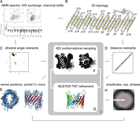
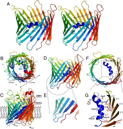
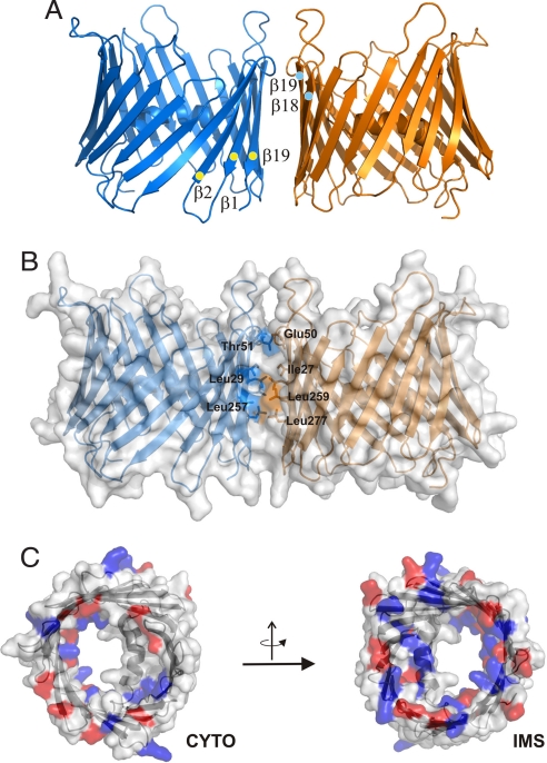
The voltage-dependent anion channel (VDAC), also known as mitochondrial porin, is the most abundant protein in the mitochondrial outer membrane (MOM). VDAC is the channel known to guide the metabolic flux across the MOM and plays a key role in mitochondrially induced apoptosis. Here, we present the 3D structure of human VDAC1, which was solved conjointly by NMR spectroscopy and x-ray crystallography. Human VDAC1 (hVDAC1) adopts a beta-barrel architecture composed of 19 beta-strands with an alpha-helix located horizontally midway within the pore. Bioinformatic analysis indicates that this channel architecture is common to all VDAC proteins and is adopted by the general import pore TOM40 of mammals, which is also located in the MOM.


Conflict of interest statement

The authors declare no conflict of interest.

Figures

Fig. 1.
Fig. 1.
Flow chart of the conjoint NMR and x-ray structure determination. The 3D structure of VDAC was determined by integrating NMR and x-ray data in an iterative structure calculation protocol: (A) The NMR data (H/D exchange, chemical shifts) served as a basis to determine the secondary structure of hVDAC1. In addition, 95 NOEs could be assigned, 65 of which are interstrand NOEs that framed the register of neighboring β-strands. (B) A 2D topology model was derived from the secondary structure, the interstrand NOEs and paramagnetic relaxation enhancements (PRE) of 18 spin-labeled single-cysteine mutants (β19 is shown twice to indicate that the barrel is closed). (C) From the chemical shifts, main-chain dihedral angle restraints for 184 φ/ψ-pairs were predicted with the program TALOS (14). The SHIFTOR server predicted 170 additional side-chain χ-angle restraints (15). (D) The NOEs, the inter strand hydrogen bonding pattern and the strand assignments were converted into distance restraints. The NOEs provided 95 distance restraints between amide protons. These were supplemented with 150 hydrogen bonds inferred from the topology model. In addition, we imposed 370 distance restraints that restrain the intrastrand Cα-distances to multiples of 3.4 Å. (E) Positional restraints were provided by four seleno-methionines (M10 (L in WT), M129, M155, M184 (V in WT), and an eight-stranded partial Cα-trace that was built manually into the well defined regions of the electron density. The Se-Met positions were used to dock the model into the density map and to structurally align the model and the Cα-trace. (F) Using the distance, dihedral angle and positional restraints as input an initial structure ensemble was generated with the ISD software (16). (G) The highest-probability conformation served as a starting structure in a subsequent reciprocal space refinement with BUSTER-TNT (17). In addition to the structure factor amplitudes and experimental phases from SHARP (18) (H), secondary structure distance restraints (D) were imposed in all BUSTER-TNT refinements. (I) The structure calculation proceeded by cycling between ensemble generation and crystallographic refinement. In the conformational sampling, the positional restraints (E) were used only in the initial iteration. In subsequent calculations, the last structure obtained by crystallographic refinement served as a template (positional restraints to all Cα-coordinates). The distance and dihedral angle restraints were imposed in all ISD structure calculations.
Fig. 2.
Fig. 2.
3D structure of hVDAC1. (A) Stereo representation of the hVDAC1 structure in a ribbon presentation color coded from the N to the C terminus (N and C). Some β-strands are marked with their respective numbers. All images were prepared using PYMOL (20). (B, C) Structure superposition of OmpC from E. coli (PDB ID code 2J1N) onto the refined hVDAC1 (115 Cα-atoms were superposed with an rmsd of 2.5 Å) viewed from top (B) and side (C), respectively. The view shown in B indicates a similar cork architecture of two different structural elements (AH, N-terminal helix of hVDAC; L3, constricting loop of porin). The horizontal dimensions of hVDAC1 are given (3.5 × 3.1 nm). Residues in the C-terminal half (β10–β19) of the two barrel proteins (in C marked with NVDAC, CVDAC, and COmpC) match well (matching elements carry yellow points for better visibility), whereas the N-terminal region differs in the relative inclination. The approximate membrane dimension surrounding the protein in the MOM is indicated by a “theoretical” lipid layer. (D) Side view of hVDAC1 in a ribbon presentation color coded from the N to the C terminus (N and C). The 19 β-strands are marked (β1–β19), whereas the N-terminal helix is folded into the pore interior. (E) Close-up of the C terminus (C) with the two terminal β-strands (β1 and β19) forming the parallel arrangement. (F) Top view onto the hVDAC1 barrel with the α-helix enclosed by and attached to the barrel walling.(G) Closeup of the N-terminally located α-helical element. Residues contacting the barrel wall or pointing toward the interior of the pore are marked (in black: Tyr7-Phe18) as well as residues on the barrel wall (in red). The secondary structure of the barrel is marked β12–β16.
Fig. 3.
Fig. 3.
Distribution of charged residues and dimer model of hVDAC1. (A) Ribbon representation of the protein dimer and strands involved in dimerization are marked. (B) Surface representation of the dimer with residues at the dimer interface color coded accordingly and marked. (C) Surface representations of hVDAC1 visualized from both sides of the membrane with the charged residues colored in red (Glu, Asp) and blue (Lys, Arg). CYTO, cytoplasmic side; IMS, intermembrane space side.
Fig. 4.
Fig. 4.
Conformational instability of the N-terminal region of the hVDAC1 barrel. (A) Comparison of amide proton exchange in wt (red) and E73V (black) hVDAC1 at 37°C, pH 6.8. Shown are signal intensity ratios of hVDAC1 in D2O and H2O. The NMR measurements were completed 2 hours after transfer of the proteins into 100% D2O. The location of the 19 β-strands and the helix are shown on top. Mutants that affect the voltage gating of scVDAC (Asp-16, Lys-20, Lys-46, Lys-61, Lys-65, Lys-84, Q154, Q282) are indicated by stars (30). Residues, which on the basis of channel conductance measurements were proposed to remain in the wall of the closed-channel pore of VDAC (31), are indicated by circles. The position of E73V is highlighted by a green square and arrow. (B) Probability for disorder in hVDAC1 as predicted by the DisProt web server (32). Based on the primary sequence of hVDAC1 a higher disorder propensity is predicted for the N-terminal part of the β-barrel including strands β1-β4 that NMR shows to be conformationally unstable. (C) Ribbon representation of WT hVDAC1 (Left) and E73V hVDAC1 (Right) in a side view and rotated by 90° resulting in a top view. Absolute signal intensities in the H/D exchange TROSY were visualized by using a continuous blue scale (light blue, <105; dark blue, >4×10⁵). Residues without detectable signal in the exchange TROSY are shown in red for wt hVDAC1 and gray for E73V hVDAC1. Unassigned residues are colored in light pink. Residues that affected the voltage gating of scVDAC (30) are shown with side chains. They are labeled by the one letter amino acid code in the top view of hVDAC1.

References

    1. Neupert W, Herrmann JM. Translocation of proteins into mitochondria. Annu Rev Biochem. 2007;76:723–749. - PubMed
    1. Hill K, et al. Tom40 forms the hydrophilic channel of the mitochondrial import pore for preproteins [see comment] Nature. 1998;395:516–521. - PubMed
    1. Wiedemann N, et al. Machinery for protein sorting and assembly in the mitochondrial outer membrane. Nature. 2003;424:565–571. - PubMed
    1. Schein SJ, Colombini M, Finkelstein A. Reconstitution in planar lipid bilayers of a voltage-dependent anion-selective channel obtained from paramecium mitochondria. J Membr Biol. 1976;30:99–120. - PubMed
    1. Blachly-Dyson E, Forte M. VDAC channels. IUBMB Life. 2001;52:113–118. - PubMed

Publication types

MeSH terms

Substances

Associated data