. 2008 Nov;19(11):2069-75.
doi: 10.1681/ASN.2008020162. Epub 2008 Oct 2.

Podocyte-specific loss of functional microRNAs leads to rapid glomerular and tubular injury

Affiliations

Podocyte-specific loss of functional microRNAs leads to rapid glomerular and tubular injury

Jacqueline Ho et al. J Am Soc Nephrol. 2008 Nov.

Abstract

MicroRNAs (miRNAs) are in a class of endogenous, small, noncoding RNAs that exert their effects through posttranscriptional repression of specific target mRNAs. Although miRNAs have been implicated in the regulation of diverse biologic processes, little is known about miRNA function in the kidney. Here, mice lacking functional miRNAs in the developing podocyte were generated through podocyte-specific knockout of Dicer, an enzyme required for the production of mature miRNAs (Nphs2-Cre; Dicer(flx/flx)). Podocyte-specific loss of miRNAs resulted in significant proteinuria by 2 wk after birth, rapid progression of marked glomerular and tubular injury beginning at 3 wk, and death by 4 wk. Expression of the slit diaphragm proteins nephrin and podocin was decreased, and expression of the transcription factor WT1 was relatively unaffected. To identify miRNA-mRNA interactions that contribute to this phenotype, we profiled the glomerular expression of miRNAs; three miRNAs expressed in glomeruli were identified: mmu-miR-23b, mmu-miR-24, and mmu-miR-26a. These results suggest that miRNA function is dispensable for the initial development of glomeruli but is critical to maintain the glomerular filtration barrier.


Figures

Figure 1.
Figure 1.
Podocyte-specific loss of mature miRNAs results in marked proteinuria and glomerular and tubular injury. (A) Histologic analyses of control or NPHS2-cre;DicerFlx/Flx mice at 3 wk of age (H&E, hematoxylin and eosin stain; PAS, periodic acid-Schiff). Affected glomeruli from mutant kidneys display cellular crescents with glomerular tuft collapse. The renal tubules are mildly dilated with proteinaceous material. Bar = 50 μm. Electron microscopy demonstrates focal foot process effacement and wrinkling of the glomerular basement membrane. (B) Coomassie-stained SDS-PAGE gel of urine from control or mutant NPHS2-cre;DicerFlx/Flx mice. There is marked albuminuria (66 kD) in the mutant mice. MW, molecular weight. (C) Urinary albumin (mg/L), creatinine (mg/dl), and albumin-to-creatinine ratios (ACR) for control and mutant mice at 2 and 3 wk of age. Onset of proteinuria occurs by 2 wk of age in the mutant mice. Magnification, ×4800.
Figure 2.
Figure 2.
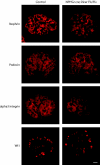
Immunofluorescence staining of podocyte markers for control (left) and NPHS2-cre;DicerFlx/Flx (right) kidneys. Expression of the slit diaphragm proteins nephrin and podocin is decreased and occurs in a speckled pattern. In contrast, there is an apparent segmental loss of α3 integrin expression in affected glomeruli. Wt1 expression is relatively preserved. Bar = 25 μm.
Figure 3.
Figure 3.
Immunofluorescence staining for PECAM, desmin, and α-smooth muscle actin for control (top) and NPHS2-cre;DicerFlx/Flx (bottom) kidneys. PECAM staining is unchanged in the mutant glomeruli, whereas desmin and α-smooth muscle actin display increased expression in the mesangium. Bar = 25 μm.
Figure 4.
Figure 4.
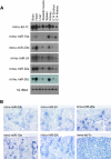
Expression of miRNAs in the glomerulus. (A) Northern blots for mmu-let-7c, mmu-miR-10a, mmu-miR-23b, mmu-miR-24, mmu-miR-26a, and mmu-miR-30c in adult and embryonic tissues confirm the expression of these miRNAs in the adult kidney. 5S rRNA, loading control. (B) Glomerular expression of mmu-miR-23b, mmu-miR-24, and mmu-miR-26a by LNA ISH at 2 to 3 wk of age. In contrast, mmu-miR-10a and mmu-miR-30a are expressed in a subset of renal tubules, and mmu-let-7c is ubiquitously expressed in the kidney. Bar = 100 μm.

Comment in

  • Dicer cuts the kidney.
    Ho JJ, Marsden PA. Ho JJ, et al. J Am Soc Nephrol. 2008 Nov;19(11):2043-6. doi: 10.1681/ASN.2008090986. Epub 2008 Oct 15. J Am Soc Nephrol. 2008. PMID: 18923053 Review. No abstract available.

References

    1. Kloosterman WP, Plasterk RH: The diverse functions of microRNAs in animal development and disease. Dev Cell 11: 441–450, 2006 - PubMed
    1. Grimson A, Farh KK, Johnston WK, Garrett-Engele P, Lim LP, Bartel DP: MicroRNA targeting specificity in mammals: Determinants beyond seed pairing. Mol Cell 27: 91–105, 2007 - PMC - PubMed
    1. Rajewsky N: microRNA target predictions in animals. Nat Genet 38[Suppl]: S8–S13, 2006 - PubMed
    1. Hammond SM: Dicing and slicing: The core machinery of the RNA interference pathway. FEBS Lett 579: 5822–5829, 2005 - PubMed
    1. Calabrese JM, Seila AC, Yeo GW, Sharp PA: RNA sequence analysis defines Dicer's role in mouse embryonic stem cells. Proc Natl Acad Sci U S A 104: 18097–18102, 2007 - PMC - PubMed

Publication types

MeSH terms