. 2008 Oct 14;105(41):15997-6002.
doi: 10.1073/pnas.0808084105. Epub 2008 Oct 2.

Parkinsonism and impaired axonal transport in a mouse model of frontotemporal dementia

Affiliations

Parkinsonism and impaired axonal transport in a mouse model of frontotemporal dementia

Lars M Ittner et al. Proc Natl Acad Sci U S A. .

Abstract

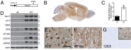
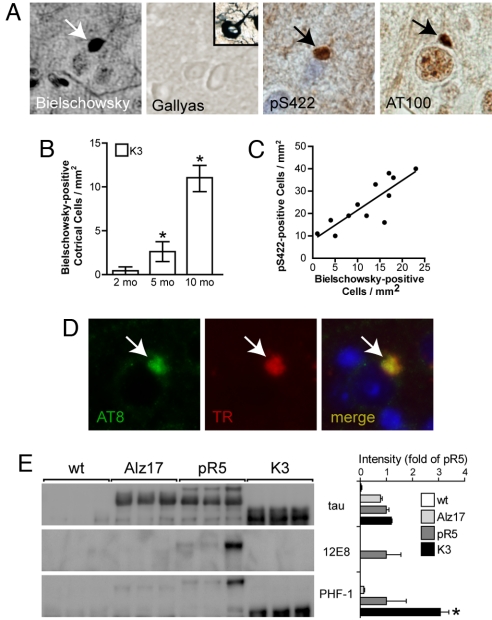
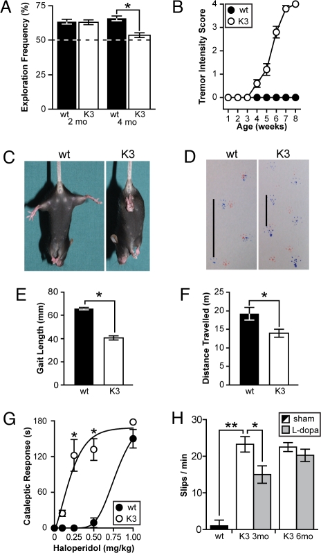
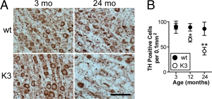
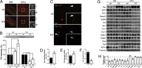
Frontotemporal dementia (FTD) is characterized by cognitive and behavioral changes and, in a significant subset of patients, Parkinsonism. Histopathologically, FTD frequently presents with tau-containing lesions, which in familial cases result from mutations in the MAPT gene encoding tau. Here we present a novel transgenic mouse strain (K3) that expresses human tau carrying the FTD mutation K369I. K3 mice develop a progressive histopathology that is reminiscent of that in human FTD with the K369I mutation. In addition, K3 mice show early-onset memory impairment and amyotrophy in the absence of overt neurodegeneration. Different from our previously generated tau transgenic strains, the K3 mice express the transgene in the substantia nigra (SN) and show an early-onset motor phenotype that reproduces Parkinsonism with tremor, bradykinesia, abnormal gait, and postural instability. Interestingly, motor performance of young, but not old, K3 mice improves upon L-dopa treatment, which bears similarities to Parkinsonism in FTD. The early-onset symptoms in the K3 mice are mechanistically related to selectively impaired anterograde axonal transport of distinct cargos, which precedes the loss of dopaminergic SN neurons that occurs in aged mice. The impaired axonal transport in SN neurons affects, among others, vesicles containing the dopamine-synthesizing enzyme tyrosine hydroxylase. Distinct modes of transport are also impaired in sciatic nerves, which may explain amyotrophy. Together, the K3 mice are a unique model of FTD-associated Parkinsonism, with pathomechanistic implications for the human pathologic process.


Conflict of interest statement

The authors declare no conflict of interest.

Figures

Fig. 1.
Fig. 1.
K3 mice model tau hyperphosphorylation of FTD with the K369I mutation. (A) K3 mice express human tau (htau43) carrying the pathogenic K369I mutation under control of the neuron-specific murine (m)Thy1.2 promoter. (B) Immunohistochemistry for human tau reveals widespread expression of transgenic tau in brain (brown) in a 3-month-old K3 mouse. No staining is observed in WT controls (not shown). (C) K3 mice show 2.94 ± 0.28-fold higher total cerebral tau level than WT controls, as determined by quantification of Western blots, using the total tau-specific tau-5 antibody (*, P < 0.0001). (D) Western blotting reveals that in the cortex of K3 mice transgenic tau (asterisk) is over-expressed compared with endogenous tau (open diamonds) as shown with tau-5. WT littermate controls are included for comparison. Transgenic tau is identified by human tau-specific antibody HT7. Whereas transgenic tau is phosphorylated at epitopes AT8, AT180, AT270, and pS422, the 12E8 epitope is not. (E–G) Transgenic tau in cortical neurons is phosphorylated at multiple sites, including AT8 and AT180 (brown). As in human PiD, the 12E8 site of tau is not phosphorylated in K3 mice. (Inset) 12E8-positive staining of a cortical neuron from a P301L tau transgenic pR5 mouse. (Scale bar, 100 μm.)
Fig. 2.
Fig. 2.
K3 mice show a progressive histopathology reminiscent of FTD with the K369I mutation. (A) The intraneuronal ovoid inclusions are Bielschowsky silver-positive (black; arrow), and Gallyas silver-negative (Inset, Gallyas-positive human NFT). The round Pick body-like lesions are also reactive with antibodies pS422 and AT100 for pathologically phosphorylated tau. The inclusions are 12E8-negative (not shown). No staining is found in controls (not shown). (B) Numbers of Bielschowsky-positive cells in K3 mice increase with age (*, P < 0.01 vs. previous age-group). (C) Numbers of Bielschowsky- and pS422-positive cells correlate in K3 brains (P < 0.001). (D) Staining with the AT8 antibody (green) and Thiazin red (TR) for fibrillar deposits co-localized (yellow) in intraneuronal lesions in K3 brains. (E) Comparison of sarkosyl-insoluble tau in brains of WT, WT tau-expressing ALZ17, P301L mutant tau-expressing pR5, and K3 mice. Western blotting reveals insoluble tau in ALZ17, pR5, and K3 mice. Insoluble tau is phosphorylated at the 12E8 epitope only in pR5 mice, whereas phosphorylation at the PHF-1 epitope is most abundant in K3 mice (*, P < 0.05).
Fig. 3.
Fig. 3.
K3 mice present with early-onset memory deficits and Parkinsonism-like motor deficits that can be partially ameliorated with L-dopa. (A) At 2 months of age, K3 mice (white bars) explore novel objects with the same preference as WT littermates (black bars). At 4 months of age, their memory is impaired as indicated by an equal exploration of both the known and the novel object (*, P < 0.001). (B) At 4 weeks of age, K3 mice (white dots) present with a slight tremor that becomes progressively more intense, until at 8 weeks of age all K3 mice suffer from severe tremor. No tremor is observed in WT littermates (black dots). Scoring of tremor is described in Methods. (C) Already at 6 weeks of age, K3 mice, but not WT littermates, clasp their hind limbs when lifted by the tail. (D and E) Abnormal gait shown by coloring of paws. Bars indicate foot step length that is 38.3% shortened in K3 mice versus WT littermates (*, P < 0.0001). (F) Already at 2 months of age, the locomotor activity of K3 mice (white bar) is reduced as they travel less than WT littermates (black bar) in the open field arena. This difference persists throughout age (*, P < 0.05). (G) Experimental induction of catalepsy with the dopamine antagonist haloperidol (administered i.p.) in the bar test reveals a four-times-increased sensitivity of the dopaminergic system in K3 mice versus WT littermates (*, P < 0.001). (H) The challenging beam task shows a pronounced postural instability of K3 mice compared with WT littermates, as indicated by repeated slipping when crossing a narrow beam (**, P < 0.0001). Single doses of L-dopa ameliorate the balance deficit of 3-month-old K3 mice (gray bar; *, P < 0.05). The L-dopa responsiveness is lost when K3 mice reach 6 months of age.
Fig. 4.
Fig. 4.
Progressive loss of dopaminergic SN neurons in K3 mice. (A) Immunohistochemistry of dopaminergic TH (brown)-containing neurons reveals that, not at 3, but at 24 months of age, significantly fewer cells are TH-positive in K3 than in WT SN. (Scale bar, 50 μm.) (B) At 3 months of age, numbers of TH-positive cells on serial sagittal sections are equal in WT and K3 SN; however, at 12 and 24 months of age, numbers of TH-positive cells are significantly reduced in K3 mice (*, P < 0.01; **, P < 0.0001).
Fig. 5.
Fig. 5.
TH transport is impaired in the nigrostriatal projection of K3 mice and in primary SN neuronal cultures established from them. (A) Staining for TH (red) on sagittal sections of 3-month-old K3 and WT mice reveals that TH intensity (I) is increased in the SN pars compacta (broken lines), and reduced in synaptic boutons of the striatum (CPu). Numbers of TH-positive synaptic boutons are unaltered in K3 mice (Right). (B) Quantification of immunohistochemical and Western blot (WB) data reveals that TH accumulates in K3 SN neurons (*, P < 0.0005). Consequently, TH is decreased in the K3 CPu (*, P < 0.0001), indicating that TH is not delivered properly to the axonal terminals of SN neurons in the K3 striatum. (C) Tau-expressing primary cultured K3 SN neurons (arrowhead) show a markedly decreased TH staining of axonal growth cones (Inset; open arrow) compared with WT neurons (arrow; Inset). This occurs in the absence of overt morphological alterations. (Scale bar, 50 μm.) (D) K3 SN neurons have 1.82-fold higher total tau levels than WT neurons (*, P < 0.01). (E) Quantification of fluorescence intensities shows increased TH in the soma of primary SN neurons of K3 mice compared with WT neurons (*, P < 0.05). (F) In contrast, TH fluorescence intensity is markedly decreased in growth cones of K3 compared with WT neurons (*, P < 0.0001). (G) SN and striatal (CPu) extracts from WT and K3 mice analyzed in parallel show a marked reduction of selective proteins in the CPu of K3 mice. Changes are found exclusively for motor proteins and markers of distinct vesicles and mitochondria (see Results). For comparison, protein levels are not reduced in the SN. (H) Quantification of protein levels in the CPu of K3 mice shown as expression (in fold) compared with WT (dashed line; *, P < 0.05; **, P < 0.01).

References

    1. Ballatore C, Lee VM, Trojanowski JQ. Tau-mediated neurodegeneration in Alzheimer's disease and related disorders. Nat Rev Neurosci. 2007;8:663–672. - PubMed
    1. Baker M, et al. Mutations in progranulin cause tau-negative frontotemporal dementia linked to chromosome 17. Nature. 2006;442:916–919. - PubMed
    1. Cruts M, et al. Null mutations in progranulin cause ubiquitin-positive frontotemporal dementia linked to chromosome 17q21. Nature. 2006;442:920–924. - PubMed
    1. Gotz J, Ittner LM. Animal models of Alzheimer's disease and frontotemporal dementia. Nat Rev Neurosci. 2008;9:532–544. - PubMed
    1. Alonso AC, Grundke-Iqbal I, Iqbal K. Alzheimer's disease hyperphosphorylated tau sequesters normal tau into tangles of filaments and disassembles microtubules. Nat Med. 1996;2:783–787. - PubMed

Publication types