. 2008 Nov;173(5):1295-310.
doi: 10.2353/ajpath.2008.071177. Epub 2008 Oct 2.

Overexpression of mIGF-1 in keratinocytes improves wound healing and accelerates hair follicle formation and cycling in mice

Affiliations

Overexpression of mIGF-1 in keratinocytes improves wound healing and accelerates hair follicle formation and cycling in mice

Ekaterina Semenova et al. Am J Pathol. 2008 Nov.

Abstract

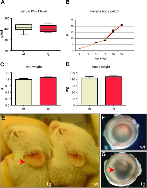
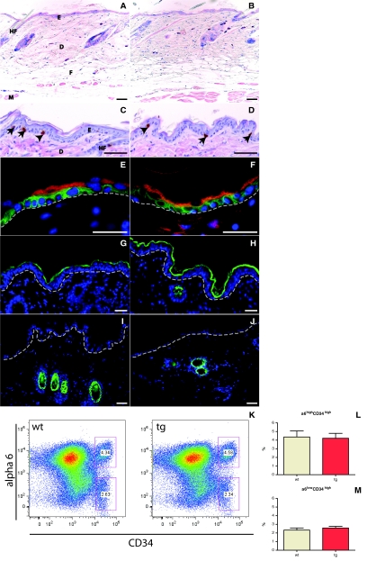
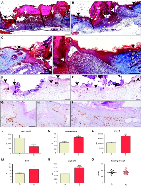
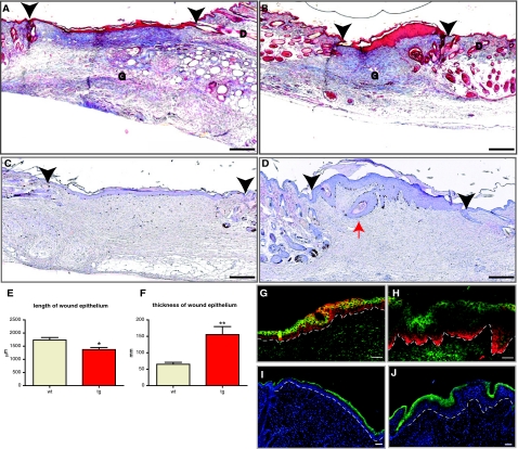
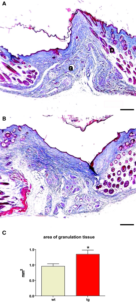
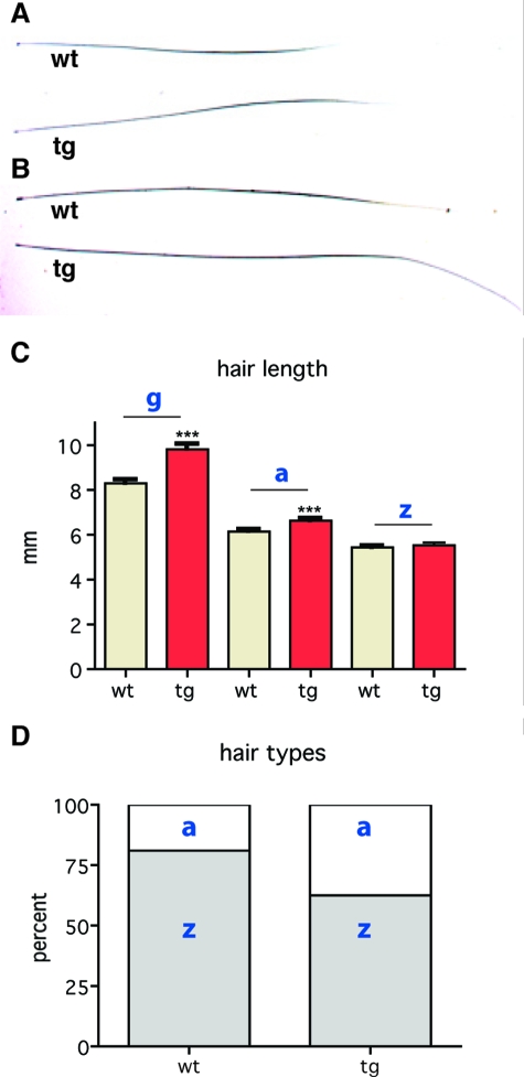
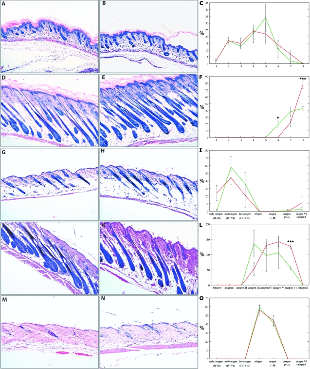
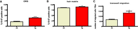
Insulin-like growth factor 1 (IGF-1) is an important regulator of growth, survival, and differentiation in many tissues. It is produced in several isoforms that differ in their N-terminal signal peptide and C-terminal extension peptide. The locally acting isoform of IGF-1 (mIGF-1) was previously shown to enhance the regeneration of both muscle and heart. In this study, we tested the therapeutic potential of mIGF-1 in the skin by generating a transgenic mouse model in which mIGF-1 expression is driven by the keratin 14 promoter. IGF-1 levels were unchanged in the sera of hemizygous K14/mIGF-1 transgenic animals whose growth was unaffected. A skin analysis of young animals revealed normal architecture and thickness as well as proper expression of differentiation and proliferation markers. No malignant tumors were formed. Normal homeostasis of the putative stem cell compartment was also maintained. Healing of full-thickness excisional wounds was accelerated because of increased proliferation and migration of keratinocytes, whereas inflammation, granulation tissue formation, and scarring were not obviously affected. In addition, mIGF-1 promoted late hair follicle morphogenesis and cycling. To our knowledge, this is the first work to characterize the simultaneous, stimulatory effect of IGF-1 delivery to keratinocytes on two types of regeneration processes within a single mouse model. Our analysis supports the use of mIGF-1 for skin and hair regeneration and describes a potential cell type-restricted action.


Figures

Figure 1
Figure 1
K14/mIGF-1 construct and transgene expression. A: Schematic representation of the of K14/mIGF-1 expression cassette. B: Northern blot analysis of total skin RNA samples using a rat mIGF-1 cDNA probe shows the transgene-derived mRNA (red arrow) in the highest expressing line tg C and in the lower expressing line tg F. No transgene-derived transcript is detectable in the wild-type skin sample. C: Northern blot analysis of total RNA from skin (sk), liver (liv), and heart (ht), using the human K14 poly A probe, shows that expression of the transgene is skin-specific in both transgenic lines C and F. D and E: Nonradioactive in situ hybridization of sections from adult back skin of a transgenic animal (line C) with a rat mIGF-1 cDNA probe shows expression of the transgene in the basal layer of the epidermis (b) and in the bulge region (bg) and the outer root sheath of the hair follicle (outer root sheath). Original magnifications, ×40.
Figure 2
Figure 2
Initial phenotypic characterization of K14/mIGF-1 mice. A: Serum levels of IGF-1, measured by IGF-1 immunoenzymometric assay, are similar (P = 0.69) in wild-type (n = 8) and transgenic mice (n = 9). B: Growth curves of wild-type (yellow) (n = 16) and transgenic (red) (n = 14) animals show a similar pattern. C and D: Liver and heart weights are similar (P = 0.1 and P = 0.4, respectively) in wild-type and transgenic mice. E: Transgenic mice (10 dpp) have enlarged ears (red arrow). F: Normal eye in a wild-type mouse (G) and eye from a transgenic mouse with a cataract (red arrow).
Figure 3
Figure 3
Histological and flow cytometric analysis of skin from wild-type and K14/mIGF-1 transgenic animals. A and B: Giemsa-stained back skin sections from wild-type mice (A) and transgenic (B) littermates at 7 weeks after birth indicate a similar general skin architecture and thickness. C and D: BrdU-stained back skin sections from wild-type (C) and transgenic (D) mice show a similar number and localization of proliferating cells (arrows). Sections were counterstained with H&E. E and F: Keratin 14 (green) and keratin 10 (red) are expressed properly in basal or suprabasal layers, respectively, of skin from wild-type (E) and transgenic (F) animals. G and H: Loricrin (green) is expressed in the granular and cornified layers in skin samples from both wild-type (G) and transgenic (H) mice. Green staining in the dermis (H) is a nonspecific staining of the hair shaft. I and J: Keratin 6 (green) expression is restricted to the hair follicles in the skin of both wild-type (I) and transgenic (J) animals. K–M: CD34 and α6 integrin flow cytometric surface expression analysis of 110,000 keratinocytes 47 dpp back skin keratinocytes, revealing two distinct putative stem cell populations (α6high/CD34high, α6low/CD34high) in wild-type and transgenic animals (K). The percentage of α6high/CD34high (L) and α6low/CD34high (M) cells in 47 dpp back skin keratinocytes from wild-type and transgenic mice is shown. DAPI (blue). E, epidermis; D, dermis; HF, hair follicle; M, muscle (panniculus carnosus); F, fatty tissue. Dotted line in E–J indicates the position of the basement membrane. Scale bars: 50 μm (A–D); 25 μm (E–J). Original magnifications: ×10 (A, B); × 40 (C, D, H–J); ×100 (E, F).
Figure 4
Figure 4
Accelerated wound closure in K14/mIGF-1 transgenic mice is driven by an increase in keratinocyte proliferation and migration. Ten wild-type animals (9 wounds) and eleven transgenic animals (13 wounds) were analyzed. A and B: Masson’s trichrome-stained sections from the center of 5-day wounds of wild-type (A) and transgenic (B) animals are shown. C and D: Enlarged images of Masson trichrome-stained sections taken from the center of 5-day wounds of wild-type (C) and transgenic (D) mice, showing the hyperproliferative epithelium (HE). E and F: BrdU-stained sections from the center of 5-day wounds of wild-type (E) and transgenic (F) animals. G--I: Enlarged images of the hyperproliferative epithelium from BrdU-stained wound sections of wild-type (G and H) and transgenic (I) animals. BrdU-stained sections were counterstained with H&E. J: Average diameter of the open wound (P = 0.0207); K: percent wound closure (P = 0.0124); L: area of HE (P < 0.0001); M: number of BrdU-positive cells within the hyperproliferative epithelium (HE) (P = 0.0414); and N: length of the wound epithelium (WE) (P < 0.0001) are shown. O: Wound bursting strength is plotted as a negative force in mmHg required to break the wound. Eleven wild-type animals (35 wounds) and ten transgenic animals (36 wounds) were assayed in the wound-bursting strength experiment. G, granulation tissue; D, dermis; Es, eschar. White Arrows indicate the foremost tips of the epithelial tongues. Black Arrowheads indicate the wound edges. Scale bars: 200 μm (A–F); 100 μm (G–I). Original magnifications: ×10 (A, B, E, F); ×20 (C, D, G–I).
Figure 5
Figure 5
Reduced size, hyperthickened epithelium, and normal epidermal differentiation in 14-day excisional wounds. Six wild-type and six transgenic animals (nine wounds for each genotype) were analyzed. A and B: Masson trichrome-stained sections from the middle of representative 14-day excisional wounds of wild-type (A) and transgenic (B) animals are shown. Granulation tissue (G) and dermis (D) are indicated. C and D: BrdU-stained sections from the middle of representative 14-day wounds of wild-type (C) and transgenic (D) animals are shown. Black arrowheads indicate the edges of the original wound. The red arrow points to an epidermal cyst. E: Graphic representation of the average length of wound epithelium (*P = 0.0099). F: Graphic representation of the average thickness of wound epithelium (**P = 0.0014). G and H: Immunofluorescence staining of the 14-day wound epithelium in wild-type (G) and transgenic (H) mice, using a keratin 14 antibody (red) and a keratin 10 antibody (green). I and J: Immunofluorescence staining of 14-day wound epithelium from wild-type (I) and transgenic (J) mice, stained with a loricrin antibody (green) and DAPI (blue). Dotted line identifies the location of a basement membrane. Scale bars: 200 μm (A–D); 50 μm (G–J). Original magnifications: ×10 (A–D); ×20 (G–J).
Figure 6
Figure 6
Normal epithelial thickness and general granulation tissue architecture in 21-day excisional wounds. A and B: Masson trichrome staining of representative sections from the middle of 21-day excisional wounds taken from wild-type (A) and transgenic (B) mice. Granulation tissue (G) and dermis (D) are indicated. Five wild-type animals (10 wounds) and five transgenic animals (9 wounds) were analyzed. C: The area of granulation tissue (*P = 0.0205) is shown. Scale bars = 200 μm. Original magnifications, ×10.
Figure 7
Figure 7
The effect of K14-driven mIGF-1 expression on the hair coat. A and B: Awl and guard hair shafts are shown. C: Transgenic guard hairs (g) are elongated by 20% (***P > 0.0001), transgenic awl hairs (a) are elongated by 10% (***P > 0.001), whereas zigzag hair (z) length is not changed. D: The frequencies of zigzag (z) and awl (a) hairs in wild-type mice are 79% and 21%, respectively, and in transgenic mice they are 60% and 40%, respectively.
Figure 8
Figure 8
mIGF-1 accelerates late hair follicle morphogenesis and anagen development. Giemsa staining of back skin sections from wild-type (A, D, G, J, M) and transgenic (B, E, H, K, N) mice is shown. C, F, I, L, and O: Quantitative analysis of hair follicles from wild-type (green) and transgenic (red) mice in various stages of morphogenesis and cycling. *P ≥ 0.05 and ***P ≥ 0.001. A–C: One dpp, early morphogenesis. D–F: Eight dpp, late morphogenesis. G–I: Seventeen dpp, early catagen. J–L: Twenty-eight dpp, anagen. M–O: Forty-nine dpp, telogen. X-axes in C and F identify nine distinct hair follicle morphogenesis stages. X-axes in I, L, and O identify distinct stages of hair follicle cycling. Y-axes show the percentage of hair follicles in each distinct stage of hair follicle morphogenesis or cycling. Original magnifications, ×10.
Figure 9
Figure 9
The effect of mIGF-1 on hair follicle keratinocyte proliferation in vivo and primary keratinocyte migration in vitro. A: The percentage of Ki-67-positive keratinocytes in the outer root sheath of the anagen hair follicle on day 28 (***P < 0.0001) is shown. B: The percentage of Ki-67-positive keratinocytes in the matrix of anagen hair follicle bulbs on day 28 (*P < 0.05) is shown. C: The average number of cells, migrated through a transwell insert (6.5 mm in diameter) within 4 hours (mean ± SEM; *P < 0.05) is shown.

References

    1. Dupont J, Holzenberger M. Biology of insulin-like growth factors in development, Birth Defects Res C Embryo Today. 2003;69:257–271. - PubMed
    1. Edmondson SR, Thumiger SP, Werther GA, Wraight CJ. Epidermal homeostasis: the role of the growth hormone and insulin-like growth factor systems. Endocr Rev. 2003;24:737–764. - PubMed
    1. Liu JP, Baker J, Perkins AS, Robertson EJ, Efstratiadis A. Mice carrying null mutations of the genes encoding insulin-like growth factor I (Igf-1) and type 1 IGF receptor (Igf1r). Cell. 1993;75:59–72. - PubMed
    1. Lange M, Thulesen J, Feldt-Rasmussen U, Skakkebaek NE, Vahl N, Jorgensen JO, Christiansen JS, Poulsen SS, Sneppen SB, Juul A. Skin morphological changes in growth hormone deficiency and acromegaly. Eur J Endocrinol. 2001;145:147–153. - PubMed
    1. Lurie R, Ben-Amitai D, Laron Z. Laron syndrome (primary growth hormone insensitivity): a unique model to explore the effect of insulin-like growth factor 1 deficiency on human hair. Dermatology. 2004;208:314–318. - PubMed

Publication types

MeSH terms