. 2008 Nov;173(5):1540-50.
doi: 10.2353/ajpath.2008.080292. Epub 2008 Oct 2.

Focal adhesion kinase-related proline-rich tyrosine kinase 2 and focal adhesion kinase are co-overexpressed in early-stage and invasive ErbB-2-positive breast cancer and cooperate for breast cancer cell tumorigenesis and invasiveness

Affiliations

Focal adhesion kinase-related proline-rich tyrosine kinase 2 and focal adhesion kinase are co-overexpressed in early-stage and invasive ErbB-2-positive breast cancer and cooperate for breast cancer cell tumorigenesis and invasiveness

Emy Behmoaram et al. Am J Pathol. 2008 Nov.

Abstract

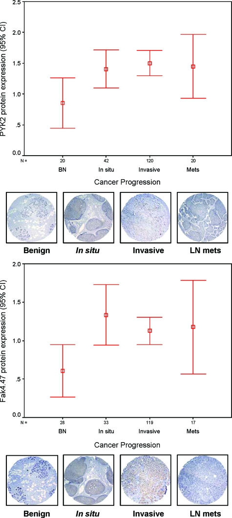
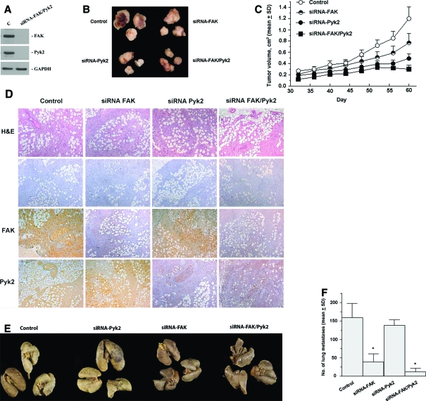
Early cancer cell migration and invasion of neighboring tissues are mediated by multiple events, including activation of focal adhesion signaling. Key regulators include the focal adhesion kinase (FAK) and FAK-related proline-rich tyrosine kinase 2 (Pyk2), whose distinct functions in cancer progression remain unclear. Here, we compared Pyk2 and FAK expression in breast cancer and their effects on ErbB-2-induced tumorigenesis and the potential therapeutic utility of targeting Pyk2 compared with FAK in preclinical models of breast cancer. Pyk2 is overexpressed in tissues from early and advanced breast cancers and overexpressed with both FAK and epidermal growth factor receptor-2 (ErbB-2) in a subset of breast cancer cases. Down-regulation of Pyk2 in ErbB-2-positive, FAK-proficient, and FAK-deficient cells reduced cell proliferation, which correlated with reduced mitogen-activated protein kinase (MAPK) activity. In contrast, Pyk2 silencing had little impact on cell migration and invasion. In vivo, Pyk2 down-regulation reduced primary tumor growth induced by a metastatic variant of ErbB-2-positive MDA 231 breast cancer cells but had little effect on lung metastases in contrast to FAK down-regulation. Dual reduction of Pyk2 and FAK expression resulted in strong inhibition of both primary tumor growth and lung metastases. Together, these data support the cooperative function of Pyk2 and FAK in breast cancer progression and suggest that dual inhibition of FAK and Pyk2 is an efficient therapeutic approach for targeting invasive breast cancer.


Figures

Figure 1
Figure 1
Summary of Pyk2 and FAK expression in progressive breast cancer tissue array specimens. Pyk2 and FAK protein expression in a TMA of benign breast tissues versus carcinoma in situ, invasive, and lymph node metastasis, expressed in percentages. Confidence intervals (95%) show normalized mean protein intensity units of Pyk2 and FAK as determined by quantitative evaluation of immunohistochemistry; scale bars, ±SE. BN, benign; In situ, in situ breast carcinoma; Invasive, invasive breast cancer; Mets, lymph node breast metastasis. Error bars are representative cores of immunostaining of Pyk2 and FAK expression in various stages of breast cancer progression using TMA. Benign breast epithelium showed negative or very low intensity compared with different stages of breast cancer. *P < 0.05 in situ, invasive versus benign.
Figure 2
Figure 2
Overexpression and activation of ErbB-2 receptors in FAK-proficient and -deficient cells. Cells at 70% confluence were serum-starved for 24 hours and kept unstimulated [control (C)] or stimulated with 5 ng/ml HRG for 10 minutes. Cell lysates were immunoprecipitated with anti-ErbB, and the blots were probed using anti-phosphotyrosine antibody and reprobed with the corresponding ErbB-specific antibody. Note the constitutive autophosphorylation of ErbB-2 in ErbB-2 expressors and the enhanced ErbB-2 and ErbB-3 phosphorylation after stimulation with HRG.
Figure 3
Figure 3
ErbB-2/3 receptor overexpression partially rescues tumorigenesis defects of FAK−/− cells. A: Cell proliferation was measured using 3-(4,5-dimethylthiazo-2-yl)-2,5-diphenyltetrazolium bromide metabolic assay on exponentially growing cells after 48 hours of HRG stimulation and nonstimulated controls. Cell migration was determined by the phagokinetic track assay, where cells were allowed to migrate on gold colloidal particle-covered coverslips in the presence or absence of HRG. Total cell motility was quantified by processing the average area free of gold colloidal particles in square millimeters ± SD. Cell invasion was evaluated by culturing cells on Boyden chambers coated with matrigel and stimulating them for 48 hours using HRG as chemotactic agent in the lower chamber as described in Materials and Methods. The number of invading and hematoxylin stained cells were counted, and the percentage of invading cells was determined. Values are means ± SD from three independent experiments (P < 0.05, ErbB-2/3 versus control). *P < 0.01, ErbB-2/3 FAK−/− versus control FAK−/−; P < 0.001, ErbB-2/3 FAK+/+ versus control FAK+/+ B: Exponentially growing cells (1.0 × 10⁶) were injected subcutaneously into the flank of SCID mice (left). Tumor growth was monitored over time as indicated in Materials and Methods. Each point represents the average of 8 animals ± SEM. Tumor volumes are not applicable for FAK+/+ at day 60 because animals were sacrificed on day 29 because of debilitating large tumors. Exponentially growing cells (1.0 × 10⁶) were inoculated intravenously to SCID mice (right). Mice were sacrificed on day 42 after inoculation, and lungs were fixed in 10% Bouin’s fixative. Lung surface metastases were counted using a stereomicroscope. Each bar represents the average lung metastases (n = 8) ± SEM. *P < 0.005, ErbB-2/3 FAK−/− versus ErbB-2/3 FAK+/+.
Figure 4
Figure 4
Impact of Pyk2 on ErbB-induced cell proliferation and cell invasion. A: A representative Western blot analysis on cells stably expressing Pyk2 siRNA showing more than 80% inhibition compared with control cells. Relative FAK expression is also shown. Shown are parental FAK−/− cells, ErbB-2/3 FAK−/− cells, ErbB-2/3 FAK−/− cells expressing PSR, and ErbB-2/3 FAK−/− cells expressing Pyk2 siRNA. Control and ErbB-2/3 FAK+/+ cells are shown as positive controls. Glyceraldehyde-3-phosphate dehydrogenase was used as internal control. Cell proliferation of control and ErbB-expressing FAK−/− cells and their matched pairs where Pyk2 was inhibited by siRNA (top). Cells were seeded on 96 wells, serum-starved for 24 hours and then stimulated with 5 ng/ml HRG. Cell proliferation was examined on day 5 using the 3-(4,5-dimethylthiazo-2-yl)-2,5-diphenyltetrazolium bromide assay as described in Materials and Methods (*P < 0.001, ErbB-2/3 FAK−/− siRNA Pyk2 versus ErbB-2/3 FAK+/+ siRNA Pyk2). Cell invasion was determined on the Boyden chamber of control and ErbB-expressing FAK−/− cells and their matched pair where Pyk2 was inhibited by siRNA (bottom). Cells were seeded on Boyden chambers coated with matrigel and stimulated for 48 hours using heregulin as chemotactic agent in the lower chamber as described in Materials and Methods. Values reported are means ± SD from at least three independent experiments (*P < 0.01, ErbB-2/3 FAK+/+ PSR versus ErbB-2/3 FAK+/+ siRNA Pyk2). B: Immunofluorescence staining showing effective overexpression of ErbB-2 in control and ErbB-2-transformed MDA 231 cells (top). Green fluorescent protein (GFP) and 4,6-diamidino-2-phenylindole (Dapi) staining are used as cellular controls. Knockdown of Pyk2 and FAK expression in MDA 231 ErbB-2 cells demonstrated in a representative Western blot analysis (middle). Cells stably expressed specific Pyk2 siRNA and FAK siRNA resulting in effective down-regulation of Pyk2 and FAK expression, respectively, compared with control cells and cells expressing PSR empty vector. Glyceraldehyde-3-phosphate dehydrogenase was used as internal control. Cell proliferation and cell invasion experiments were performed on control cells, cells expressing PSR, cells expressing FAK siRNA, and cells expressing Pyk2 siRNA (bottom). Cell proliferation was evaluated after 5 days using the 3-(4,5-dimethylthiazo-2-yl)-2,5-diphenyltetrazolium bromide metabolic assay. Cell invasion was determined using the Boyden chamber invasion assay and HRG as a chemoattractant for 48 hours as described in Materials and Methods. *P < 0.005, compared with PSR controls.
Figure 5
Figure 5
Pyk2 regulates ErbB-induced activation of MAPKs in FAK-deficient cells. A: Cell extracts from parental FAK−/− cells, ErbB-2/3-FAK−/− cells, and their matched pairs expressing PSR or siRNA Pyk2. ErbB-2/3 FAK+/+ cells are shown as a control. Cells were serum-starved for 24 hours and then stimulated with HRG for 10 minutes. Total cell extracts were then collected and used for Western blot analysis to monitor the levels of phospho-Erk1/2. *P < 0.005, compared with control FAK−/−. Total Erk1/2 was used as internal control. Pyk2 expression is also shown to demonstrate efficiency of siRNA knockdown. B: Cell extracts from cells transiently transfected with increasing amount of siRNA Pyk2 were used to examine the levels of phospho-Erk following the same conditions as in A. Western blot analysis for Pyk2 expression shows efficiency of dose-dependent knockdown. Each experiment was repeated at least in triplicate. Error bars represent the average phosphor-Erk/Erk ± SD. *P < 0.01, siRNA Pyk2 25 nmol/L/50 nmol/L FAK−/− compared with PSR control; **P < 0.005, siRNA Pyk2 100 nmol/L FAK−/− compared with PSR control.
Figure 6
Figure 6
Inhibition of Pyk2 and FAK in the MDA 231-M breast cancer model leads to distinct inhibition of tumor growth versus lung metastases. A: A representative Western blot analysis of cells demonstrating dual inhibition of both FAK and Pyk2 through stable expression of FAK and Pyk2 siRNA. Cells expressing siRNA show more than 90% inhibition compared with control cells. B: Representative appearance of primary tumors after implantation of cells into the mammary fat pad of Scid mice. C: Tumor growth kinetics monitored over time as indicated in Materials and Methods. Each point represents the mean of eight mice ± SD. D: Immunohistochemistry on primary tumor tissue from control, siRNA FAK, siRNA Pyk2, and siRNA FAK/Pyk2. Control represents tumors induced by cells expressing PSR plasmid. Tumors were fixed in formalin, embedded in paraffin, and used for immunohistochemical analysis for FAK and Pyk2 or stained with H&E as described in Materials and Methods. −, negative control where primary antibodies were omitted. E: Incidence of lung metastases from the same group of animals. Representative lungs from mice inoculated with the various cell types. F: Quantification of lung surface metastases was determined after tissue fixation with Bouin. Results are mean surface lung metastases (n = 8) ± SD. *P < 0.001, compared with control.

Similar articles

Cited by

References

    1. Du QS, Ren XR, Xie Y, Wang Q, Mei L, Xiong WC. Inhibition of PYK2-induced actin cytoskeleton reorganization: PYK2 autophosphorylation and focal adhesion targeting by FAK. J Cell Sci. 2001;114:2977–2987. - PubMed
    1. Chen HC, Appeddu PA, Parsons JT, Hildebrand JD, Schaller MD, Guan JL. Interaction of focal adhesion kinase with cytoskeletal protein talin. J Biol Chem. 1995;270:16995–16999. - PubMed
    1. Vadlamudi RK, Adam L, Nguyen D, Santos M, Kumar R. Differential regulation of components of the focal adhesion complex by heregulin: role of phosphatase SHP-2. J Cell Physiol. 2002;190:189–199. - PubMed
    1. Giancotti FG, Ruoslahti E. Integrin signaling. Science. 1999;285:1028–1032. - PubMed
    1. Sieg DJ, Hauck CR, Ilic D, Klingbeil CK, Schaefer E, Damsky CH, Schlaepfer DD. FAK integrates growth-factor and integrin signals to promote cell migration. Nat Cell Biol. 2000;2:249–256. - PubMed

Publication types

MeSH terms