. 2008 Oct 15;181(8):5313-22.
doi: 10.4049/jimmunol.181.8.5313.

PD-1-dependent mechanisms maintain peripheral tolerance of donor-reactive CD8+ T cells to transplanted tissue

Affiliations

PD-1-dependent mechanisms maintain peripheral tolerance of donor-reactive CD8+ T cells to transplanted tissue

Brent H Koehn et al. J Immunol. .

Abstract

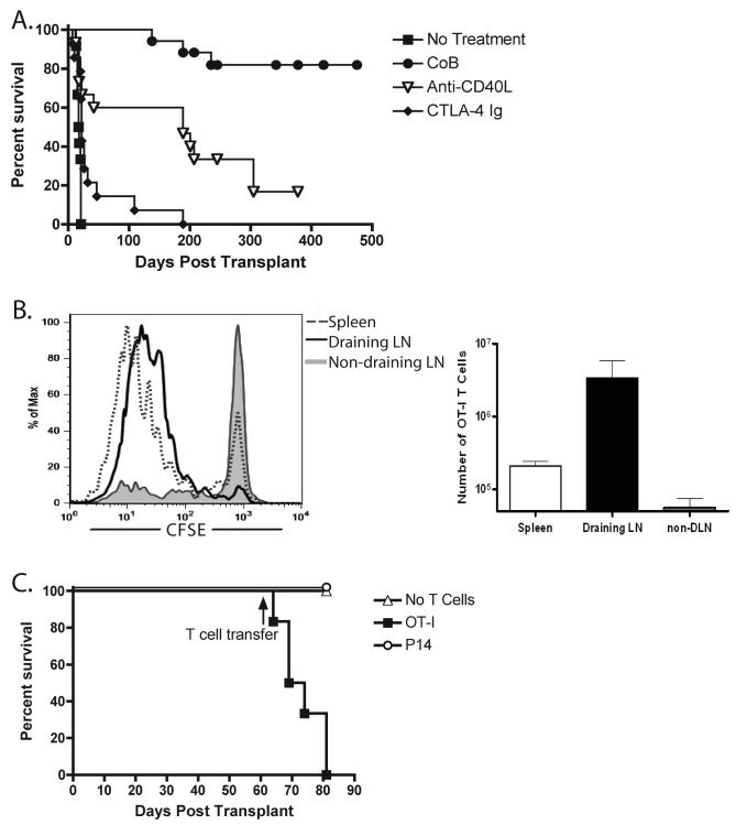
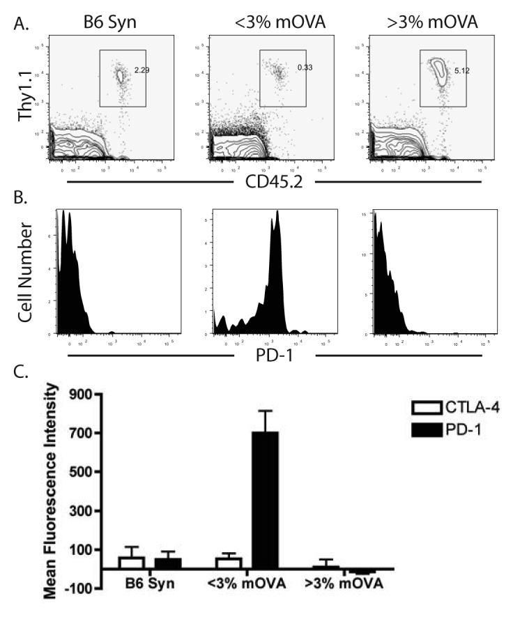
Peripheral mechanisms of self-tolerance often depend on the quiescent state of the immune system. To what degree such mechanisms can be engaged in the enhancement of allograft survival is unclear. To examine the role of the PD-1 pathway in the maintenance of graft survival following blockade of costimulatory pathways, we used a single-Ag mismatch model of graft rejection where we could track the donor-specific cells as they developed endogenously and emerged from the thymus. We found that graft-specific T cells arising under physiologic developmental conditions at low frequency were actively deleted at the time of transplantation under combined CD28/CD40L blockade. However, this deletion was incomplete, and donor-specific cells that failed to undergo deletion up-regulated expression of PD-1. Furthermore, blockade of PD-1 signaling on these cells via in vivo treatment with anti-PD-1 mAb resulted in rapid expansion of donor-specific T cells and graft loss. These results suggest that the PD-1 pathway was engaged in the continued regulation of the low-frequency graft-specific immune response and thus in maintenance of graft survival.


Figures

Figure 1
Figure 1. Long-term graft survival in costimulation blockade-treated recipients was maintained despite the presence of actively presented antigen
(A) mOVA skin grafts were placed onto B6 recipients, which were then treated on day 0 and 2 with 0.25 mg anti-CD40L (MR-1), 0.25 mg CTLA-4 Ig or the combination of both (CoB) (n ≥ 10 mice per group). Treatment with CoB resulted in long-term graft survival with an MST of >500 days (. (B) 5×10⁶ CFSE-labeled OT-I T cells were transferred i.v. to mOVA skin grafted B6 recipients (healed >60 days) (n=3 mice per group). 48 hours after transfer, OT-I accumulation and proliferation were assessed in the spleen, draining inguinal LN, or non-draining contralateral inguinal LN. Dividing OT-I T cells were detected in the spleen and draining LN, but not in the contralateral non-draining LN. (C) 5×10⁶ CFSE labeled OT-I T cells or irrelevant P14 T cells were transferred i.v. to mOVA skin grafted B6 recipients (healed >60 days) (n=5 mice per group). Data demonstrate that adoptive transfer of naïve OT-I, but not P14, T cells precipitated graft rejection in mOVA skin graft recipients.
Figure 2
Figure 2
Treatment of low but not high frequency OT-I chimeras with costimulation blockade following mOVA skin graft placement resulted in long-term graft survival. (A) B6 mice were treated with busulfan and mixed donor bone marrow derived from WT B6 and OT-I TCR tg mice as described in Materials and Methods. The three left panels depict the gating strategy to identify OVA257-264/Kb-specific OT-I T cells as they develop in these recipients, and the right panel depicts the kinetics of the appearance of CD8+ Thy1.1+ Vα2+ OT-I T cells in the peripheral blood following bone marrow transplant. Each line represents an individual mouse. (B) mOVA skin grafts were placed onto OT-I chimeric mice harboring the indicated frequencies of peripheral blood OT-I T cells (0.1-1% n=6, 1-3% n=5, 3-5% n=8, 5-12% n=8). Mice were treated days 0 and 2 with CTLA-4 Ig and αCD40L. Results indicated that recipients bearing >3% peripheral OT-I T cells rapidly rejected their grafts in the presence of costimulation blockade, mice bearing <3% peripheral OT-I cells enjoyed long-term graft survival. (C) Peripheral blood frequencies of OT-I (Thy1.1+) T cells after skin grafting are depicted (n ≥ 8 mice/group). The frequencies of OT-I T cells in mice with an initial OT-I frequency of <3% declined following mOVA skin graft and costimulation blockade. (E) Frequencies of donor-BM derived non-T cell chimerism after skin grafting are depicted (n ≥ 8 mice/group). Constant levels of non-T cell donor chimerism were observed in these animals.
Figure 3
Figure 3. Maintained graft survival was associated with PD-1 expression on donor-reactive T cells
The phenotype of OT-I T cells was analyzed in B6 OT-I chimeras that received either a syngeneic B6 graft or an mOVA graft. (A) The frequencies of Thy1.1+ CD45.2+ (OT-I) T cells in recipients of syngeneic or mOVA skin grafts. Plots are gated on CD8+ lymphocytes. (B) The level of PD-1 expression OT-I T cells from these mice was determined. Plots shown are gated OT-I T cells, and demonstrate that high PD-1 expression is observed on T cells from low frequency OT-I chimeras receiving an mOVA skin graft and costimulation blockade. (C) Peripheral OT-I T cells from OT-I chimeric mice receiving syngeneic B6 skin grafts (n=5), low frequency OT-I chimeric mice receiving mOVA skin grafts (n=16), and high frequency OT-I chimeric mice receiving mOVA skin grafts (n=3) were analyzed for the presence of PD-1 and CTLA-4. The mean fluorescence intensity of these markers on CD8+ Thy1.1+ Vα2+ cells reveal that while CTLA-4 was not upregulated in any of the groups, low frequency OT-I chimeras receiving mOVA skin grafts consistently expressed PD-1 (p<0.05).
Figure 4
Figure 4. In vivo PD-1 blockade precipitated graft loss and induced donor-reactive T cell expansion
Low frequency (<3%) OT-I chimeric animals with mOVA skin grafts surviving >90 days were treated with indicated antibody at day 0 (>120 days post transplant). Anti-PD-1 (J43) or hamster IgG isotype control were given as 500 µg on day 0 and 250 µg every other day; rat anti-mouse PD-L1 (10F.9G2) or rat IgG2b isotype control was given at 200 µg every third day. Both regimens were discontinued after 2 weeks. Anti-CD25 was given at 500 µg on days 0, 2, 4, and 6. n ≥ 8 mice per group. (A), Treatment with anti-PD-1 or anti-PDL-1, but not isotype control or anti-CD25, precipitated graft rejection in recipients of long-surviving skin grafts. (B) The frequency of OT-I CD8+ T cells in low frequency OT-I chimeric animals increased following in vivo PD-1 blockade (day 120). (C), Representative histograms of PD-1 expression on CD8+ Thy1.1+ Vα2+ T cells prior to (day 97) and post in vivo PD-L1 blockade (day 150). PD-1 is down regulated on CD8+ Thy1.1+ Vα2+ T cells following anti-PDL-1 treatment and subsequent graft loss, but is maintained on CD8+ Thy1.1+ Vα2+ T cells in recipients treated with rat IgG2b isotype control.
Figure 5
Figure 5. OT-I T cells regained proliferative capacity and cytokine effector function post PD-1 blockade
Low frequency OT-I chimeric animals with surviving mOVA skin grafts for >60 days were treated with anti-PD-1 or control hamster Ig. At days 6 and 7 post-PD-1 blockade, mice were given 1mg BrdU in PBS i.p. (n=3-4 mice per group). At day 8 following treatment, spleen and LN were harvested and interrogated for (A) BrdU uptake or (B) the ability to make IFN-γ after 5 hours of SIINFEKL restimulation. Results indicated that low frequency OT-I chimeras treated with anti-PD-1 exhibited an increased frequency of BrdU+ cycling cells (A, p=0.0073) and IFN-γ+ cells (B, p=0.0373), as compared to mice receiving hamster isotype control antibody.

References

    1. Jenkins MK. The ups and downs of T cell costimulation. Immunity. 1994;1:443–446. - PubMed
    1. Turka LA, Linsley PS, Lin H, Brady W, Leiden JM, Wei R, Gibson ML, Zheng X, Myrdal S, Gordon D, Bailey T, Bolling SF, Thompson CB. T-cell activation by the CD28 ligand B7 is required for cardiac allograft rejection in vivo. Proc. Natl. Acad. Sci. USA. 1992;89:11102–11105. - PMC - PubMed
    1. Pearson TC, Alexander DZ, Corbascio M, Hendrix R, Ritchie SC, Linsley PS, Faherty D, Larsen CP. Analysis of the B7 costimulatory pathway in allograft rejection. Transplantation. 1997;63:1463–1469. - PubMed
    1. Pearson TC, Alexander DZ, Winn KJ, Linsley PS, Lowry RP, Larsen CP. Transplantation tolerance induced by CTLA4-Ig. Transplantation. 1994;57:1701–1706. - PubMed
    1. Sayegh MH, Akalin E, Hancock WW, Russell ME, Carpenter CB, Linsley PS, Turka LA. CD28-B7 blockade after alloantigenic challenge in vivo inhibits Th1 cytokines but spares Th2. J. Exp. Med. 1995;181:1869–1874. - PMC - PubMed