. 2008 Oct 15;181(8):5545-50.
doi: 10.4049/jimmunol.181.8.5545.

Interleukin-10 promotes Mycobacterium tuberculosis disease progression in CBA/J mice

Affiliations

Interleukin-10 promotes Mycobacterium tuberculosis disease progression in CBA/J mice

Gillian L Beamer et al. J Immunol. .

Abstract

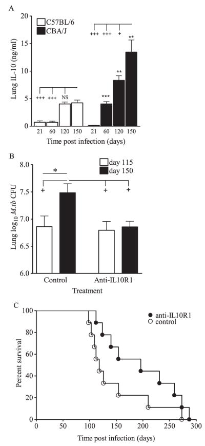
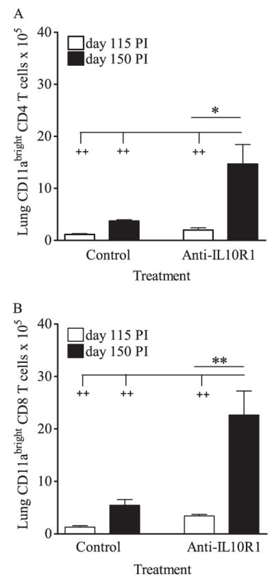
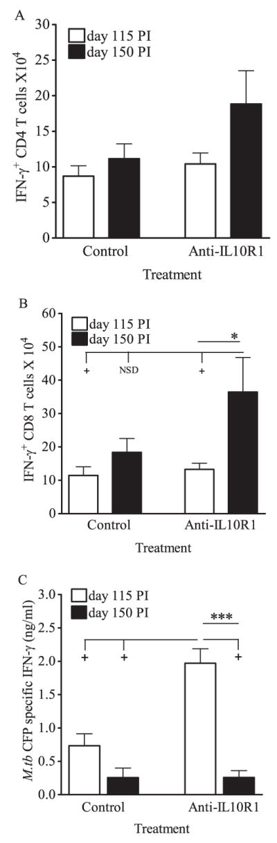
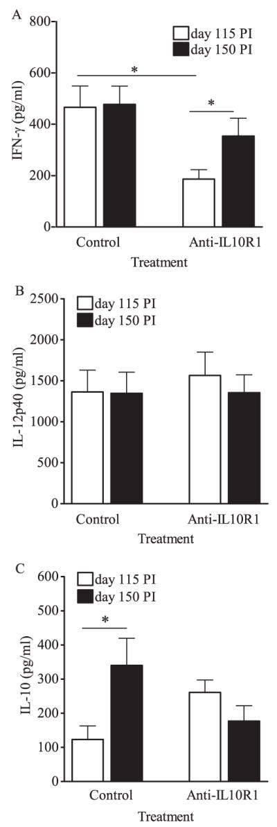
IL-10 is a potent immunomodulatory cytokine that affects innate and acquired immune responses. The immunological consequences of IL-10 production during pulmonary tuberculosis (TB) are currently unknown, although IL-10 has been implicated in reactivation TB in humans and with TB disease in mice. Using Mycobacterium tuberculosis-susceptible CBA/J mice, we show that blocking the action of IL-10 in vivo during chronic infection stabilized the pulmonary bacterial load and improved survival. Furthermore, this beneficial outcome was highly associated with the recruitment of T cells to the lungs and enhanced T cell IFN-gamma production. Our results indicate that IL-10 promotes TB disease progression. These findings have important diagnostic and/or therapeutic implications for the prevention of reactivation TB in humans.


Figures

FIGURE 1
FIGURE 1
A role for IL-10 in driving disease progression in CBA/J mice. A, CBA/J and C57BL/6 mice were aerogenically infected with M.tb Erdman, euthanized at 21, 60, 120, and 150 days postinfection, lungs were homogenized, and IL-10 was detected by ELISA in clarified homogenates. Data are the means ± SD of one experiment with four mice per strain per time point analyzed by Student’s t test (**, p < 0.01 and ***, p < 0.001) at each time point and by one-way ANOVA with Tukey’s posttest for multigroup comparisons over time (+, p < 0.05 and +++, p < 0.001). B, CBA/J mice were aerogenically infected with M.tb Erdman and injected weekly with anti-IL-10R1 or control Ab starting at day 90 postinfection. Mice were euthanized at days 115 and 150, lungs homogenized and plated onto 7H11 agar, and M.tb CFU were determined after 21 days incubation at 37°C. Data are the means ± SEM of five independent experiments each with three to six mice per treatment group per time point, analyzed pairwise by Student’s t test (*, p < 0.05) and by one-way ANOVA with Tukey’s posttest for multigroup comparisons (+, p < 0.05). Total numbers of mice: control group 115 days, n = 26; control group 150 days, n = 25; anti-IL-10R group 115 days, n = 22; anti-IL-10R group day 150, n = 22. For survival, separate groups of anti-IL-10R1-treated mice and controls were euthanized when signs of morbidity developed (C) and statistical significance was determined by log-rank analysis. Survival data are combined from two independent experiments.
FIGURE 2
FIGURE 2
Anti-IL-10R1 treatment increased lung cellularity during chronic M.tb infection in CBA/J mice. Mice were aerogenically infected with M.tb Erdman, injected weekly with anti-IL-10R1 or control Ab starting at day 90 postinfection, and euthanized at days 115 and 150. Isolated lung cells were counted, fixed, and labeled with anti-CD3, anti-CD4, and anti-CD8 for flow cytometric analysis. Total number of cells isolated (A), number of CD4 T cells (B), and number of CD8 T cells (C) are the means ± SEM of five independent experiments, each with three to six mice per group per time point, analyzed by Student’s t test for pairwise comparisons and one-way ANOVA with Tukey’s posttest for multigroup comparisons. Total numbers of mice: control group 115 days, n = 27; control group 150 days, n = 24; anti-IL-10R group 115 days, n = 24; anti-IL-10R group day 150, n = 22.
FIGURE 3
FIGURE 3
Anti-IL-10R1 treatment of CBA/J mice increased the numbers of CD11abright T cells in the lung during chronic M.tb infection. Mice were aerogenically infected with M.tb Erdman, injected weekly with anti-IL-10R1 or control Ab starting at day 90 postinfection, and euthanized at days 115 and 150. Isolated lung cells were counted, fixed, and labeled with anti-CD3, anti-CD4, anti-CD8, and anti-CD11a for flow cytometric analysis. Numbers of CD11abright CD4 (A) and CD8 (B) T cells are the means ± SEM and are representative of two independent experiments, each with four to five mice per group per time point, with pairwise analyses using Student’s t test (*, p < 0.05 and **, p < 0.001) or multigroup analysis using one-way ANOVA with Tukey’s posttest (+, p < 0.05; ++, p < 0.01; +++, p < 0.001).
FIGURE 4
FIGURE 4
Anti-IL-10R1 treatment improved IFN-γ production during chronic M.tb infection of CBA/J mice. Mice were aerogenically infected with M.tb Erdman, injected weekly with anti-IL-10R1 or control Ab starting at day 90 postinfection, and euthanized at days 115 and 150. Isolated lung cells were stimulated with anti-CD3 and anti-CD28 for 4 h, fixed, labeled with anti-TCRβ, anti-CD4, and anti-CD8, permeabilized, and labeled with anti-IFN-γ for intracellular flow cytometry. Numbers of IFN-γ+ CD4 T cells (A) and IFN-γ+ CD8 T cells (B) are the means ± SEM of four independent experiments, each with three to six mice per group per time point, with pairwise analyses using Student’s t test (*, p < 0.05) and multigroup comparisons using one-way ANOVA (+, p < 0.05; NSD, no significant difference). Total numbers of mice: control group 115 days, n = 19; control group 150 days, n = 16; anti-IL-10R group 115 days, n = 20; anti-IL-10R group day 150, n = 18. C, Isolated lung cells were stimulated ex vivo with M.tb culture filtrate protein, and secreted Ag-specific IFN-γ in supernatants was quantified by ELISA. Data are the means ± SEM of two independent experiments, each with four to five mice per treatment group per time point, with pairwise analyses by Student’s t test (***, p < 0.001) and multigroup comparisons using one-way ANOVA (+, p < 0.05). Total numbers of mice: control group 115 days, n = 9; control group 150 days, n = 8; anti-IL-10R group 115 days, n = 9; anti-IL-10R group day 150, n = 8.
FIGURE 5
FIGURE 5
Cytokines in lung tissue following anti-IL-10R1 treatment during chronic M.tb infection. CBA/J mice were aerogenically infected with M.tb Erdman and injected weekly with anti-IL-10R1 or control Ab starting at day 90 postinfection. Mice were euthanized at days 115 and 150, lungs homogenized, and IFN-γ (A), IL-12p40 (B), and IL-10 (C) quantified by ELISA in clarified supernatants. Data shown are the means ± SEM of three (A and C) or four (B) independent experiments, each with three to six mice per treatment group per time point, and analyzed in pairs by Student’s t test (*, p < 0.05). Total numbers of mice (A and C): control group 115 days, n = 17; control group 150 days, n = 17; anti-IL-10R group 115 days, n = 14; anti-IL-10R group day 150, n = 14. Total numbers of mice (B): control group 115 days, n = 22; control group 150 days, n = 18; anti-IL-10R group 115 days, n = 22; anti-IL-10R group day 150, n = 17.

References

    1. Moore KW, de Waal Malefyt R, Coffman RL, O’Garra A. Interleukin-10 and the interleukin-10 receptor. Annu Rev Immunol. 2001;19:683–765. - PubMed
    1. de Waal Malefyt R, Abrams J, Bennett B, Figdor C, de Vries J. Interleukin 10 (IL-10) inhibits cytokine synthesis by human monocytes: an autoregulatory role of IL-10 produced by monocytes. J Exp Med. 1991;174:1209 –1220. - PMC - PubMed
    1. Fiorentino D, Zlotnik A, Mosmann T, Howard M, O’Garra A. IL-10 inhibits cytokine production by activated macrophages. J Immunol. 1991;147:3815–3222. - PubMed
    1. Bogdan C, Vodovotz Y, Nathan C. Macrophage deactivation by interleukin 10. J Exp Med. 1991;174:1549 –1555. - PMC - PubMed
    1. Koppelman B, Neefjes JJ, de Vries JE, de Waal Malefyt R. Interleukin-10 down-regulates MHC class II alphabeta peptide complexes at the plasma membrane of monocytes by affecting arrival and recycling. Immunity. 1997;7:861– 871. - PubMed

Publication types