Multicenter Study
. 2008 Oct 15;181(8):5768-74.
doi: 10.4049/jimmunol.181.8.5768.

B cells from patients with Graves' disease aberrantly express the IGF-1 receptor: implications for disease pathogenesis

Affiliations
Multicenter Study

B cells from patients with Graves' disease aberrantly express the IGF-1 receptor: implications for disease pathogenesis

Raymond S Douglas et al. J Immunol. .

Abstract

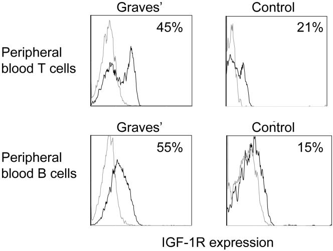
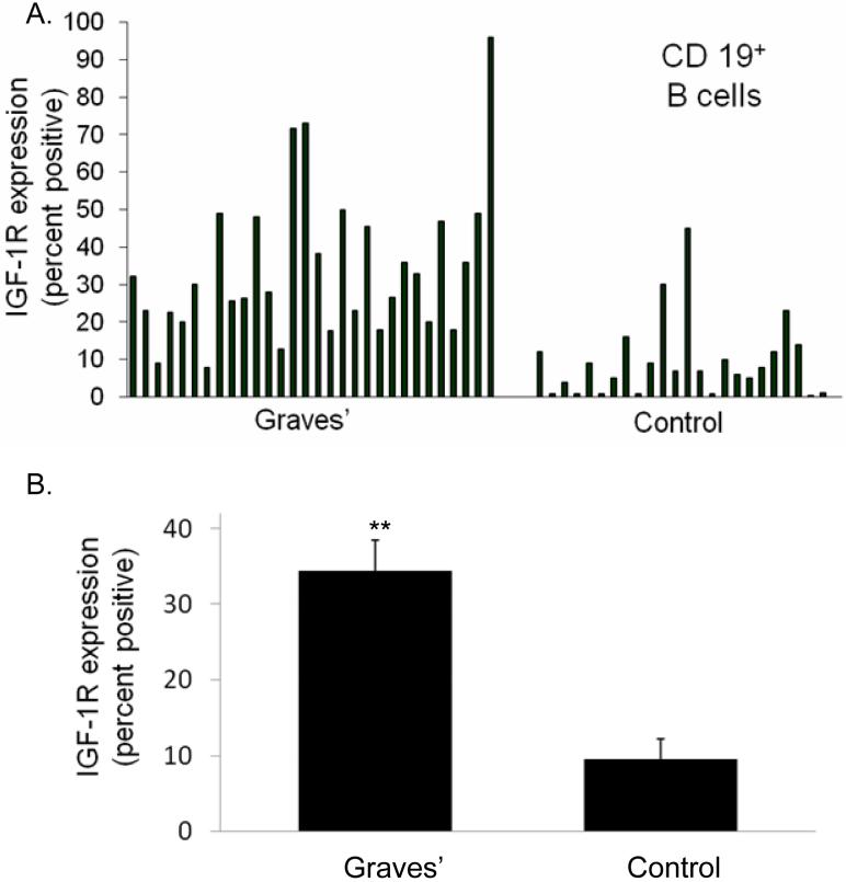
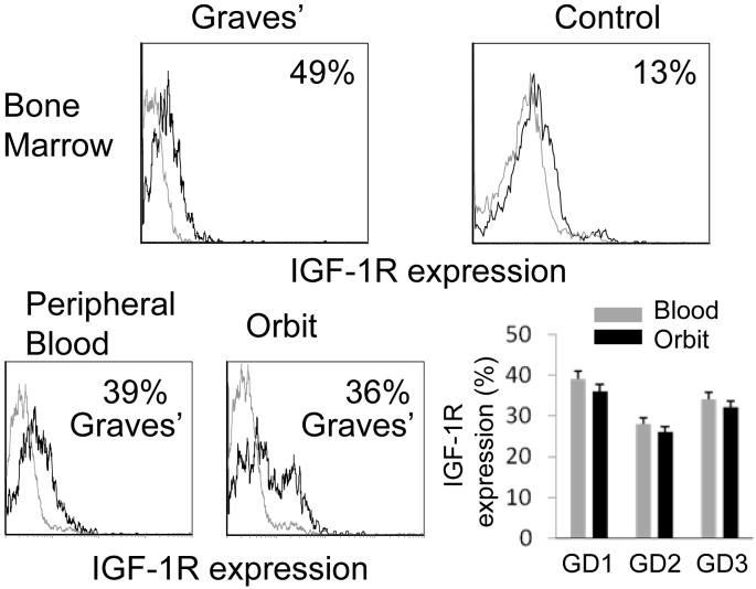
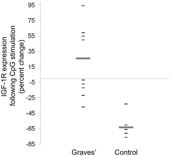
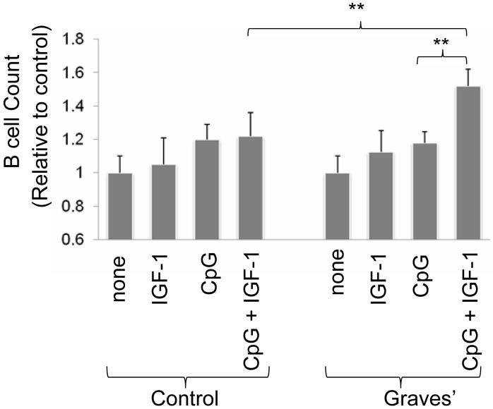
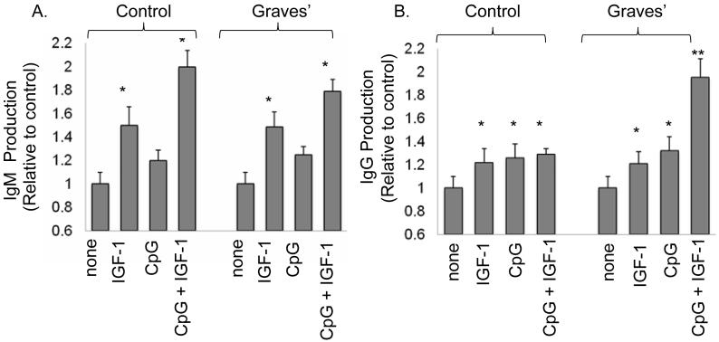
Graves' disease (GD) is an autoimmune process involving the thyroid and connective tissues in the orbit and pretibial skin. Activating anti-thyrotropin receptor Abs are responsible for hyperthyroidism in GD. However, neither these autoAbs nor the receptor they are directed against have been convincingly implicated in the connective tissue manifestations. Insulin-like growth factor-1 receptor (IGF-1R)-bearing fibroblasts overpopulate connective tissues in GD and when ligated with IgGs from these patients, express the T cell chemoattractants, IL-16, and RANTES. Disproportionately large fractions of peripheral blood T cells also express IGF-1R in patients with GD and may account, at least in part, for expansion of IGF-1R(+) memory T cells. We now report a similarly skewed B cell population exhibiting the IGF-1R(+) phenotype from the blood, orbit, and bone marrow of patients with GD. This expression profile exhibits durability in culture and is maintained or increased with CpG activation. Moreover, IGF-1R(+) B cells produce pathogenic Abs against the thyrotropin receptor. In lymphocytes from patients with GD, IGF-1 enhanced IgG production (p < 0.05) and increased B cell expansion (p < 0.02) in vitro while those from control donors failed to respond. These findings suggest a potentially important role for IGF-1R display by B lymphocytes in patients with GD in supporting their expansion and abnormal Ig production.


Figures

Figure 1
Figure 1
Increased fraction of peripheral blood T and B cells from a patient with GD display IGF-1R compared to those from a control donor. PBMCs were stained with anti-CD3, anti-CD19 and anti-IGF-1R Abs as described in “Methods” and subjected to multi-parameter flow cytometry. The grey open histograms represent staining with isotype control Abs.
Figure 2
Figure 2
A disproportionate fraction of peripheral blood B cells from 30 patients with GD express IGF-1R compared with that found in 24 control donors (A) individual data sets demonstrating fractional IGF-1R+ B cells. (B) Analysis of IGF-1R display in B cells as an aggregate of multiple patients with GD vs control donors. (34 ± 4% IGF-1R+ B cells (mean ± SE, n=30) vs 9 ± 3% IGF-1R+ control B cells (n=24)). Data are expressed as mean ± standard error (**p< 1 × 10⁻⁶).
Figure 3
Figure 3
Increased fraction of bone marrow- and orbit-derived B cells from patients with GD express IGF-1R. Bone marrow aspirate (upper panels) from a patient with GD demonstrates increased fraction of IGF-1R+ B cells compared to a control aspirate. B cells isolated from the orbit and peripheral blood of a patient with GD (lower panels) display increased fraction of IGF-1+ B cells (representative flow cytometry histograms of three patients). Isotype Ab staining is shown as an open grey histogram. (lower right) IGF-1R expression by B cells from the peripheral blood and orbit, each sample pair obtained from one of 3 separate patients. .
Figure 4
Figure 4
Abundance of IGF-1R+ B cells from patients with GD is maintained or increased with CpG activation compared to those from control donors. PBMCs were isolated and cultured in the presence of CpG (2 μg/ml). IGF-1R display was assessed by flow cytometry at 24 h and is expressed as percent change from un-stimulated cultures. Each datum point represents a single patient’s sample. Broad horizontal bars indicate mean values for each group (GD vs control, p< 0.002).
Figure 5
Figure 5
IGF-1 potentiates B cell expansion when added together with a concentration of CpG yielding a sub-maximal response. B cell number was assessed as described in “Methods” after 5 days in culture with CpG (2 μg/ml), IGF-1 (10ng.ml) as single agents or in combination. Data derive from 5 independent experiments (mean ± SE, ** denotes differences at p<0.02).
Figure 6
Figure 6
IGF-1 potentiates IgM and IgG production, both in B cells from patients with GD and in those from control donors. IgM and IgG were assayed after 12 days in culture stimulated with CpG and/or IGF-1. (A) IGF-1 (10 ng/ml) alone or in combination with CpG (2 μg/ml), significantly increase IgM production by GD and control B cells. (B) CpG and IGF-1, used as single agents enhanced IgG production by B cells from both sources (*p<0.05 vs US). The combination of CpG and IGF-1 significantly increases IgG production only in B cells from patients with GD compared with treatment with either single agent (**p<0.01). Results are expressed as the mean ± SE of 5 independent experiments.

References

    1. Dorner T, Lipsky PE. Signalling pathways in B cells: implications for autoimmunity. Curr Top Microbiol Immunol. 2006;305:213–240. - PubMed
    1. Martinez-Gamboa L, Brezinschek HP, Burmester GR, Dorner T. Immunopathologic role of B lymphocytes in rheumatoid arthritis: rationale of B cell-directed therapy. Autoimmun Rev. 2006;5:437–442. - PubMed
    1. Prabhakar BS, Bahn RS, Smith TJ. Current perspective on the pathogenesis of Graves’ disease and ophthalmopathy. Endocr Rev. 2003;24:802–835. - PubMed
    1. Tandon N, Metcalfe RA, Barnett D, Weetman AP. Expression of the costimulatory molecule B7/BB1 in autoimmune thyroid disease. Quart J Med. 1994;87:231–236. - PubMed
    1. Sato S, Fujimoto M, Hasegawa M, Takehara K, Tedder TF. Altered B lymphocyte function induces systemic autoimmunity in systemic sclerosis. Mol Immunol. 2004;41:1123–1133. - PubMed

Publication types

MeSH terms