Comparative Study
. 2008 Oct 15;181(8):5791-802.
doi: 10.4049/jimmunol.181.8.5791.

Subsets of myeloid-derived suppressor cells in tumor-bearing mice

Affiliations
Comparative Study

Subsets of myeloid-derived suppressor cells in tumor-bearing mice

Je-In Youn et al. J Immunol. .

Abstract

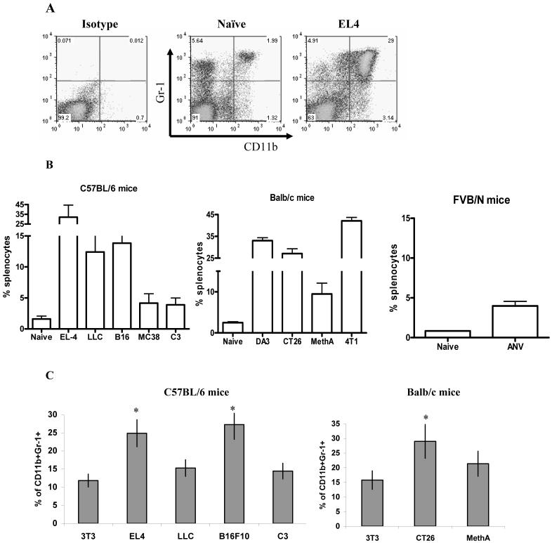
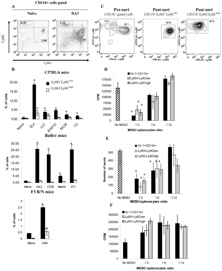
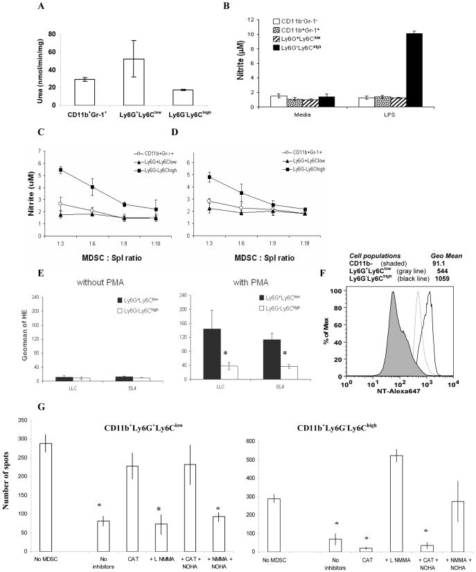
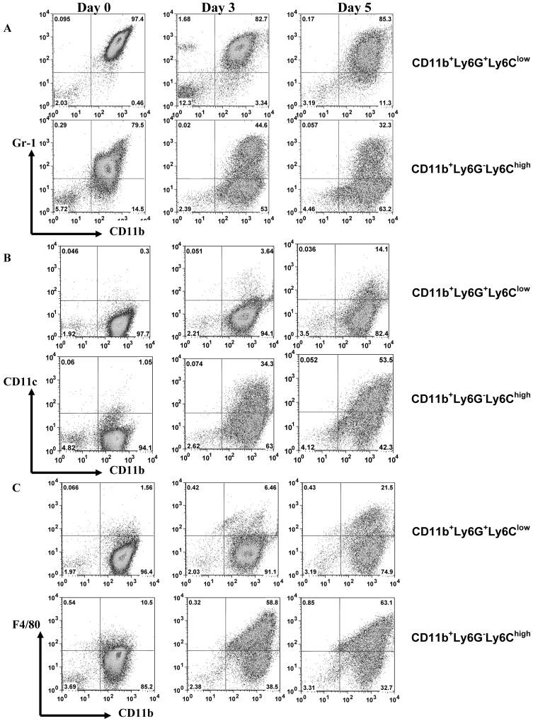
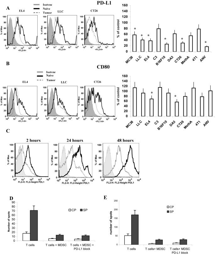
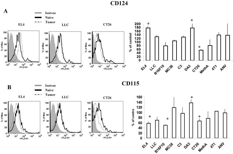
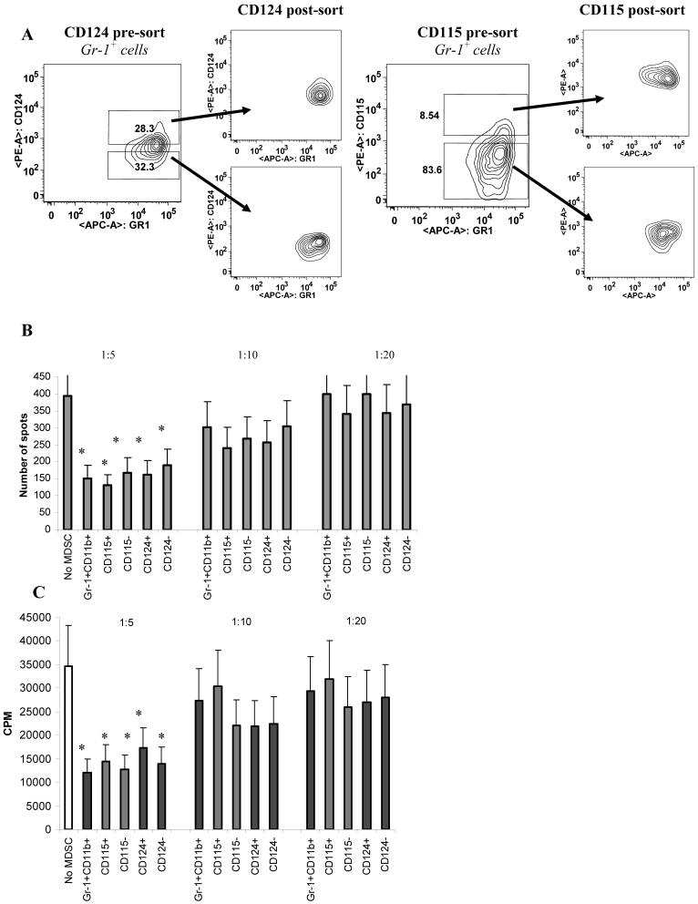
Myeloid-derived suppressor cells (MDSC) are a heterogeneous group of cells that play a critical role in tumor associated immune suppression. In an attempt to identify a specific subset of MDSC primarily responsible for immunosuppressive features of these cells, 10 different tumor models were investigated. All models showed variable but significant increase in the population of MDSC. Variability of MDSC expansion in vivo matched closely the effect of tumor cell condition medium in vitro. MDSC consists of two major subsets of Ly6G(+)Ly6C(low) granulocytic and Ly6G(-)Ly6C(high) monocytic cells. Granulocytic MDSC have increased level of reactive oxygen species and undetectable level of NO whereas monocytic MDSC had increased level of NO but undetectable levels of reactive oxygen species. However, their suppressive activity per cell basis was comparable. Almost all tumor models demonstrated a preferential expansion of granulocytic subset of MDSC. We performed a phenotypical and functional analysis of several surface molecules previously suggested to be involved in MDSC-mediated suppression of T cells: CD115, CD124, CD80, PD-L1, and PD-L2. Although substantial proportion of MDSC expressed those molecules no differences in the level of their expression or the proportion, positive cells were found between MDSC and cells from tumor-free mice that lack immune suppressive activity. The level of MDSC-mediated T cell suppression did not depend on the expression of these molecules. These data indicate that suppressive features of MDSC is caused not by expansion of a specific subset but more likely represent a functional state of these cells.


Figures

Figure 1
Figure 1. Accumulation of Gr-1+CD11b+ cells in spleens of tumor-bearing mice
Splenocytes from naïve or tumor-bearing mice (3 weeks after tumor inoculation, tumor size 1.5 cm in diameter) were stained with anti-CD11b and anti-Gr-1 antibodies. A. Typical example of flow cytometry analysis. B. The percentage of Gr-1+CD11b+ cells in spleen from naïve or tumor-bearing mice on the C57BL/6, BALB/c, or FVB/N backgrounds as indicated. Each group included from 3 to 8 mice. Mean and standard deviation are shown. Differences between proportion of Gr-1+CD11b+ cells in spleens from naïve and tumor-bearing mice were statistically significant for all tumor models (p<0.05). C. Bone marrow cells from naïve mice were cultured for 5 d with 10 ng/ml GM-CSF and IL-4 in the presence of control (3T3) or conditioned media from indicated tumor cell lines. Cells were collected, labeled with anti-Gr-1 and anti-CD11b Abs, and analyzed by flow cytometry. * - statistically significant differences from control (3T3) level (p<0.05).
Figure 2
Figure 2. The presence and functional activity of granulocytic and monocytic subsets of MDSC in tumor-bearing mice
Splenocytes from naïve and tumor-bearing mice were stained with CD11b, Ly6G and ly6C antibodies. A. Typical example of flow cytometry analysis. B. The percentage of Ly6G+Ly6Clow or Ly6G-Ly6Chigh cells in spleen from naïve or tumor-bearing mice. * - statistically significant differences between naïve and tumor-bearing mice (p<0.05). Gr-1+CD11b+ total MDSC, CD11b+Ly6G+Ly6Clow granulocytic, and CD11b+Ly6G-Ly6Chigh monocytic MDSC were sorted from spleens of EL-4 tumor-bearing mice using FACSAria cell sorter. C. An example of cell sort of granulocytic and monocytic subsets of MDSC. D, E. Sorted MDSC subsets were cultured at different ratios with 2×10⁵ splenocytes from OT-1 mice in the presence of control or specific peptides. Cell proliferation (D) and IFN-γ production (E) were measured using 3H-thymidine uptake and ELISPOT assay as described in Material in Methods. Each experiment was performed in triplicates. Three experiments with similar results were performed. The values of T-cell activity in the presence of control peptide were subtracted from the values obtained in the presence of specific peptide. F. Splenocytes from C57BL/6 mice were cultured with anti-CD3/CD28 antibody in the presence of different ratios of MDSC subsets. Cell proliferation was measured in triplicates and Mean ± SD are shown.
Figure 3
Figure 3. The mechanisms of suppressive activity of different MDSC subsets
Gr-1+CD11b+ total MDSC, CD11b+Ly6G+Ly6Clow granulocytic, and CD11b+Ly6G-Ly6Chigh monocytic MDSC were sorted from spleens of EL-4 tumor-bearing mice using FACSAria cell sorter. A. Arginase activity of different cell populations was measured in triplicates as described in Material and Methods. Mean ± SD are shown. B. Sorted populations of MDSC were stimulated with 1 μg/ml LPS for 24-hr, supernatants were collected and nitrite concentration was measured as described in Material and Methods. Experiments were performed in triplicates. C. OT-1 splenocytes (105 cells per well) were stimulated in triplicates for 48 hr with specific peptide in the presence of different ratios of sorted indicated subsets of MDSC and NO level was measured in supernatants. Mean ± SD are shown. D. Splenocytes from naïve C57BL/6 mice were stimulated from 48 hr with anti-CD3/CD28 antibodies in the presence of different ratios of indicated MDSC subsets. NO level was measured in supernatants as described in Material and Methods. Mean ± SD are shown. E. The level of ROS in MDSC subsets was measured using HE staining and flow cytometry as described in Material and Methods. * - statistically significant differences between groups (p<0.05). F. Expression of nitrotyrosine (NT) in MDSC subsets. Splenocytes from EL4 tumor bearing mice were stained with CD11b, Ly6G, Ly6C, and intracellular NT. Expression of NT was evaluated in CD11b- cells (shade area), CD11b+Ly6G+Ly6Clow (gray line), CD11b+Ly6G-Ly6Chigh (black line). G. Indicated populations of MDSC were sorted from spleens of naive or tumor-bearing mice and incubated in the presence of 10 μg/ml specific peptide SIINFEKL at 1:3 ratio with 106 splenocytes from OT-1 mice. After 24 hr cells were collected and labeled with anti-CD8 and anti-NT antibodies. CD8+ cells were gated and the proportion of NT positive cells was calculated. I. Suppressive activity of indicated MDSC subsets was evaluated as described in Fig. 3B. Sorted granulocytic and monocytic subsets of MDSC were cultured at 1:4 ratio with OT-1 splenocytes in the presence of specific or control peptide. L-NMMA (0.5 mM), nor-NOHA (0.5 mM), or catalase (1000 U/mL) were added at the beginning of the culture. The number of IFN-γ producing cells was evaluated in ELISPOT assay performed as described earlier (28). The numbers of spots were counted in triplicates and calculated using an automatic ELISPOT counter (Cellular Technology, Ltd). The values of IFN-γ production in the presence of control peptide were subtracted from the values obtained in the presence of specific peptide. *- statistically significant difference from splenocytes cultured without MDSC (p<0.05).
Figure 4
Figure 4. Differentiation of MDSC subsets in vitro
CD11b+Ly6G+Ly6Clow granulocytic MDSC and CD11b+Ly6G-Ly6Chigh monocytic MDSC were sorted from spleens of EL-4 tumor-bearing mice as described in Fig. 2. Cells were culutured in the presence of 10 ng/ml GM-CSF for 3 and 5 days and phenotype of cells was evaluated as indicated. A. Staining with anti-Gr-1 and Cd11b antibodies, B. Staining with anti-CD11c and Cd11b antibodies, C. Staining with anti-F4/80 and CD11b antibodies.
Figure 5
Figure 5. PD-L1 and CD80 expression on MDSC from tumor-bearing mice
Splenocytes from naïve or tumor-bearing mice were stained with anti-Gr-1-APC, anti-CD11b-PE-Cy7 and anti-PD-L1-PE (A) or anti-CD80-FITC (B) antibodies. Expression of PD-L1 or CD80 within the population of Gr-1+ CD11b+ MDSC was measured and calculated as percentage of change from the level of control mice with matched haplotype. Each tumor model included at least 4 mice. * - statistically significant differences from control (p<0.05). C. Gr-1+CD11b+ MDSC sorted from spleens of EL-4 tumor-bearing mice were cultured for 2, 24, or 48 h with 1 μg/ml of control IgG or specific anti-PD-L1 antibody (kind gift from Dr. L. Chen, John Hopkins University) in complete medium. Cells were stained with either isotype control (shaded area), or anti-PD-L1 antibody. PD-L1 in cells pre-treated with control IgG is shown as a black line, pre-treated with anti-PD-L1 antibody as a grey line. D. MDSC isolated from spleens of EL-4 tumor-bearing mice were pre-treated for 30 min on ice with control IgG or anti-PD-L1 antibody. Excess of antibody was washed and MDSC were cultured at 1:4 ratio with OT-1 splenocytes in the presence of control (CP) or specific (SP) peptides. IFN-γ production was evaluated in quadruplicates after 36 hr of culture suing ELISPOT assay. Mean ± SD of the number of spots per 5×10⁴ splenocytes is shown. E. CD8+ T-cell tolerance was induced in mice as described previously (18, 35). OT-1 T cells (5×10⁶) were injected i.v. into naïve C57BL/6 recipients. Two days later 3×10⁶ MDSC isolated from EL-4 tumor-bearing mice were injected i.v. followed by immunization with 100 μg of specific (SIINFEKL) peptide in IFA. Control IgG or anti-PD-L1 antibody (100 μg/mouse) were injected i.p. three times: 2 days prior to cell transfer and immunization, 2 days after immunization of mice and 3 days later. Ten days after immunization mice were sacrificed and lymph node cells were re-stimulated with control (CP) or specific (SP) peptides and the number of IFN-γ producing cells was evaluated in quadruplicates in ELISPOT assay. Mean ± SD of the number of spots per 2×10⁵ lymph node cells are shown.
Figure 6
Figure 6. Expression of CD124 (IL-4Rα) and CD115 (M-CSFR) on MDSC from tumor-bearing mice
Splenocytes from naïve or tumor-bearing mice were stained with anti-Gr-1-APC, anti-CD11b-PE-Cy7 and anti-CD124-PE (A) or anti-CD115-PE (B) antibodies. Expression of CD124 or CD115 within the population of Gr-1+CD11b+ MDSC was measured and calculated as percentage of change from the level of control mice with matched haplotype. Each tumor model included at least 4 mice. * - significant differences from control (p<0.05).
Figure 7
Figure 7. Functional activity of different populations of MDSC
Five populations of cells were sorted from spleens of EL-4 tumor-bearing mice: Gr-1+CD11b+; Gr-1+CD11b+CD115+; Gr-1+CD11b+CD115-; Gr-1+CD11b+CD124+; Gr-1+CD11b+CD124-. A. Typical example of sorting gates. B. Myeloid cells were added at indicated ratios to OT-1 splenocytes and cultured for 36 hr in the presence of control or specific peptides. IFN-γ production was measured in quadruplicates in ELISPOT assay. The number of spots per 105 splenocytes are shown. The experimental values obtained in the presence of control peptide were subtracted from the values obtained in the presence of specific peptide. * - statistically significant differences in the number of spots from spleens cultured without MDSC. C. Experiments were set up as described in Fig. 7B. Cell proliferation was measured after 4 days of culture using 3H-thymidine assay.

References

    1. Sica A, Bronte V. Altered macrophage differentiation and immune dysfunction in tumor development. J Clin Invest. 2007;117:1155–1166. - PMC - PubMed
    1. Kusmartsev S, Gabrilovich DI. Role Of Immature Myeloid Cells in Mechanisms of Immune Evasion In Cancer. Cancer Immunol Immunother. 2006;55:237–245. - PMC - PubMed
    1. Rabinovich GA, Gabrilovich D, Sotomayor EM. Immunosuppressive strategies that are mediated by tumor cells. Annu Rev Immunol. 2007;25:267–296. - PMC - PubMed
    1. Talmadge JE. Pathways mediating the expansion and immunosuppressive activity of myeloid-derived suppressor cells and their relevance to cancer therapy. Clin Cancer Res. 2007;13:5243–5248. - PubMed
    1. Delano MJ, Scumpia PO, Weinstein JS, Coco D, Nagaraj S, Kelly-Scumpia KM, O’Malley KA, Wynn JL, Antonenko S, Al-Quran SZ, Swan R, Chung CS, Atkinson MA, Ramphal R, Gabrilovich DI, Reeves WH, Ayala A, Phillips J, Laface D, Heyworth PG, Clare-Salzler M, Moldawer LL. MyD88-dependent expansion of an immature GR-1(+)CD11b(+) population induces T cell suppression and Th2 polarization in sepsis. J Exp Med. 2007;204:1463–1474. - PMC - PubMed

Publication types

MeSH terms