. 2008 Nov 19;27(22):2977-87.
doi: 10.1038/emboj.2008.202. Epub 2008 Oct 2.

A novel disulphide switch mechanism in Ero1alpha balances ER oxidation in human cells

Affiliations

A novel disulphide switch mechanism in Ero1alpha balances ER oxidation in human cells

Christian Appenzeller-Herzog et al. EMBO J. .

Abstract

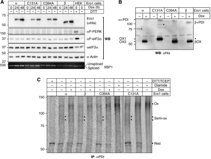
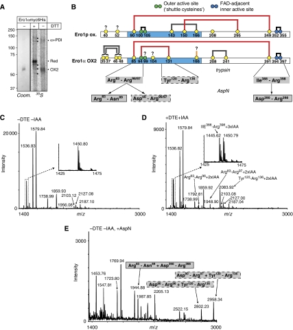
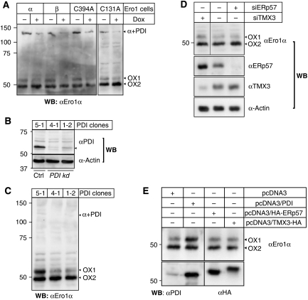
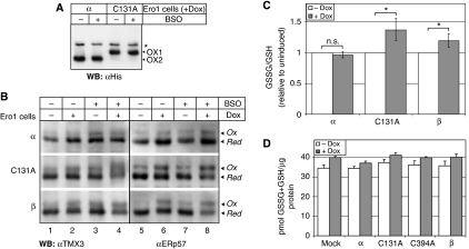
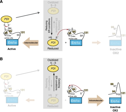
Oxidative maturation of secretory and membrane proteins in the endoplasmic reticulum (ER) is powered by Ero1 oxidases. To prevent cellular hyperoxidation, Ero1 activity can be regulated by intramolecular disulphide switches. Here, we determine the redox-driven shutdown mechanism of Ero1alpha, the housekeeping Ero1 enzyme in human cells. We show that functional silencing of Ero1alpha in cells arises from the formation of a disulphide bond-identified by mass spectrometry--between the active-site Cys(94) (connected to Cys(99) in the active enzyme) and Cys(131). Competition between substrate thiols and Cys(131) creates a feedback loop where activation of Ero1alpha is linked to the availability of its substrate, reduced protein disulphide isomerase (PDI). Overexpression of Ero1alpha-Cys131Ala or the isoform Ero1beta, which does not have an equivalent disulphide switch, leads to augmented ER oxidation. These data reveal a novel regulatory feedback system where PDI emerges as a central regulator of ER redox homoeostasis.


Figures

Figure 1
Figure 1
Characterization of inducible Ero1 cell lines. (A) Ero1α (α), Ero1α-C131A (C131A), Ero1α-C394A (C394A) and Ero1β (β) cells were induced with doxycyclin (Dox) for 0, 24 or 48 h, the cell lysates normalized for protein content, separated by SDS–PAGE under reducing conditions and analysed by western blotting using the indicated antibodies. The distribution of spliced and unspliced XBP1 mRNA was assessed by RT–PCR and analysed on an agarose gel. As positive and negative controls for the phosphorylation of PERK and eIF2α, and the splicing of XBP1 mRNA, HEK293 cells (HEK) were treated or not with the known ER stress inducer DTT (2 mM, 1 h). (B) Ero1 cells were incubated with or without doxycyclin (Dox), treated with NEM to quench post-lysis thiol–disulphide exchange reactions, and the oxidation state of exogenous Ero1 variants was visualized by western blotting using αHis after non-reducing SDS–PAGE. The gel mobilities of monomeric Ero1α variants and Ero1β are indicated as OX1, OX2 and βOX, respectively. α+PDI and β+PDI denote the mixed disulphides between Ero1α/Ero1β and PDI. Note that compared with the endogenous protein (see, e.g., Figure 3A), the epitope-tagged Ero1α proteins migrated slower by SDS–PAGE. Asterisk, background band; X, His-tagged 75 kDa molecular weight marker (leakage from neighbouring lane); filled circle, unidentified mixed-disulphide complex preferentially formed with Ero1α-C131A (our unpublished data). (C) The in vivo redox state of the two active sites in PDI (a and a′) in uninduced (−Dox) versus doxycyclin-induced (+Dox) Ero1 cells (α, β, C394A, C131A) was determined as described previously (Appenzeller-Herzog and Ellgaard, 2008a). After in situ modification of free cysteines with NEM, metabolically labelled PDI was immunoprecipitated, and disulphide-bonded cysteines were reduced and then modified with mPEG-mal. Differentially alkylated PDI was analysed by SDS–PAGE and phosphorimaging. Samples reduced with DTT and TCEP, or oxidized with diamide ahead of NEM-modification, served as mobility markers. Red, both active sites reduced; Ox, both active sites oxidized; Semi-ox, semi-oxidized PDI (owing to the faint appearance of the two semi-oxidized forms mainly present in the uninduced lanes, arrowheads mark their mobility in the autoradiograms). Note that mPEG-mal-modified bands are not sharp in appearance because of the large size dispersity of the reagent.
Figure 2
Figure 2
The OX2 redox species of Ero1α is molecularly defined by the regulatory Cys94–Cys131 disulphide. (A) Ero1αmyc6His was two-step purified from unlabelled or 35S-Cys-labelled, doxycyclin-induced and NEM-treated Ero1α cells. A Coomassie-stained gel (Coom.) and a phosphorimager scan (35S) from the unlabelled and labelled protein batch, respectively, are shown. Ero1α OX2 was shifted to the reduced (Red) form upon treatment of the sample with DTT. α+PDI, mixed-disulphide complex between Ero1α and PDI; asterisks, background bands. (B) Schematic representation of the cysteine connectivities in oxidized (ox.) Ero1p (Gross et al, 2004) and Ero1α OX2 (this study). The cysteines are shown as yellow, green (outer active site) or blue (inner active site) circles with amino-acid numbering, and disulphides as thick grey (likely structural), black (active site) or red (reported or inferred regulatory function; Sevier et al, 2007 and this study) lines. The thick orange line at Cys166 indicates the connection to a likely (but unidentified) disulphide partner. The question marks denote cysteines with unresolved oxidation state. The flexible loop regions are coloured in light blue. The braces for Cys35/37 and Cys46/48 indicate the two possible connections that could not be distinguished from our data. The three disulphide-linked peptides after trypsin cleavage and their trimming products upon further digestion with AspN are shown in grey boxes where the solid and dashed borders reflect their connectivity. Note that the alternative tryptic cleavage at Arg96 or Arg97 gave rise to two peptides that were both disulphide-linked to the Tyr120–Arg136 peptide (see panel E). (C, D) Mass spectra of HPLC fraction B before (C) and after (D) reduction and alkylation with DTE and IAA (±DTE±IAA), respectively. The monoisotropic masses at m/z 1445.62 (insets), 1792.81, 1948.90 and 2083.92 appear only in the reduced and alkylated sample and correspond to the four IAA-modified peptides labelled in the spectrum (see also Table I). (E) The monoisotropic masses at m/z 1944.88, 2802.83 and 2958.34 upon AspN digestion of fraction B correspond to the indicated disulphide-linked di-peptides (see also Table I). Bordering patterns of boxes match those in panel B.
Figure 3
Figure 3
The redox state of Cys94–Cys131 is modulated by the availability of PDI. (A) Uninduced (−Dox) or induced (+Dox) Ero1 cells (α, C131A, C394A, β) were treated with NEM, solubilized in lysis buffer and the lysates were depleted of exogenous, His-tagged Ero1α using TALON beads. The supernatant was subjected to precipitation with ConA-sepharose, and the glycoprotein fraction was analysed by western botting using αEro1α following non-reducing SDS–PAGE. Stripping and reprobing the same membrane with αPDI identified the ∼130 kDa band as the mixed-disulphide complex between endogenous Ero1α and PDI (α+PDI, data not shown). (B) Lysates of a control (Ctrl) shRNA clone (5-1) and two PDI knockdown (kd) clones (4-1 and 1-2) (Ou and Silver, 2006) were analysed by western blotting using αPDI to monitor knockdown efficiency and α-Actin as loading control. (C) 5-1, 4-1 and 1-2 cells were treated with NEM, lysed and the ConA-sepharose-precipitated glycoprotein fraction was subjected to αEro1α western blot analysis. The three independent samples were not normalized for protein content, meaning that no conclusions about the relative abundance of Ero1α between lanes can be made. (D) HeLa cells left untreated or treated with siRNAs against TMX3 (siTMX3) or ERp57 (siERp57) were harvested and lysed in the presence of NEM. Lysates were directly analysed either by western blotting with the indicated antibodies or, for the detection of Ero1α, after ConA-precipitation to concentrate the protein. Similar results were obtained with HEK293 cells (not shown). The hairline indicates where a lane has been removed. (E) HEK293 cells were transfected with the indicated cDNAs, harvested and lysed in the presence of NEM. Lysates were either directly subjected to western blot analysis using αPDI and αHA (lower panels) or incubated with ConA-sepharose to concentrate the glycoprotein fraction ahead of western blot detection of endogenous Ero1α (upper panel).
Figure 4
Figure 4
Increased in vivo activity of Ero1α-C131A and Ero1β is buffered by GSH. (A) Ero1α and C131A cells were induced (+Dox), treated with or without BSO and processed in the presence of NEM before αHis western blot analysis. (B) Ero1 cells were treated with doxycyclin (Dox) and/or BSO as indicated, and the in vivo redox states of TMX3 and ERp57 trapped by differential alkylation with NEM and AMS, followed by western blotting. Treatment with BSO reduced GSSG+GSH to 20% of control (data not shown). The mobilities of the reduced (Red) and oxidized (Ox) species, as verified by control samples using lysates from DTT- or diamide-treated cells (not shown), are indicated. Note that the mobility shift of the ERp57 band reflects the oxidation state of the a′ domain (Supplementary Figure S6). The results are representative of three independent experiments. (C) Cellular GSSG levels increase upon overexpression of Ero1α-C131A or Ero1β. Ero1 cells (α, C131A, β) were left untreated or induced with doxycyclin for 24 h and analysed for the intracellular levels of GSSG and GSH. The relative changes of GSSG/GSH caused by Ero1 overexpression along with 95% confidence intervals as calculated by using a linear model to correct for variation between experimental batches are plotted (n=12; for the raw data, refer to Supplementary Figure S7). Asterisks indicate statistical significance (P<0.03). n.s., not significant. (D) GSSG+GSH and total protein was determined in samples obtained from the indicated cell lines with (+Dox) or without (−Dox) doxycyclin induction for 24 h. GSSG+GSH (in pmol) was normalized to protein content (in μg) (mean±s.d., n=3).
Figure 5
Figure 5
Model for homoeostatic control of the PDI redox state by Cys131-dependent feedback regulation of Ero1α. The thiol–disulphide composition in the ER depends on the load of nascent polypeptides—many of which are substrates for PDI-mediated oxidation—as well as on the redox distribution of ER-localized glutathione. As both of these terms vary under physiological conditions, the redox state of the thiol–disulphide system in the ER can fluctuate (reducing versus oxidizing ER conditions). As detailed in the text, such fluctuations are likely mirrored in the steady-state distribution between oxidized and reduced PDI (grey box). Panels (A) and (B) depict the two extreme cases where the outer active-site disulphide in Ero1α is exclusively attacked by a PDI- or Cys131-derived thiolate anion. In the cell, these two reaction pathways compete with each other. (A) Under reducing ER conditions, PDI (for simplicity, only one active site in PDI is depicted) accumulates in the reduced form. Reduced PDI is a substrate for oxidation by Ero1α, which is initiated through the intermolecular nucleophilic attack by an active-site cysteine in PDI on Cys94 in the oxidized outer active site of Ero1α. The competing intramolecular reaction of Cys131 with the Cys94–Cys99 disulphide is disfavoured by the abundance of reduced PDI. A thiol–disulphide exchange reaction between Ero1α (in the active form) and PDI then results in the formation of oxidized PDI. By this mechanism, the activity of Ero1α counterbalances reducing ER conditions. (B) Under oxidizing ER conditions, PDI is predominantly oxidized and the outer active-site disulphide of Ero1α will, therefore, preferentially react with Cys131. The resulting OX2 configuration of Ero1α is inactive because the outer active Cys94 is covalently blocked by Cys131. The mechanism of reactivation of Ero1α OX2 remains unclear. In principle, it could involve a nucleophilic attack of Cys99 on Cys94 to resolve Cys94–Cys131, but the oxidation state of Cys99 in OX2 is not known (indicated by the question mark).

Comment in

References

    1. Anelli T, Alessio M, Mezghrani A, Simmen T, Talamo F, Bachi A, Sitia R (2002) ERp44, a novel endoplasmic reticulum folding assistant of the thioredoxin family. EMBO J 21: 835–844 - PMC - PubMed
    1. Appenzeller-Herzog C, Ellgaard L (2008a) In vivo reduction–oxidation state of protein disulfide isomerase: the two active sites independently occur in the reduced and oxidized forms. Antioxid Redox Signal 10: 55–64 - PubMed
    1. Appenzeller-Herzog C, Ellgaard L (2008b) The human PDI family: versatility packed into a single fold. Biochim Biophys Acta 1783: 535–548 - PubMed
    1. Benham AM, Cabibbo A, Fassio A, Bulleid N, Sitia R, Braakman I (2000) The CXXCXXC motif determines the folding, structure and stability of human Ero1-Lalpha. EMBO J 19: 4493–4502 - PMC - PubMed
    1. Bertoli G, Simmen T, Anelli T, Molteni SN, Fesce R, Sitia R (2004) Two conserved cysteine triads in human Ero1alpha cooperate for efficient disulfide bond formation in the endoplasmic reticulum. J Biol Chem 279: 30047–30052 - PubMed

Publication types

MeSH terms

LinkOut - more resources