. 2008 Oct 22;27(20):2712-24.
doi: 10.1038/emboj.2008.194. Epub 2008 Oct 2.

Hsp104, Hsp70 and Hsp40 interplay regulates formation, growth and elimination of Sup35 prions

Affiliations

Hsp104, Hsp70 and Hsp40 interplay regulates formation, growth and elimination of Sup35 prions

James Shorter et al. EMBO J. .

Abstract

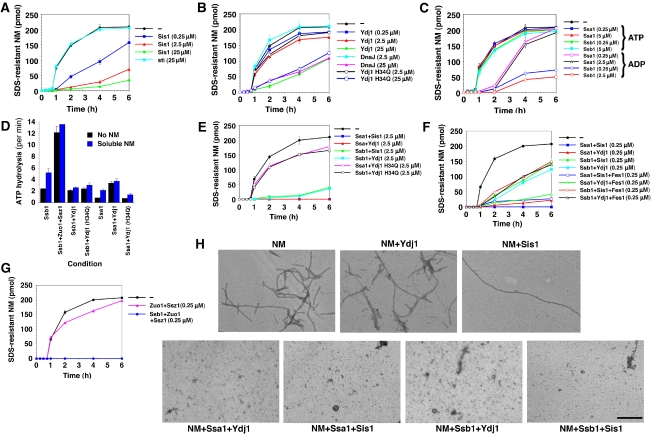
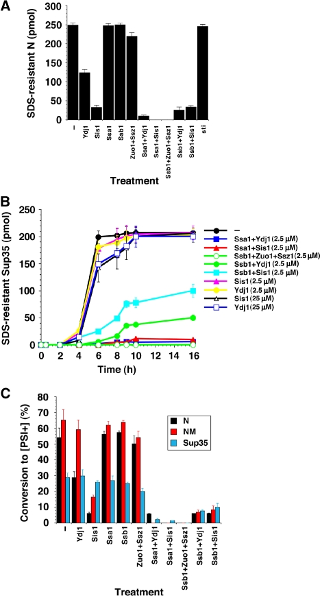
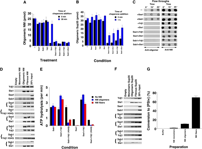
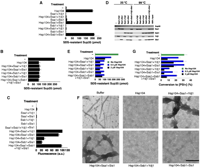
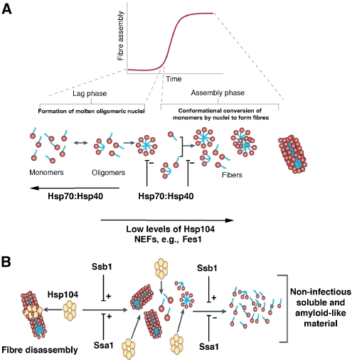
Self-templating amyloid forms of Sup35 constitute the yeast prion [PSI(+)]. How the protein-remodelling factor, Hsp104, collaborates with other chaperones to regulate [PSI(+)] inheritance remains poorly delineated. Here, we report how the Ssa and Ssb components of the Hsp70 chaperone system directly affect Sup35 prionogenesis and cooperate with Hsp104. We identify the ribosome-associated Ssb1:Zuo1:Ssz1 complex as a potent antagonist of Sup35 prionogenesis. The Hsp40 chaperones, Sis1 and Ydj1, preferentially interact with Sup35 oligomers and fibres compared with monomers, and facilitate Ssa1 and Ssb1 binding. Various Hsp70:Hsp40 pairs block prion nucleation by disassembling molten oligomers and binding mature oligomers. By binding fibres, Hsp70:Hsp40 pairs occlude prion recognition elements and inhibit seeded assembly. These inhibitory activities are partially relieved by the nucleotide exchange factor, Fes1. Low levels of Hsp104 stimulate prionogenesis and alleviate inhibition by some Hsp70:Hsp40 pairs. At high concentrations, Hsp104 eliminates Sup35 prions. This activity is reduced when Ssa1, or enhanced when Ssb1, is incorporated into nascent prions. These findings illuminate several facets of the chaperone interplay that underpins [PSI(+)] inheritance.


Figures

Figure 1
Figure 1
Effects of Hsp70 and Hsp40 on spontaneous NM fibrillization. (AC) Kinetics of unseeded, rotated (80 r.p.m.) NM (2.5 μM) fibrillization in the presence or absence of either soybean trypsin inhibitor (sti), Sis1 (A), Ydj1, DnaJ (0.25–25 μM) (B), Ssa1 or Ssb1 (0.25–5 μM) (C). All reactions contained ATP (5 mM), except for some reactions in (C) that contained ADP (5 mM). Fibrillization was monitored by the amount of SDS-resistant NM. Values represent means±s.d. (n=3). (D) Ssb1 or Ssa1 (1 μM) were incubated for 30 min at 25°C with or without Ydj1 or Ydj1 H34Q (1 μM) in the presence or absence of soluble NM (2.5 μM). The amount of ATP hydrolysis was then determined. Values represent means±s.d. (n=3). (EG) NM (2.5 μM) fibrillization as in (A) in the presence or absence of either Ssa1:Ydj1 H34Q, Ssb1:Ydj1 H34Q (2.5 μM) (E), Ssa1:Sis1, Ssa1:Ydj1, Ssb1:Sis1, Ssb1:Ydj1, (0.25-2.5 μM) (E, F), Ssa1:Sis1:Fes1, Ssa1:Ydj1:Fes1, Ssb1:Sis1:Fes1, Ssb1:Ydj1:Fes1 (0.25 μM) (F), Zuo1:Ssz1 or Ssb1:Zuo1:Ssz1 (0.25 μM) (G). Values represent means±s.d. (n=3). (H) EM of unseeded, rotated (80 r.p.m.) NM (2.5 μM) fibrillization after 6 h in the absence or presence of either Ydj1, Sis1, Ssa1:Sis1, Ssa1:Ydj1, Ssb1:Sis1 or Ssb1:Ydj1 (2.5 μM). Bar, 0.5 μm.
Figure 2
Figure 2
Effects of Hsp70 and Hsp40 on spontaneous N, NM and Sup35 prionogenesis. (A) Unseeded, rotated (80 rpm) His–N (2.5 μM) fibrillization with ATP (5 mM) after 20 min in the presence or absence of sti, Ydj1, Sis1, Ssa1, Ssb1, Ssa1:Sis1, Ssa1:Ydj1, Ssb1:Sis1 or Ssb1:Ydj1 (2.5 μM). Fibrillization was monitored by the amount of SDS-resistant N. Values represent means±s.d. (n=3). (B) Kinetics of unseeded, rotated (80 r.p.m.) Sup35 (2.5 μM) fibrillization with ATP (5 mM) in the presence or absence of Ydj1, Sis1 (2.5–25 μM), Ssa1:Sis1, Ssa1:Ydj1, Ssb1:Zuo1:Ssz1, Ssb1:Sis1 or Ssb1:Ydj1 (2.5 μM). Fibrillization was monitored by the amount of SDS-resistant Sup35. Values represent means±s.d. (n=3). (C) His–N (2.5 μM) was assembled for 20 min, or NM (2.5 μM) or Sup35 (2.5 μM) were assembled for 6 h in the presence or absence of Ydj1, Sis1, Ssa1:Sis1, Ssa1:Ydj1, Ssb1:Zuo1:Ssz1, Ssb1:Sis1 or Ssb1:Ydj1 (2.5 μM). Reaction products were concentrated and transformed into [psi] cells. The proportion of [PSI+] colonies was then determined. Values represent means±s.d. (n=3).
Figure 3
Figure 3
Kinetic sensitivity of NM and Sup35 fibrillization to Hsp70 and Hsp40. (A) Unseeded, rotated (80 r.p.m.) NM (2.5 μM) fibrillization reactions were performed with ATP (5 mM), and at the indicated times, reactions were either terminated or treated with buffer, Hsp104, Sis1, Ssa1:Ydj1, Ssa1:Sis1, Ssb1:Ydj1 or Ssb1:Sis1 (2.5 μM) and incubated for a total time of 6 h. Fibrillization was assessed by sedimentation analysis. Values represent means±s.d. (n=3). (B) Unseeded, rotated (80 r.p.m.) Sup35 (2.5 μM) fibrillization reactions were performed with ATP (5 mM) and at the indicated times reactions were either terminated or treated with buffer, Hsp104, Sis1, Ssa1:Ydj1, Ssa1:Sis1, Ssb1:Ydj1 or Ssb1:Sis1 (2.5 μM), and incubated for a total time of 6 h. Fibrillization was assessed by ThT fluorescence. Values represent means±s.d. (n=3).
Figure 4
Figure 4
Hsp70 and Hsp40 target NM oligomers and fibres to inhibit fibrillization. (A) NM (2.5 μM) with ATP (5 mM) was rotated for 5 min (80 r.p.m.) in the presence or absence of Ssa1, Ssb1, Ydj1, Sis1, Ssa1:Ydj1, Ssa1:Sis1, Ssb1:Ydj1 or Ssb1:Sis1 (2.5 μM) (black bars). Alternatively, NM (2.5 μM) with ATP (5 mM) alone was rotated (80 r.p.m.) for 30 min and then Ssa1, Ssb1, Ydj1, Sis1, Ssa1:Ydj1, Ssa1:Sis1, Ssb1:Ydj1 or Ssb1:Sis1 (2.5 μM) were added. This reaction was continued for 10 min (blue bars). Oligomeric NM was recovered by centrifugation at 436 000 g for 30 min and the amount in the pellet fraction determined. Values represent means±s.d. (n=3). (B) Sup35 (2.5 μM) with ATP (5 mM) was rotated for 1 h (80 r.p.m.) in the presence or absence of either Ssa1, Ssb1, Ydj1, Sis1, Ssa1:Ydj1, Ssa1:Sis1, Ssb1:Ydj1 or Ssb1:Sis1 (2.5 μM) (black bars). Alternatively, Sup35 (2.5 μM) with ATP (5 mM) alone was rotated (80 r.p.m.) for 1 h and then Ssa1, Ssb1, Ydj1, Sis1, Ssa1:Ydj1, Ssa1:Sis1, Ssb1:Ydj1 or Ssb1:Sis1 (2.5 μM) were added. This reaction was continued for 10 min (blue bars). Oligomeric Sup35 was recovered by centrifugation at 436 000 g for 30 min and the amount in the pellet fraction determined. Values represent means±s.d. (n=3). (C) NM (2.5 μM) with ATP (5 mM) was rotated (80 r.p.m.) for 0–50 min. At 0, 15 and 45 min, His–tagged Ssa1, Ssb1, Ydj1, Sis1, Ssa1:Ydj1, Ssa1:Sis1, Ssb1:Ydj1 or Ssb1:Sis1 were added. At 50 min, reactions were depleted of chaperones using Ni-NTA agarose. Depleted reactions were spotted onto nitrocellulose and probed with anti-oligomer or anti-NM antibody. (D) Ssa1, Ssb1, Ydj1, Sis1, Ssa1:Ydj1, Ssa1:Ydj1 H34Q, Ssa1:Sis1, Ssb1:Ydj1, Ssb1:Ydj1 H34Q or Ssb1:Sis1 (2.5 μM) were incubated in the presence of ATP (5 mM) with Ni-NTA beads (empty) or beads bound to NM–His monomers, oligomers or fibres. Washed beads were eluted and eluates were fractionated by SDS–PAGE and Coomassie-stained or processed for immunoblot (to detect Sis1). (E) Ssb1 or Ssa1 (1 μM) were incubated for 30 min at 25°C with or without Ydj1 or Ydj1 H34Q (1 μM) in the presence or absence of NM oligomers or fibres (2.5 μM). The amount of ATP hydrolysis was then determined. Values represent means±s.d. (n=3). (F) Ssa1, Ssb1, Ydj1, Sis1, Ssa1:Ydj1, Ssa1:Sis1, Ssb1:Ydj1 or Ssb1:Sis1 (2.5 μM) were incubated in the presence of ATP (5 mM) with Ni-NTA beads (empty) or beads bound to His–Sup35 monomers, oligomers or fibres. Washed beads were eluted, and eluates were fractionated by SDS–PAGE and Coomassie-stained. (G) NM–His monomers, oligomers or fibres were transformed into [psi] cells. The proportion of [PSI+] colonies was then determined. Values represent means±s.d. (n=3).
Figure 5
Figure 5
Hsp70 and Hsp40 inhibit seeded NM fibrillization and occlude prion recognition elements. (A) Seeded (2% wt/wt) NM (2.5 μM) fibrillization with ATP (5 mM) after 16 h in the presence or absence of sti, Ydj1, Sis1, Ssa1, Ssb1, Fes1, Ssa1:Ydj1, Ssa1:Sis1, Ssb1:Ydj1, Ssb1:Sis1, Ssa1:Ydj1:Fes1, Ssa1:Sis1:Fes1, Ssb1:Ydj1:Fes1 or Ssb1:Sis1:Fes1 (2.5 μM). Fibrillization was monitored by the amount of SDS-resistant NM. Values represent means±s.d. (n=3). (B) Seeded (2% wt/wt) Sup35 (2.5 μM) fibrillization with ATP (5 mM) after 16 h in the presence or absence of sti, Ydj1, Sis1, Ssa1, Ssb1, Fes1, Ssa1:Ydj1, Ssa1:Sis1, Ssb1:Ydj1, Ssb1:Sis1, Ssa1:Ydj1:Fes1, Ssa1:Sis1:Fes1, Ssb1:Ydj1:Fes1 or Ssb1:Sis1:Fes1 (2.5 μM). Fibrillization was monitored by ThT fluorescence. Values represent means±s.d. (n=3). (C) Excimer fluorescence of NM G38C-pyrene (black) or G96C-pyrene (blue) (2.5 μM) after 16 h seeded (2% wt/wt) assembly with ATP (5 mM) in the absence or presence of sti, Ydj1, Sis1, Ssa1, Ssb1, Ssa1:Ydj1, Ssa1:Sis1, Ssb1:Ydj1 or Ssb1:Sis1 (2.5 μM). The ratio of excimer fluorescence to non-excimer fluorescence (I465 nm/I375 nm) is plotted. Values represent means±s.d. (n=3).
Figure 6
Figure 6
Hsp70 and Hsp40 modulate the prion-remodelling activities of Hsp104. (A) Unseeded, rotated (80 r.p.m.) Sup35 (2.5 μM) fibrillization with ATP (5 mM) after 2 h in the presence or absence of Hsp104 (0.03 μM) plus or minus Ssa1:Ydj1, Ssa1:Sis1, Ssb1:Ydj1 or Ssb1:Sis1 (0.25 μM). Fibrillization was assessed by the amount of SDS-resistant Sup35. Values represent means±s.d. (n=3). (B) Sup35 fibres (2.5 μM monomer) were incubated with or without the indicated combination of Hsp104, Ssa1, Ssb1, Ydj1 and Sis1 (2 μM) plus ATP (5 mM). Disassembly was assessed by the amount of SDS-resistant Sup35. Values represent means±s.d. (n=3). (C) Disaggregation of heat-aggregated GFP (0.45 μM) in the presence of the indicated combination of Hsp104, Ssa1, Ssb1, Ydj1 and Sis1 (2 μM) plus ATP (5 mM). Disaggregation was assessed by the amount of GFP fluorescence. Values represent means±s.d. (n=3). (D) Sup35 (2.5 μM) fibres were assembled with rotation (80 r.p.m.) for 8 h with or without various combinations of Ssa1:Ydj1, Ssa1:Sis1, Ssb1:Ydj1, Ssb1:Sis1 or Ssa1:Ssb1:Ydj1:Sis1 (0.15 μM) plus ATP (5 mM). Fibres were then collected by sedimentation at 436 000 g for 10 min and treated with SDS–PAGE sample buffer at either 25 or 99°C and processed for immunoblot. (EG) Sup35 (2.5 μM) fibres were assembled with rotation (80 r.p.m.) for 8 h with or without various combinations of Ssa1:Ydj1, Ssa1:Sis1, Ssb1:Ydj1, Ssb1:Sis1 or Ssa1:Ssb1:Ydj1:Sis1 (0.15 μM) plus ATP (5 mM). Hsp104 (0.5 μM or 2 μM) was then added and reactions incubated for a further 30 min. Remodelling was assessed by the amount of SDS-resistant Sup35 (E). Values represent means±s.d. (n=3). (F) Alternatively, products from 2 μM Hsp104 reactions were viewed by EM. Bar, 150 nm. (G) Other reactions were concentrated and transformed into [psi] cells. The proportion of [PSI+] colonies was then determined. Values represent means±s.d. (n=3).
Figure 7
Figure 7
Model of Hsp104, Hsp70 and Hsp40 interplay during Sup35 prionogenesis. (A) Sup35 prions assemble after a lag phase during which a dynamic ensemble of monomeric and molten oligomeric species form. The intermolecular contacts that nucleate prion assembly are likely established within molten oligomers. Once formed, fibres stimulate their own assembly by recruiting and converting monomers at their ends. The Hsp70:Hsp40 pairs, Ssa1:Ydj1, Ssa1:Sis1, Ssb1:Ydj1 and Ssb1:Sis1 inhibit this process by disassembling molten oligomers and by binding mature nuclei to occlude prion recognition elements and prevent seeded assembly. This inhibition is partially relieved by the NEF, Fes1. At low concentrations, Hsp104 stimulates prionogenesis and can relieve inhibition by Ssa1:Sis1 and Ssb1:Sis1, but not Ssa1:Ydj1 and Ssb1:Ydj1. (B) At high concentrations, Hsp104 disassembles Sup35 fibres by a fragmentation process that is stimulated by the incorporation of Ssa1 and Ssb1 (plus Hsp40) into Sup35 fibres. In the absence of Hsp70 and Hsp40, excessive disassembly by Hsp104 ultimately converts Sup35 fibres to a collection of soluble and non-infectious amyloid-like conformers. This step is promoted by Ssb1 and inhibited by Ssa1.

References

    1. Albanese V, Yam AY, Baughman J, Parnot C, Frydman J (2006) Systems analyses reveal two chaperone networks with distinct functions in eukaryotic cells. Cell 124: 75–88 - PubMed
    1. Allen KD, Wegrzyn RD, Chernova TA, Muller S, Newnam GP, Winslett PA, Wittich KB, Wilkinson KD, Chernoff YO (2005) Hsp70 chaperones as modulators of prion life cycle: novel effects of Ssa and Ssb on the Saccharomyces cerevisiae prion [PSI+]. Genetics 169: 1227–1242 - PMC - PubMed
    1. Bagriantsev SN, Gracheva EO, Richmond JE, Liebman SW (2008) Variant-specific [PSI+] infection is transmitted by Sup35 polymers within [PSI+] aggregates with heterogeneous protein composition. Mol Biol Cell 19: 2433–2443 - PMC - PubMed
    1. Chernoff YO, Lindquist SL, Ono B, Inge-Vechtomov SG, Liebman SW (1995) Role of the chaperone protein Hsp104 in propagation of the yeast prion-like factor [PSI+]. Science 268: 880–884 - PubMed
    1. Chernoff YO, Newnam GP, Kumar J, Allen K, Zink AD (1999) Evidence for a protein mutator in yeast: role of the Hsp70-related chaperone ssb in formation, stability, and toxicity of the [PSI+] prion. Mol Cell Biol 19: 8103–8112 - PMC - PubMed

Publication types

MeSH terms