. 2008;3(10):1630-8.
doi: 10.1038/nprot.2008.150.

The SCX/IMAC enrichment approach for global phosphorylation analysis by mass spectrometry

Affiliations

The SCX/IMAC enrichment approach for global phosphorylation analysis by mass spectrometry

Judit Villén et al. Nat Protoc. 2008.

Abstract

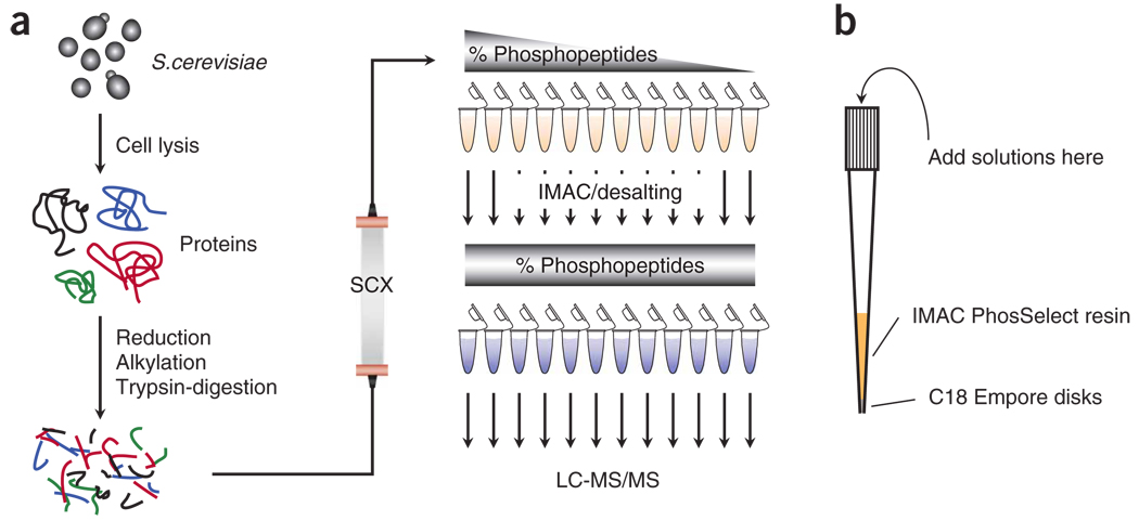
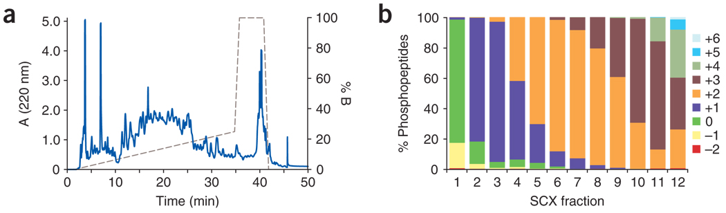
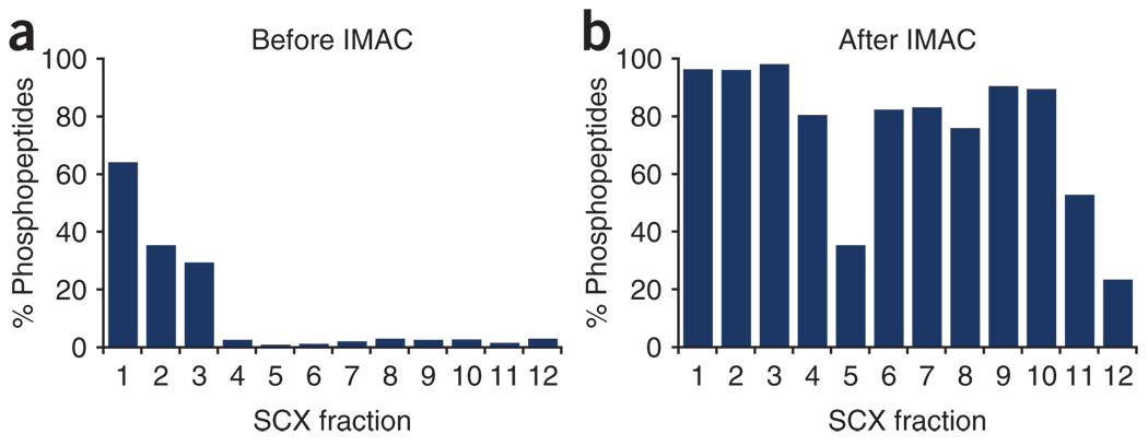
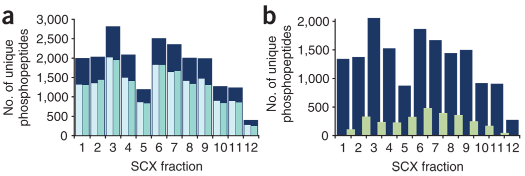
The success in profiling the phosphoproteome by mass spectrometry-based proteomics has been intimately related to the availability of methods that selectively enrich for phosphopeptides. To this end, we describe a protocol that combines two sequential enrichment steps. First, strong cation exchange (SCX) chromatography separates peptides by solution charge. Phosphate groups contribute to solution charge by adding a negative charge at pH 2.7. Therefore, at that pH, phosphopeptides are expected to elute earlier than their nonphosphorylated homologs. Second, immobilized metal affinity chromatography (IMAC) takes advantage of phosphate's affinity for metal ions such as Fe(3+) to uniformly enrich for phosphopeptides from the previously collected SCX fractions. We have successfully employed the SCX/IMAC enrichment strategy in the exploration of phosphoproteomes from several systems including mouse liver and Drosophila embryos characterizing over 5,500 and 13,000 phosphorylation events, respectively. The SCX/IMAC enrichment protocol requires 2 days, and the entire procedure from cells to a phosphorylation data set can be completed in less than 10 days.


Figures

Figure 1
Figure 1. Diagrams illustrating the protocol
(a) Scheme of the procedure for phosphopeptide enrichment and analysis. Protein extract is in-solution reduced, alkylated and digested with trypsin. Peptides are desalted and separated by SCX chromatography. Twelve fractions are collected, desalted, enriched by IMAC and analyzed by LC-MS/MS techniques. (b) Illustration of the StageTips used for combined IMAC enrichment and phosphopeptide desalting, where the IMAC resin, after incubation with peptide mixtures is finished, is packed on the top of C18 Empore disks. In the absence of ACN, phosphopeptides eluting from the IMAC resin are bound to the C18 disks, and salts can be removed before organic elution.
Figure 2
Figure 2. Performance of semipreparative SCX chromatography
(a) Chromatogram of the SCX separation of peptides. (b) Solution charge separation of nonredundant phosphopeptides identified from each fraction.
Figure 3
Figure 3. Enrichment obtained after each step represented as the fraction of phosphopeptides over all the peptides identified
(a) SCX chromatography alone enriches for phosphopeptides in early fractions. (b) Further IMAC enrichment over SCX fractions produces >75% phosphopeptides in most samples.
Figure 4
Figure 4. Phosphopeptide distributions
(a) Duplicate analyses of each fraction (pale blue and green) significantly increases the number of unique phosphopeptides identified (dark blue). (b) Overlap between consecutive SCX fractions (green) as compared with the number of unique phosphopeptides identified from one replicate of each fraction (dark blue).
Figure 5
Figure 5. Contribution of mass accuracy to phosphopeptide identification rates
(a) Distribution of mass deviations for all searched spectra. Matches to the forward database (blue) and those matching to the reverse database (red) are shown binned by 0.2 p.p.m. By searching with a larger precursor ion mass tolerance window than the distribution of true positives, many incorrect matches, which are uniformly distributed throughout the mass deviation window, can be removed. (b) Distribution of XCorr values of all searched spectra binned in 0.2 XCorr units. Matches to reversed sequences are shown in red. This represents one-half of all false-positives. Matches to the forward database with the number of reverse hits subtracted (true-positives) are shown in blue. (c) Same distribution as in b allowing only matches with a mass deviation between −2.5 to +2.5 p.p.m. By utilizing the known mass accuracy, most incorrect matches (in red) are removed from the data set. Additional filtering parameters such as dCn′ and solution charge can be used also to reduce the FDR; they will accept many correct answers with relatively lower XCorr values.

References

    1. Hastie CJ, McLauchlan HJ, Cohen P. Assay of protein kinases using radiolabeled ATP: a protocol. Nat. Protoc. 2006;1:968–971. - PubMed
    1. Collins MO, Yu L, Choudhary JS. Analysis of protein phosphorylation on a proteome-scale. Proteomics. 2007;7:2751–2768. - PubMed
    1. Hunter T, Cooper JA. Protein-tyrosine kinases. Annu. Rev. Biochem. 1985;54:897–930. - PubMed
    1. Rush J, et al. Immunoaffinity profiling of tyrosine phosphorylation in cancer cells. Nat. Biotechnol. 2005;23:94–101. - PubMed
    1. Andersson L, Porath J. Isolation of phosphoproteins by immobilized metal (Fe3+) affinity chromatography. Anal. Biochem. 1986;154:250–254. - PubMed

Publication types

LinkOut - more resources