. 2008 Sep 30;6(9):e234.
doi: 10.1371/journal.pbio.0060234.

Capturing hammerhead ribozyme structures in action by modulating general base catalysis

Affiliations

Capturing hammerhead ribozyme structures in action by modulating general base catalysis

Young-In Chi et al. PLoS Biol. .

Abstract

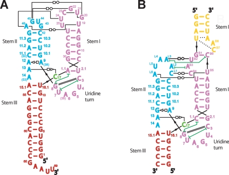
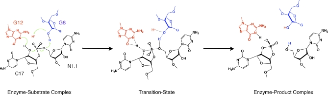
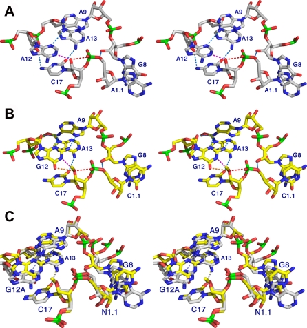
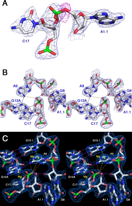
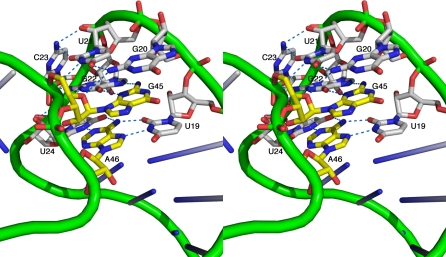
We have obtained precatalytic (enzyme-substrate complex) and postcatalytic (enzyme-product complex) crystal structures of an active full-length hammerhead RNA that cleaves in the crystal. Using the natural satellite tobacco ringspot virus hammerhead RNA sequence, the self-cleavage reaction was modulated by substituting the general base of the ribozyme, G12, with A12, a purine variant with a much lower pKa that does not significantly perturb the ribozyme's atomic structure. The active, but slowly cleaving, ribozyme thus permitted isolation of enzyme-substrate and enzyme-product complexes without modifying the nucleophile or leaving group of the cleavage reaction, nor any other aspect of the substrate. The predissociation enzyme-product complex structure reveals RNA and metal ion interactions potentially relevant to transition-state stabilization that are absent in precatalytic structures.


Conflict of interest statement

Competing interests. The authors have declared that no competing interests exist.

Figures

Figure 1
Figure 1. Two Classes of Full-Length Hammerhead Ribozymes
Secondary and schematic tertiary structural representations of the sTRSV hammerhead (A) and the Schistosoma hammerhead [12] (B), depicting the two classes [6] of hammerhead ribozyme tertiary contacts.
Figure 2
Figure 2. The Hammerhead Ribozyme Self-Cleavage Reaction
Schematic diagram of the enzyme–substrate, transition-state, and enzyme–product complexes of an unmodified hammerhead active site, interpolated from the 2GOZ structure in which G12 (red) is positioned to function as a general base in the cleavage reaction, and G8 (blue) is positioned consistent with a possible role in acid catalysis. To function as a general base, the N1 of G12 must be deprotonated (as shown), and it can then abstract the 2′-H from C17 (in black) to generate the nucleophile. The 2′-OH of G8 (in blue) is positioned to donate a proton to the 5′-O of residue N1.1, the leaving-group in the self-cleavage reaction. Green arrows represent electron pairs that mediate proton transfer and covalent bond breakage and formation. The transition state consists of a trigonal bipyramidal oxyphosphorane in which the nucleophile and leaving group occupy the axial positions. Partial bond formation and breakage is indicated with dotted lines. The products of the cleavage reaction possess 2′,3′-cyclic phosphate and 5′-OH termini as shown. The 2′,3′-cyclic phosphate is not hydrolyzed by the ribozyme, and in the structure, it is found in the form of a predissociation complex. In the sTRSV hammerhead structure, the G12A modification results in a much weaker base, but one that is not protonated at N1. The nucleotide N1.1 is not conserved. In 2GOZ, it is C1.1, and in the sTRSV hammerhead, it is an A.
Figure 3
Figure 3. The Hammerhead Ribozyme Reactant and Product Active Sites
(A and B) Stereo views of two hammerhead ribozyme active sites [37]. The active site of the uncleaved G12A sTRSV hammerhead (A) with an unmodified nucleophile, and the Schistosome hammerhead 2GOZ [12] (B) with a 2′-OMe modification of the nucleophilic 2′-oxygen of C17. Hydrogen bonds are shown as light-blue dotted lines, the trajectory of bond formation is indicated as a red dotted line, and potential “active” hydrogen bonds in base catalysis are indicated as pink and orange dotted lines. (C) depicts a superposition of (A) and (B).
Figure 4
Figure 4. Hammerhead Ribozyme Cleavage in the Crystal
(A) Refining the uncleaved structure against the cleavage-product data produces a negative residual (or Fcalc − Fobs) difference peak (shown in red, contoured at 3 σ) centered on the 5′-oxygen, the leaving group of the cleavage reaction. A gap in the 2Fo-Fc map (shown in blue, contoured at 1.0 σ) is apparent, despite model bias from the uncleaved structure. This appears in both crystallographically independent molecules in the asymmetric unit. (B) The refined cleaved structure makes a better fit to the electron density. (C) A stereo view of the active site of the hammerhead ribozyme, showing potential (yellow and orange dotted lines) and actual (pink dotted lines) bonding interactions involving two Mg2+ ions (yellow spheres) and the RNA. The potential interactions may form stabilizing contacts when the scissile phosphate is in the trigonal bipyramidal oxyphosphorate transition state, helping to dissipate excess negative charge. In particular, the invariant A9 may engage in transition-state stabilization interactions in extrapolation from the product structure, as indicated by the orange dotted lines.
Figure 5
Figure 5. Hammerhead Ribozyme Tertiary Contacts
Close-up stereo view of the tertiary interactions between Stems I and II in the sTRSV hammerhead RNA. The trace of the phosphodiester backbone is represented as green tubes, and the nucleotides that participate in tertiary contacts between Stem I and Stem II are shown explicitly as atomic color-coded stick figures. Carbon atoms in the Stem I nucleotides are white, and carbon atoms in the Stem II nucleotides are yellow. Nitrogen atoms in both cases are dark blue, oxygen atoms are red, and phosphorus atoms are green. Hydrogen bonds are shown as blue dotted lines. Figure S2A and S2B depict complementary views.

Comment in

References

    1. Nelson JA, Uhlenbeck OC. When to believe what you see. Mol Cell. 2006;23:447–450. - PubMed
    1. Westhof E. A tale in molecular recognition: the hammerhead ribozyme. J Mol Recognit. 2007;20:1–3. - PubMed
    1. McKay DB. Structure and function of the hammerhead ribozyme: an unfinished story. RNA. 1996;2:395–403. - PMC - PubMed
    1. Wedekind JE, McKay DB. Crystallographic structures of the hammerhead ribozyme: relationship to ribozyme folding and catalysis. Annu Rev Biophys Biomol Struct. 1998;27:475–502. - PubMed
    1. Blount KF, Uhlenbeck OC. The structure-function dilemma of the hammerhead ribozyme. Annu Rev Biophys Biomol Struct. 2005;34:415–440. - PubMed

Publication types