Review
. 2009:49:31-56.
doi: 10.1146/annurev-pharmtox-061008-103038.

The role of Gbetagamma subunits in the organization, assembly, and function of GPCR signaling complexes

Affiliations
Review

The role of Gbetagamma subunits in the organization, assembly, and function of GPCR signaling complexes

Denis J Dupré et al. Annu Rev Pharmacol Toxicol. 2009.

Abstract

The role of Gbetagamma subunits in cellular signaling has become well established in the past 20 years. Not only do they regulate effectors once thought to be the sole targets of Galpha subunits, but it has become clear that they also have a unique set of binding partners and regulate signaling pathways that are not always localized to the plasma membrane. However, this may be only the beginning of the story. Gbetagamma subunits interact with G protein-coupled receptors, Galpha subunits, and several different effector molecules during assembly and trafficking of receptor-based signaling complexes and not simply in response to ligand stimulation at sites of receptor cellular activity. Gbetagamma assembly itself seems to be tightly regulated via the action of molecular chaperones and in turn may serve a similar role in the assembly of specific signaling complexes. We propose that specific Gbetagamma subunits have a broader role in controlling the architecture, assembly, and activity of cellular signaling pathways.


Figures
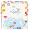
Figure 1
Figure 1
Established and more recently identified effectors that are regulated by Gβγ subunits. Gβγ subunits regulate a number of effectors at the cell surface, including adenylyl cyclase isoforms, Kir3 and voltage-gated calcium channels, and phospholipase Cβ isoforms, among others. More recently, a number of novel interacting proteins have been identified that transduce Gβγ-dependent signals in other subcellular compartments such as the Golgi apparatus [protein kinase D (PKD)], cytosol [histone deacetylase 5 (HDAC5)], and nucleus [RGS7 binding protein (R7BP), adipocyte enhancer-binding protein (AEPB1)], glucocorticoid receptor (GR), and possibly HDAC5). Whether all of these intracellular events require GPCRs or Gα subunits remains to be determined. The examples presented here are representative and do not include all of the either classical or novel effectors. For simplicity, GPCRs are shown as monomers even though they can be dimeric and may even be multimeric. See the text for more details.
Figure 2
Figure 2
GPCRs signal from distinct subcellular compartments. GPCRs have also demonstrated to signal from a number of different intracellular locations. In addition to ligand-induced signals activated at the cell surface, GPCRs have been identified with functional effects in internalizing endosomes, in the nucleus (perhaps as cleaved fragments), on the nuclear membrane, and in the ER (reviewed in 116). These receptors can be associated with unique sets of signaling partners at each of these sites as denoted by the different possible complexes. This is made even more complicated when the combinatorial power of receptor heterodimerization is taken into account. The red arrows denote potential trafficking itineraries, which could lead to the presence of particular receptors and their associated signaling machinery at distinct intracellular locations. See the text for more details.
Figure 3
Figure 3
GPCRs initially interact with their signaling partners before they leave the biosynthetic machinery. Receptor dimers or oligomers, receptor-Gβγ complexes and effector-Gβγ complexes can form in the ER even when antegrade protein trafficking from ER to Golgi is blocked with dominant negative versions of GTPases such as Sar 1 and Rab 1. The Gα subunit becomes associated with the nascent Gβγ-based complexes last, probably at ER exit sites, the ER/Golgi intermediate complex (ERGIC), or perhaps the cis-Golgi. Gβγ subunits play a central role in early interactions in the assembly of GPCR signaling complexes.
Figure 4
Figure 4
A role for Gβγ subunits in the assembly of GPCR dimers? An experiment using bioluminescence resonance energy transfer (BRET) to demonstrate that increasing amounts of the membrane-localized Gβγ scavenger, CD8-βARK-CT, but not soluble βARK-CT or CD8, can inhibit β2AR homodimer formation in a concentration-dependent manner. HEK 293 cells were transfected with recombinant plasmids to express β2AR tagged with Renilla luciferase (β2AR-RLuc), and β2AR tagged with green fluorescent protein (β2AR-GFP10). BRET was measured as described (194). Data are expressed as mean ± SEM of at least 3 different experiments and normalized by comparing with β2AR-Rluc and β2AR-GFP expressed alone. Total cDNA levels were equalized for transfection using pcDNA3. * indicates p < 0.05 compared with controls (Rluc- and GFP-tagged donor and acceptor alone) using a one-tailed Student’s t-test. The final bar represents a negative control in which CD4-Rluc is used as the BRET donor.

References

    1. Gether U, Kobilka BK. G protein-coupled receptors. II. Mechanism of agonist activation. J. Biol. Chem. 1998;273:17979–17982. - PubMed
    1. Lagerstrom MC, Schioth HB. Structural diversity of G protein-coupled receptors and significance for drug discovery. Nat. Rev. Drug Discov. 2008;7:339–357. - PubMed
    1. Shenker A, Laue L, Kosugi S, Merendino JJ, Jr, Minegishi T, Cutler GB., Jr A constitutively activating mutation of the luteinizing hormone receptor in familial male precocious puberty. Nature. 1993;365:652–654. - PubMed
    1. Robinson PR, Cohen GB, Zhukovsky EA, Oprian DD. Constitutively active mutants of rhodopsin. Neuron. 1992;9:719–725. - PubMed
    1. Parma J, Dupréz L, Van Sande J, Cochaux P, Gervy C, et al. Somatic mutations in the thyrotropin receptor gene cause hyperfunctioning thyroid adenomas. Nature. 1993;365:649–651. - PubMed

Publication types

MeSH terms

Substances

LinkOut - more resources