Review
. 2009 Feb;11(2):267-82.
doi: 10.1089/ars.2008.2115.

Connexins in vascular physiology and pathology

Affiliations
Review

Connexins in vascular physiology and pathology

Anne C Brisset et al. Antioxid Redox Signal. 2009 Feb.

Abstract

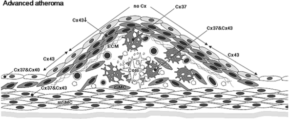
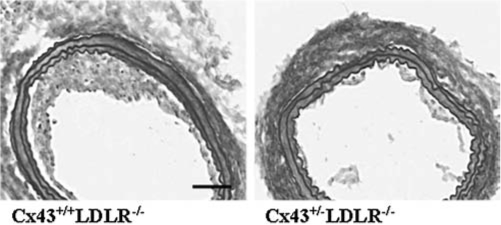
Cellular interaction in blood vessels is maintained by multiple communication pathways, including gap junctions. They consist of intercellular channels ensuring direct interaction between endothelial and smooth muscle cells and the synchronization of their behavior along the vascular wall. Gap-junction channels arise from the docking of two hemichannels or connexons, formed by the assembly of six connexins, and achieve direct cellular communication by allowing the transport of small metabolites, second messengers, and ions between two adjacent cells. Physiologic variations in connexin expression are observed along the vascular tree, with most common connexins being Cx37, Cx40, and Cx43. Changes in the level of expression of connexins have been correlated to the development of vascular disease, such as hypertension, atherosclerosis, or restenosis. Recent studies on connexin-deficient mice highlighted key roles of these communication pathways in the development of these pathologies and confirmed the need for targeted pharmacologic approaches for their prevention and treatment. The aim of this issue is to review the current knowledge on the implication of gap junctions in vascular function and most common cardiovascular diseases.


Figures

FIG. 1.
FIG. 1.
Focal planes of in situ mouse cremaster arteriole. Mouse cremaster arterioles were stained with phalloidin to mark actin in endothelium and smooth muscle and were observed under an Olympus Fluoview confocal microscope in different focal planes. (A) SMCs overlay the arteriole, with the arrow indicating orientation. (B) In the same field of view as A, ECs can be seen running perpendicular to the orientation of SMCs. (C) Another focal plane from the same arteriole in A and B, where the image is viewed transversely. In this orientation, actin can be seen linking the ECs and SMCs, presumably at the MEJ (*). With these different focal planes, one is able to detect with certainty the connexin localization within each cell type. Bar is 15 μm.
FIG. 2.
FIG. 2.
Myoendothelial junction in mouse cremaster arteriole. Usually, the use of electron microscopy is required to identify the MEJ in arterioles. In this electron micrograph, an EC extension is seen breaking through the internal elastic lamina and making contact with an SMC at the MEJ (*). Bar is 1 μm.
FIG. 3.
FIG. 3.
Expression of connexins at the arterial bifurcation. In healthy arteries, under a laminar flow, ECs express both Cx37 and Cx40. Medial SMCs (mSMC) display Cx43. Circulating monocytes express low levels of Cx37. Under low or oscillatory shear stress, ECs become dysfunctional and express Cx43. Monocytes and T lymphocytes are recruited by the dysfunctional endothelium and migrate over the EC barrier into the subendothelium. Macrophages (indicated as MO) start to express higher levels of Cx37. The dysfunctional endothelium slowly progresses over years to a fatty streak characterized by accumulation of lipids in the macrophages, which hence transform to macrophages foam cells with continued expression of Cx37.
FIG. 4.
FIG. 4.
Expression of connexins in early atheroma. Under the influence of cytokines and growth factors secreted by the dysfunctional endothelium and infiltrated inflammatory cells, medial SMCs migrate to the intima, where they proliferate. Lipids start to accumulate outside the cells and within ECM secreted by intimal SMCs. Cx43 expression is upregulated in intimal SMC (ISMC), especially in the developing fibrous cap. ECs continue to express both Cx37 and Cx40, and macrophage foam cells still express Cx37.
FIG. 5.
FIG. 5.
Expression of connexins in advanced atheroma. The fibrous cap completely covering the lesion area is composed of intimal SMCs and ECM. Extracellular lipids including cholesterol crystals (easily recognized in histology) are accumulated within the necrotic core composed of debris of apoptotic cells and degraded ECM. Macrophage foam cells close to the necrotic core express both Cx37 and Cx43. ECs still express Cx37 and Cx40 with two exceptions: ECs covering the lesion stop to express connexins, and those of shoulder regions start to express Cx43. In the fibrous cap, expression of Cx43 in intimal SMCs decreases as the lesion becomes more complicated. Medial SMCs beneath the lesion start to express Cx37.
FIG. 6.
FIG. 6.
(A) Metabolic pathway of mevalonate and cholesterol. The enzymatic reactions and their pharmacologic inhibitors are indicated. (B, C) To investigate the metabolic pathway involved in statin-induced modulation of Cx43 expression in primary human saphenous vein SMCs (C), cells were stimulated for 24 h with simvastatin (S) alone or in combination with chemical compounds affecting the downstream signalling pathway of HMG-CoA, such as mevalonate (M; 400 μM), farnesyl transferase inhibitor (FTI-277; 3 μM), or geranylgeranyl transferase inhibitor type I (GGTI-286; 10 μM), or with the isoprenoid intermediates FPP or GGPP (at 10 μM each). A typical example of a Western blot for Cx43 is shown in (C). Bar chart illustrates quantification of Cx43 expression (normalized to β-actin) in five independent experiments.
FIG. 7.
FIG. 7.
Balloon-injury in mice with reduced Cx43 expression. Representative Van Gieson-Miller staining of carotid cryosections 14 days after balloon distention injury in Cx43+/+LDLR−/− mouse (left) and in a Cx43+/−LDLR−/− mouse (right). Neointimal hyperplasia is reduced in the Cx43+/−LDLR−/− mouse as compared with its Cx43+/+LDLR−/− mouse littermate control. Bar is 100 μm.

References

    1. Abraham V. Chou ML. DeBolt KM. Koval M. Phenotypic control of gap junctional communication by cultured alveolar epithelial cells. Am J Physiol. 1999;276:L825–L834. - PubMed
    1. Aliev G. Castellani RJ. Petersen RB. Burnstock G. Perry G. Smith MA. Pathobiology of familial hypercholesterolemic atherosclerosis. J Submicrosc Cytol Pathol. 2004;36:225–240. - PubMed
    1. Beny J. Electrical coupling between smooth muscle cells and endothelial cells in pig coronary arteries. Pflugers Arch. 1997;433:364–367. - PubMed
    1. Beny JL. Information networks in the arterial wall. News Physiol Sci. 1999;14:68–73. - PubMed
    1. Berliner JA. Watson AD. A role for oxidized phospholipids in atherosclerosis. N Engl J Med. 2005;353:9–11. - PubMed

Publication types

LinkOut - more resources