. 2008 Oct 7;14(4):324-34.
doi: 10.1016/j.ccr.2008.08.012.

Plasminogen activator inhibitor-1 protects endothelial cells from FasL-mediated apoptosis

Affiliations

Plasminogen activator inhibitor-1 protects endothelial cells from FasL-mediated apoptosis

Khalid Bajou et al. Cancer Cell. .

Abstract

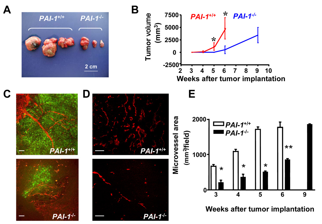
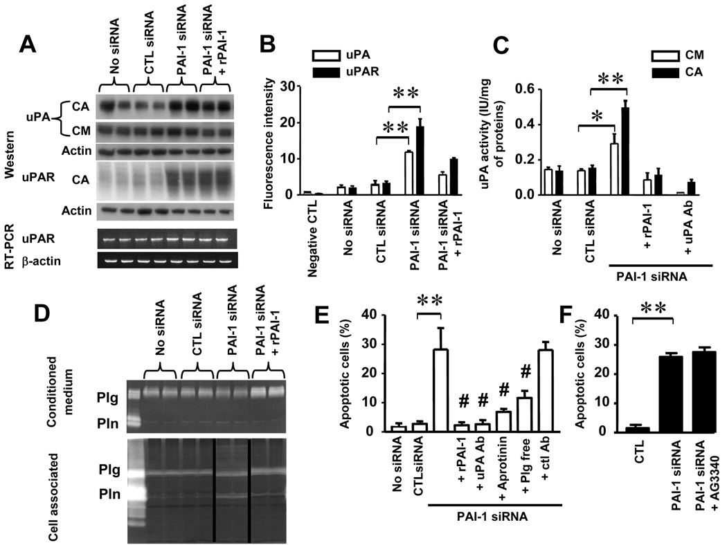
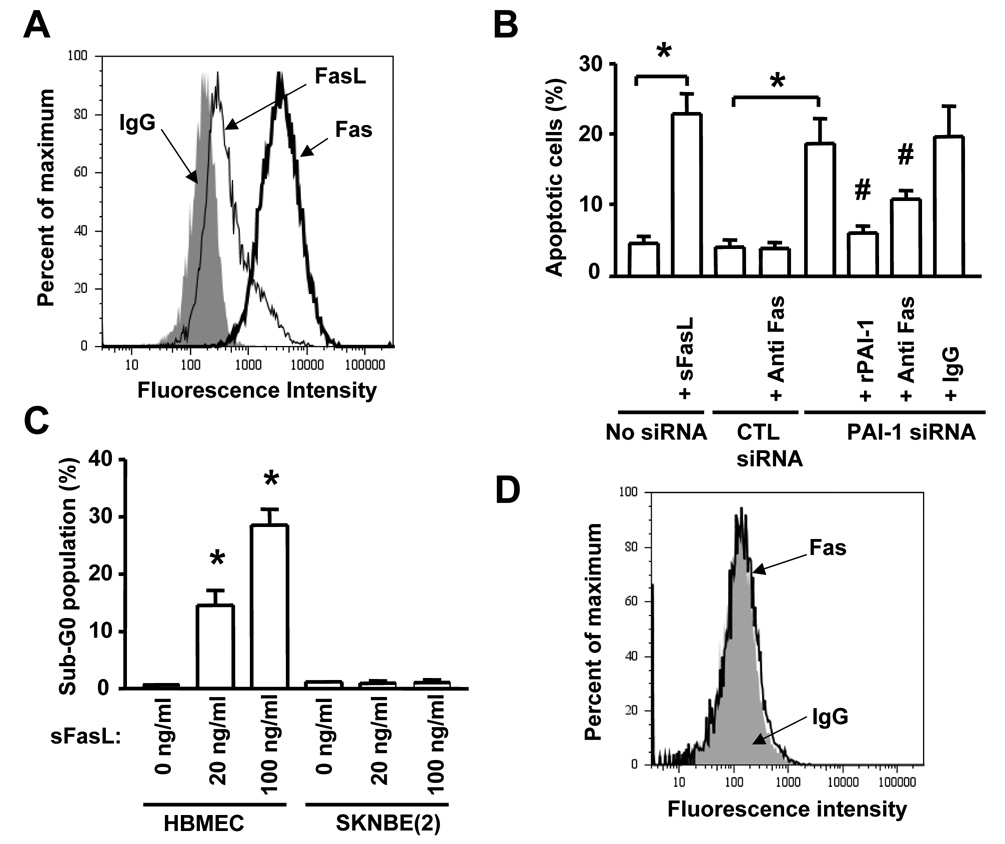
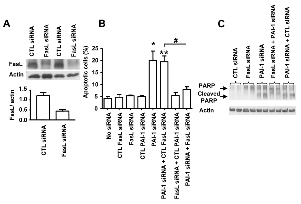
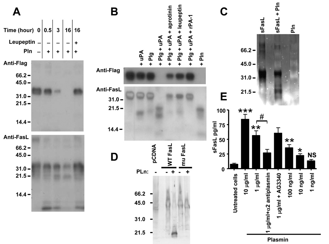
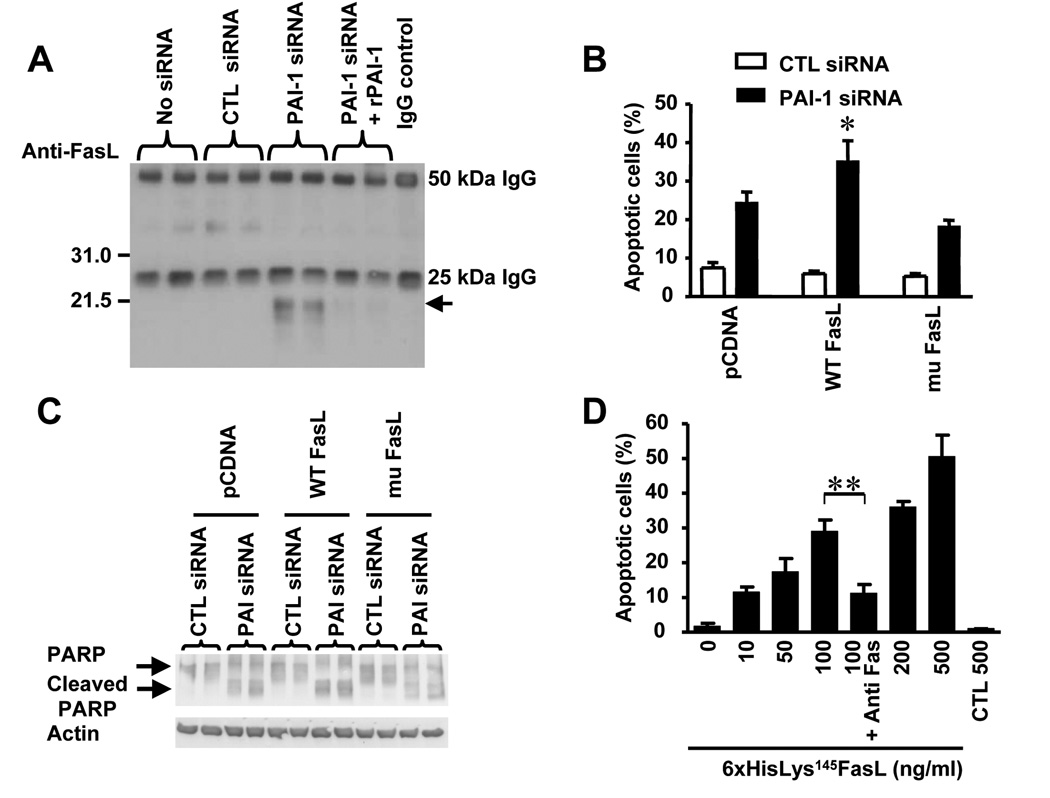
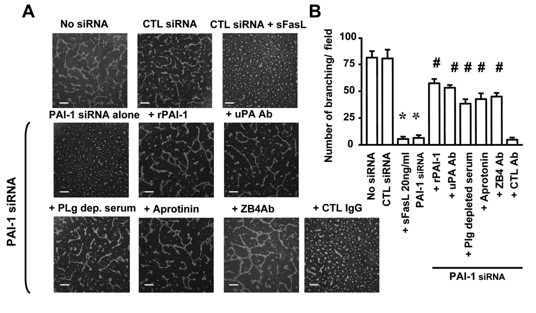
Plasminogen activator inhibitor-1 (PAI-1) paradoxically enhances tumor progression and angiogenesis; however, the mechanism supporting this role is not known. Here we provide evidence that PAI-1 is essential to protect endothelial cells (ECs) from FasL-mediated apoptosis. In the absence of host-derived PAI-1, human neuroblastoma cells implanted in PAI-1-deficient mice form smaller and poorly vascularized tumors containing an increased number of apoptotic ECs. We observed that knockdown of PAI-1 in ECs enhances cell-associated plasmin activity and increases spontaneous apoptosis in vitro. We further demonstrate that plasmin cleaves FasL at Arg144-Lys145, releasing a soluble proapoptotic FasL fragment from the surface of ECs. The data provide a mechanism explaining the proangiogenic activity of PAI-1.


Figures

Figure 1
Figure 1. Absence of Host-derived PAI-1 Reduces Human Neuroblastoma Tumor Growth and Angiogenesis
(A) Picture of representative neuroblastoma tumors collected 6 weeks after orthotopic implantation in Rag−/− PAI-1+/+ or Rag−/− PAI-1−/− mice. (B) Tumor volume in PAI-1+/+ (n= 6 to 8) and PAI-1−/− (n = 6 to 8) mice over time. Means ± standard deviation (SD) . (C) Representative images of fluorescent angiographies obtained 3 weeks after tumor implantation. Tumor cells are EGFP positive (green) and blood vessels (red) are recognized by the presence of the biotinylated tomato lectin-Texas red avidin D complex. (D) Representative microphotographs of tumor sections stained for PECAM-1/CD31, 4 weeks after tumor implantation. (E) Microvessel area values were determined on tumor sections stained for PECAM-1/CD31 at indicated time points as previously described. The data represent the means (± SD) of 6 sections examined each in 6 to 8 tumors per time point. Scale bars are 100 µm in C and D (*p<0.05, ** p<0.01 in B and E).
Figure 2
Figure 2. PAI-1 Protects EC from Apoptosis
(A) The levels of PAI-1 in the conditioned medium (CM) and in cell extracts (cell associated, CA) of EC transfected with different siRNAs were measured by ELISA. The data represent the means (± SD) of triplicate samples. (B) The percentage of apoptotic EC was determined 72 hr after transfection with siRNA by TUNEL assay. rPAI-1 (0.25 and 2.5 µg/ml) was added 24 hr after transfection. The data represent the means (± SD) of triplicate samples. (C) Caspase-3 activity in experimental conditions described in (B) was determined by colorimetric assay as described in Experimental Procedures. The data represent the means (± SD) of triplicate samples. (D) PARP cleavage was examined by Western blot analysis in the same experimental conditions as in (B). rPAI-1 was added at 2.5 µg/ml 24h after siRNA transfection. (* p<0.01 compared to CTL siRNA, # p<0.05, ## p<0.01 compared to PAI-1 siRNA, B and C).
Figure 3
Figure 3. uPA-mediated Plasminogen Activation Induces EC Apoptosis
(A) Top: Western blot analysis of cell-associated (CA) and secreted (CM) uPA and CA uPAR in EC transfected with siRNA as indicated on the top. rPAI-1 was added at 2.5 µg/ml. Bottom: RT-PCR analysis of uPAR and β-actin expression in the cell extracts obtained in the same conditions. (B) The presence of uPA and uPAR at the cell surface of cells treated as in (A) was examined by flow cytometry. The data represent the means (± SD) of triplicate samples and are representative of 3 experiments showing similar results. (C) The proteolytic activity of uPA in cell extracts (CA) and CM was determined by colorimetric assay as indicated in Experimental Procedures. The data are the means (± SD) of triplicate samples. (D) The presence of plasminogen (Plg) and plasmin (Pln) in the same experimental conditions as in (A) was examined by casein zymography in CM and cell extracts (CA). (E,F) The percentage of apoptotic EC transfected with PAI-1 siRNA and control siRNA and treated as indicated was determined by flow cytometry. The data represent the means (± SD) of triplicate dishes and are representative of 2 separate experiments showing similar results. rPAI-1 was added at 2.5 µg /ml, aprotinin at 200 µg/ml and anti uPA antibody or mouse IgG control at 7 µg/ml. AG3340 was added at 10 µg/ml (* p<0.05, ** p<0.01 for PAI-1 siRNA versus control siRNA in B,C,E and F, # p<0.01 for values versus PAI-1 siRNA in E).
Figure 4
Figure 4. Increase in FasL-mediated Apoptosis in the Absence of PAI-1
(A) The expression of Fas and FasL in HBMEC was determined by flow cytometry. (B) The percentage of apoptotic HBMEC transfected with siRNAs in the absence or presence of an anti-Fas blocking antibody or rPAI-1 (2.5 µg/ml) was determined by flow cytometry after staining with propidium iodide. The data are the means (± SD) of triplicate samples. (C) Apoptosis in HBMEC and SK-N-BE(2) cells was determined by flow cytometry as in (B) after the cells were treated for 24 hr with rsFasL at indicated concentrations. The data are the means (± SD) of triplicate samples. (D) The expression of Fas in SK-N-BE(2) tumor cells was determined by flow cytometry (* p<0.001, # p<0.01 in B and C).
Figure 5
Figure 5. FasL is Required for EC Apoptosis upon PAI-1 Downregulation
(A) HBMEC were transfected with a FasL siRNA or a scrambled siRNA sequence (control) and examined after 72 hr for the presence of FasL in the cell lysate by Western blot. A quantitative analysis of the data obtained by the scanning of 3 separate blots is shown in the lower panel. The data are the mean ratios (± SD) of FasL/actin. (B) HBMEC were transfected with PAI-1 siRNA, FasL siRNA or a combination of both siRNAs and their respective scrambled controls and examined after 72 hr for apoptosis by flow cytometry. The data represent the mean numbers (± SD) of apoptotic cells from triplicate samples (* p<0.025, ** p<0.01, # p<0.005). (C) Cell lysates of HBMEC treated as indicated in the figure were examined for PARP cleavage by Western blot analysis.
Figure 6
Figure 6. Cleavage of Recombinant and Membrane-associated FasL by Plasmin
(A,B) Western blot analysis of FLAG-FasL (10 µg/ml) treated in the conditions indicated on top using an anti-FLAG antibody (upper blots) or an anti-FasL antibody (lower blots). The concentrations used were as follows: plasmin (Pln), 1 µg/ml; plasminogen (Plg), 1 µg/ml; leupeptin, 500 µg/ml; aprotinin, 1 mg/ml; uPA, 600 IU/ml; and rPAI-1, 50 µg/ml. (C) Silver staining of a polyacrylamide gel of recombinant sFasL incubated in the presence or absence of Pln as indicated in A and B. (D) Western blot analysis with an anti-FasL antibody of membrane proteins extracted from HT1080 cells expressing WT FasL or mu FasL and incubated for 1 hr in the presence or absence of plasmin (1 µg/500 µg of lysate) at 37°C before SDS-PAGE. (E) Levels of sFasL in the serum-free culture medium of HBMEC incubated in the presence of plasmin and inhibitors as indicated. α2-antiplasmin was added at a concentration of 100 µg/ml and AG3340 at a concentration of 10 µg/ml. The data represent the mean concentrations (±SD) of sFasL in the culture medium of triplicate samples and are representative of 2 experiments showing similar results (* p<0.025, ** p<0.01, *** p<0.005 versus untreated cells, # p<0.005 versus plasmin treated cells).
Figure 7
Figure 7. The 21.5 kDa Plasmin-generated FasL is Pro-apoptotic
(A) The presence of the 21.5 kDa sFasL plasmin-generated fragment was detected by immunoprecipitation and Western blot analysis with an anti-FasL antibody in the CM of HBMEC treated as indicated on top. (B) HBMEC were transfected with a pcDNA plasmid containing either a WT FasL or a mu FasL. Stable transfected cells were selected and re-transfected with a PAI-1 siRNA or a control siRNA and tested for apoptosis by FACS analysis after 72 hr. The data represent the means (± SD) of triplicate samples (* p< 0.05). (C) Cells treated as described in (B) were examined for the presence of cleaved PARP by Western blot analysis. (D) The recombinant 6xHisLys145FasL protein that corresponds to the 21.5 kDa plasmin-generated FasL obtained as shown in supplemental data Figure 2C was added at indicated concentrations to HBMEC for 48 hr and the percentage of apoptotic cells was measured by flow cytometry. An anti-Fas antibody (ZB4) was added at 500 ng/ml. The data represent the means (±SD) from triplicate samples (** p<0.005).
Figure 8
Figure 8. FasL Inhibits EC Branching in Matrigel
(A) Representative photomicrographs of HBMEC plated on Matrigel and treated as indicated. (B) Quantitative analysis of the data. The data represent the mean (± SD) numbers of branches per microscopic field from a total of 6 fields each examined in 3 different dishes (* p<0.001, # p<0.001). Bar = 400 µm

References

    1. Aoudjit F, Vuori K. Engagement of the alpha2beta1 integrin inhibits Fas ligand expression and activation-induced cell death in T cells in a focal adhesion kinase-dependent manner. Blood. 2000;95:2044–2051. - PubMed
    1. Aoudjit F, Vuori K. Matrix attachment regulates Fas-induced apoptosis in endothelial cells: a role for c-flip and implications for anoikis. J Cell Biol. 2001;152:633–643. - PMC - PubMed
    1. Bajou K, Maillard C, Jost M, Lijnen RH, Gils A, Declerck P, Carmeliet P, Foidart JM, Noel A. Host-derived plasminogen activator inhibitor-1 (PAI-1) concentration is critical for in vivo tumoral angiogenesis and growth. Oncogene. 2004;23:6986–6990. - PubMed
    1. Bajou K, Masson V, Gerard RD, Schmitt PM, Albert V, Praus M, Lund LR, Frandsen TL, Brunner N, Dano K, Fusenig NE, Weidle U, Carmeliet G, Loskutoff D, Collen D, Carmeliet P, Foidart JM, Noel A. The plasminogen activator inhibitor PAI-1 controls in vivo tumor vascularization by interaction with proteases, not vitronectin. Implications for antiangiogenic strategies. J. Cell Biol. 2001;152:777–784. - PMC - PubMed
    1. Bajou K, Noel A, Gerard RD, Masson V, Brunner N, Holst-Hansen C, Skobe M, Fusenig NE, Carmeliet P, Collen D, Foidart JM. Absence of host plasminogen activator inhibitor 1 prevents cancer invasion and vascularization. Nat. Med. 1998;4:923–928. - PubMed

Publication types

MeSH terms