

Expression of fragile X mental retardation protein within the vocal control system of developing and adult male zebra finches
- PMID: 18835331
- PMCID: PMC2598769
- DOI: 10.1016/j.neuroscience.2008.09.005
Expression of fragile X mental retardation protein within the vocal control system of developing and adult male zebra finches
Abstract
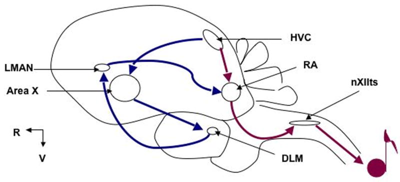
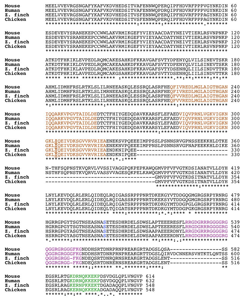
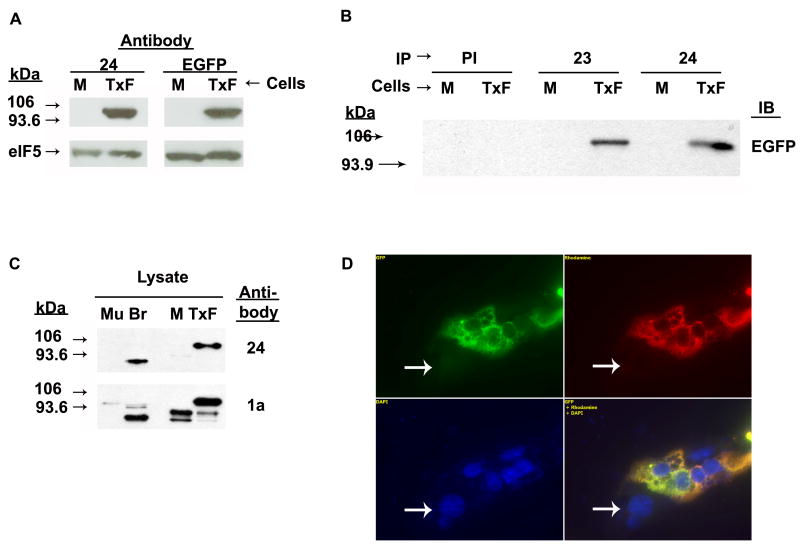
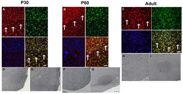
Individuals with fragile X syndrome (FXS) are cognitively impaired and have marked speech delays and deficits. Our goal was to characterize expression of fragile X mental retardation protein (FMRP), encoded by Fmr1 fragile X mental retardation 1 gene or transcript (FMR1), in an animal model that learns to vocalize, namely the zebra finch Taeniopygia guttata (Tgu). We cloned and sequenced the zebra finch ortholog of FMR1 (TguFmr1) and developed an antibody that recognizes TguFmrp specifically. TguFmrp has structural features similar to its human ortholog FMRP. Because FXS patients exhibit sensorimotor deficits, we examined TguFmrp expression prior to, during, and after sensorimotor song learning in zebra finches. We found that TguFmrp is expressed throughout the brain and in four major song nuclei of the male zebra finch brain, primarily in neurons. Additionally, prior to sensorimotor learning, we observed elevated TguFmrp expression in the robust nucleus of the arcopallium (RA) of post-hatch day 30 males, compared with the surrounding telencephalon, suggesting a preparation for this stage of song learning. Finally, we observed variable TguFmrp expression in the RA of adolescent and adult males: in some males it was elevated and in others it was comparable to the surrounding telencephalon. In summary, we have characterized the zebra finch ortholog of FMRP and found elevated levels in the premotor nucleus RA at a key developmental stage for vocal learning.
Figures


Similar articles
-
Exploring the zebra finch Taeniopygia guttata as a novel animal model for the speech-language deficit of fragile X syndrome.Results Probl Cell Differ. 2012;54:181-97. doi: 10.1007/978-3-642-21649-7_10. Results Probl Cell Differ. 2012. PMID: 22009353 Free PMC article.
-
Expression of the GABA(A) receptor gamma4-subunit gene in discrete nuclei within the zebra finch song system.Neuroscience. 2008 Nov 11;157(1):143-52. doi: 10.1016/j.neuroscience.2008.08.057. Epub 2008 Sep 6. Neuroscience. 2008. PMID: 18824085
-
A dynamic, sex-specific expression pattern of genes regulating thyroid hormone action in the developing zebra finch song control system.Gen Comp Endocrinol. 2017 Jan 1;240:91-102. doi: 10.1016/j.ygcen.2016.09.016. Epub 2016 Sep 29. Gen Comp Endocrinol. 2017. PMID: 27693816
-
Distribution of language-related Cntnap2 protein in neural circuits critical for vocal learning.J Comp Neurol. 2014 Jan 1;522(1):169-85. doi: 10.1002/cne.23394. J Comp Neurol. 2014. PMID: 23818387 Free PMC article.
-
The sensitive period for auditory-vocal learning in the zebra finch: Consequences of limited-model availability and multiple-tutor paradigms on song imitation.Behav Processes. 2019 Jun;163:5-12. doi: 10.1016/j.beproc.2017.07.007. Epub 2017 Jul 23. Behav Processes. 2019. PMID: 28743517 Free PMC article. Review.
Cited by
-
MOV10 and FMRP regulate AGO2 association with microRNA recognition elements.Cell Rep. 2014 Dec 11;9(5):1729-1741. doi: 10.1016/j.celrep.2014.10.054. Epub 2014 Nov 20. Cell Rep. 2014. PMID: 25464849 Free PMC article.
-
Rebound from Inhibition: Self-Correction against Neurodegeneration?J Clin Cell Immunol. 2017 Apr;8(2):492. doi: 10.4172/2155-9899.1000492. Epub 2017 Mar 13. J Clin Cell Immunol. 2017. PMID: 28775912 Free PMC article.
-
Avian models for brain mechanisms underlying altered social behavior in autism.Front Physiol. 2022 Oct 28;13:1032046. doi: 10.3389/fphys.2022.1032046. eCollection 2022. Front Physiol. 2022. PMID: 36388132 Free PMC article. Review.
-
Early Chronic Fluoxetine Treatment of Ts65Dn Mice Rescues Synaptic Vesicular Deficits and Prevents Aberrant Proteomic Alterations.Genes (Basel). 2024 Apr 3;15(4):452. doi: 10.3390/genes15040452. Genes (Basel). 2024. PMID: 38674386 Free PMC article.
-
A new regulatory function of the region proximal to the RGG box in the fragile X mental retardation protein.J Cell Sci. 2011 Sep 15;124(Pt 18):3060-5. doi: 10.1242/jcs.086751. Epub 2011 Aug 24. J Cell Sci. 2011. PMID: 21868366 Free PMC article.
References
-
- Abitbol M, Menini C, Delezoide AL, Rhyner T, Vekemans M, Mallet J. Nucleus basalis magnocellularis and hippocampus are the major sites of FMR-1 expression in the human fetal brain. Nat Genet. 1993;4:147–153. - PubMed
-
- Antar LN, Dictenberg JB, Plociniak M, Afroz R, Bassell GJ. Localization of FMRP-associated mRNA granules and requirement of microtubules for activity-dependent trafficking in hippocampal neurons. Genes Brain Behav. 2005;4:350–359. - PubMed
-
- Antar LN, Li C, Zhang H, Carroll RC, Bassell GJ. Local functions for FMRP in axon growth cone motility and activity-dependent regulation of filopodia and spine synapses. Moll Cell Neurosci. 2006;32:37–48. - PubMed
-
- Aronov D, Andalman AS, Fee MS. A specialized forebrain circuit for vocal babbling in the juvenile songbird. Science. 2008;320:630–634. - PubMed
Publication types
MeSH terms
Substances
Grants and funding
LinkOut - more resources
Full Text Sources
Medical
Molecular Biology Databases
