

Prenatal mild ventriculomegaly predicts abnormal development of the neonatal brain
- PMID: 18835482
- PMCID: PMC2630424
- DOI: 10.1016/j.biopsych.2008.07.031
Prenatal mild ventriculomegaly predicts abnormal development of the neonatal brain
Abstract
Background: Many psychiatric and neurodevelopmental disorders are associated with mild enlargement of the lateral ventricles thought to have origins in prenatal brain development. Little is known about development of the lateral ventricles and the relationship of prenatal lateral ventricle enlargement with postnatal brain development.
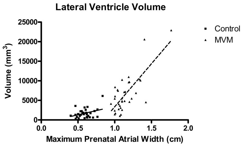
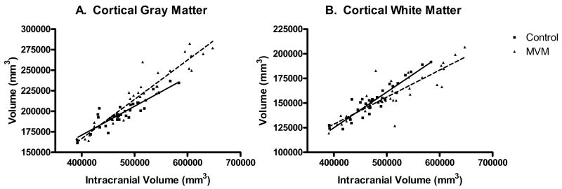
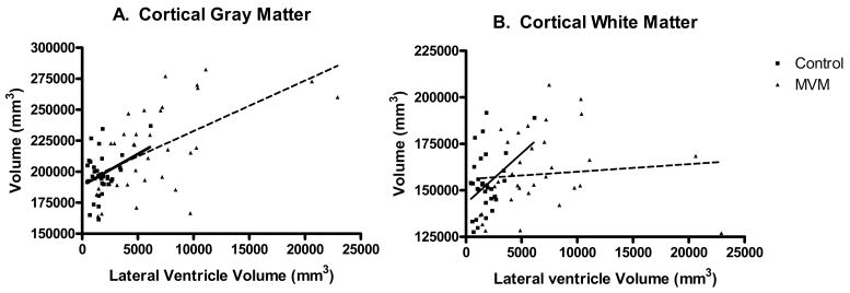
Methods: We performed neonatal magnetic resonance imaging on 34 children with isolated mild ventriculomegaly (MVM; width of the atrium of the lateral ventricle >/= 1.0 cm) on prenatal ultrasound and 34 age- and sex-matched control subjects with normal prenatal ventricle size. Lateral ventricle and cortical gray and white matter volumes were assessed. Fractional anisotropy (FA) and mean diffusivity (MD) in corpus callosum and corticospinal white matter tracts were determined obtained using quantitative tractography.
Results: Neonates with prenatal MVM had significantly larger lateral ventricle volumes than matched control subjects (286.4%; p < .0001). Neonates with MVM also had significantly larger intracranial volumes (ICV; 7.1%, p = .0063) and cortical gray matter volumes (10.9%, p = .0004) compared with control subjects. Diffusion tensor imaging tractography revealed a significantly greater MD in the corpus callosum and corticospinal tracts, whereas FA was significantly smaller in several white matter tract regions.
Conclusions: Prenatal enlargement of the lateral ventricle is associated with enlargement of the lateral ventricles after birth, as well as greater gray matter volumes and delayed or abnormal maturation of white matter. It is suggested that prenatal ventricle volume is an early structural marker of altered development of the cerebral cortex and may be a marker of risk for neuropsychiatric disorders associated with ventricle enlargement.
Conflict of interest statement
Financial Disclosures
The authors reported no biomedical financial interests or potential conflicts of interest.
Figures


References
-
- Lawrie SM, Abukmeil SS. Brain abnormality in schizophrenia. A systematic and quantitative review of volumetric magnetic resonance imaging studies. Br J Psychiatry. 1998;172:110–20. - PubMed
-
- Wright IC, Rabe-Hesketh S, Woodruff PW, David AS, Murray RM, Bullmore ET. Meta-analysis of regional brain volumes in schizophrenia. Am J Psychiatry. 2000;157:16–25. - PubMed
-
- Piven J, Arndt S, Bailey J, Havercamp S, Andreasen NC, Palmer P. An MRI study of brain size in autism. Am J Psychiatry. 1995;152:1145–1149. - PubMed
-
- Lyoo IK, Noam GG, Lee CK, Lee HK, Kennedy BP, Renshaw PF. The corpus callosum and lateral ventricles in children with attention-deficit hyperactivity disorder: a brain magnetic resonance imaging study. Biol Psychiatry. 1996;40:1060–1063. - PubMed
Publication types
MeSH terms
Grants and funding
LinkOut - more resources
Full Text Sources
Medical
