. 2008 Nov 28;283(48):33750-62.
doi: 10.1074/jbc.M803156200. Epub 2008 Oct 2.

Interaction of the N- and C-terminal autoregulatory domains of FRL2 does not inhibit FRL2 activity

Affiliations

Interaction of the N- and C-terminal autoregulatory domains of FRL2 does not inhibit FRL2 activity

Dominique C Vaillant et al. J Biol Chem. .

Abstract

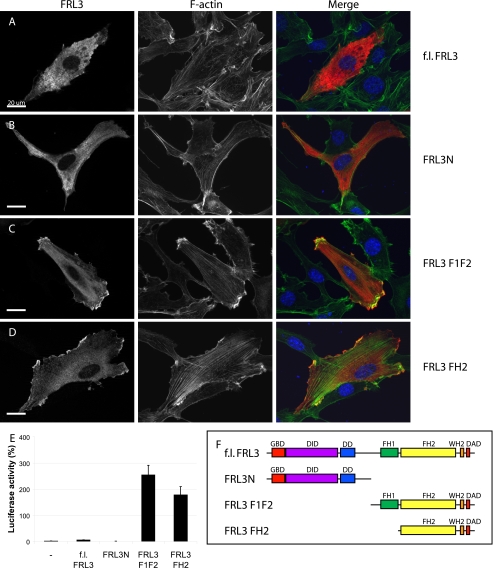
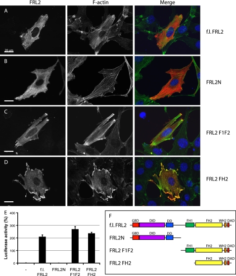
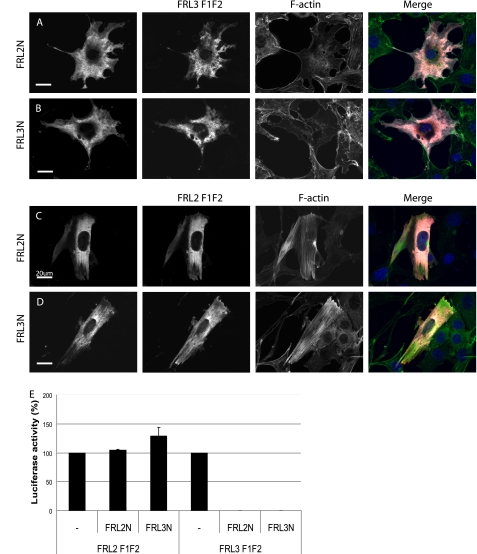
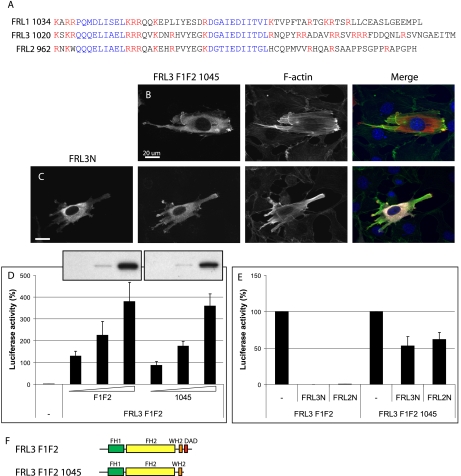
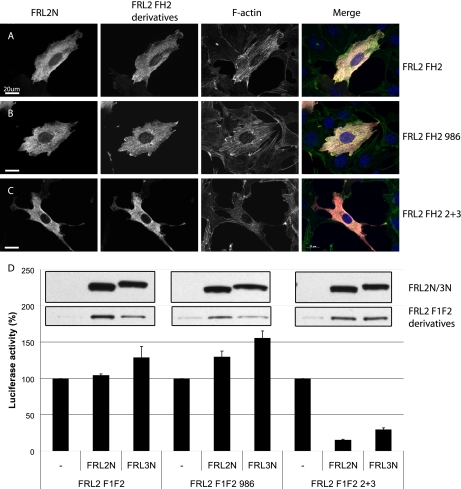
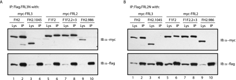
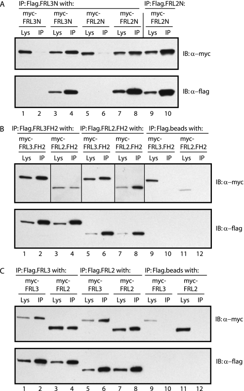
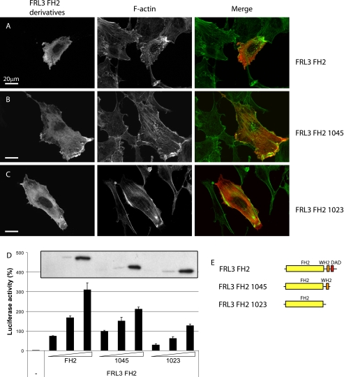
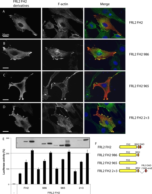
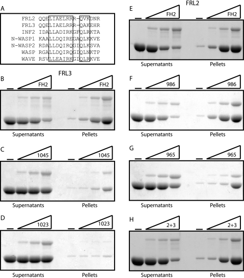
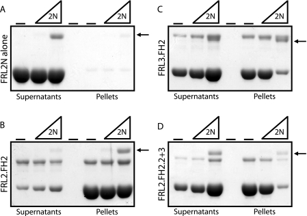
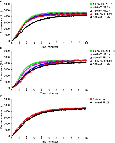
Formin homology proteins are a highly conserved family of cytoskeletal remodeling proteins best known for their ability to induce the formation of long unbranched actin filaments. They accomplish this by nucleating the de novo polymerization of F-actin and also by acting as F-actin barbed end "leaky cappers" that allow filament elongation while antagonizing the function of capping proteins. More recently, it has been reported that the FH2 domains of FRL1 and mDia2 and the plant formin AFH1 are able to bind and bundle actin filaments via distinct mechanisms. We find that like FRL1, FRL2 and FRL3 are also able to bind and bundle actin filaments. In the case of FRL3, this activity is dependent upon a proximal DAD/WH2-like domain that is found C-terminal to the FH2 domain. In addition, we show that, like other Diaphanous-related formins, FRL3 activity is subject to autoregulation mediated by the interaction between its N-terminal DID and C-terminal DAD. In contrast, the DID and DAD of FRL2 also interact in vivo and in vitro but without inhibiting FRL2 activity. These data suggest that current models describing DID/DAD autoregulation via steric hindrance of FH2 activity must be revised. Finally, unlike other formins, we find that the FH2 and N-terminal dimerization domains of FRL2 and FRL3 are able to form hetero-oligomers.


Figures

FIGURE 1.
FIGURE 1.
FRL3 is an autoregulated formin. Epitope-tagged derivatives of FRL3 corresponding to the full-length protein, N terminus, FH1 + FH2, and FH2 were expressed by transient transfection in NIH 3T3 cells (0.3 μg of DNA). Protein expression was detected with 9E10 anti-Myc monoclonal (red); F-actin was visualized with fluorescein phalloidin (green). A, full-length FRL3 is distributed diffusely throughout the cytoplasm of transfected cells (100% of cells) and fails to induce stress fiber formation (94%, n = 105). B, FRL3N distributes throughout the cytoplasm and shows apparent membrane targeting in NIH 3T3 cells (99%). FRL3N-expressing cells have reduced levels of F-actin (97%, n = 113). C, expression of FRL3.F1F2 induces robust stress fiber accumulation (93%) and dense accumulation of F-actin at the cell periphery that tightly associates with the overexpressed protein (89%, n = 102). D, FRL3.FH2 induces stress fibers (73%), dense patches of F-actin in lamellopodia-like structures at the periphery of the cell and granular patches of actin throughout the cytoplasm (86%, n = 109). E, expression of FRL3.F1F2 and FH2 (1 μg of DNA transfected) induces robust activation of an SRF reporter gene. Reporter gene activity was standardized to activation induced by expression of an SRF-VP16 control fusion protein. Error bars, S.E., n = 3. F, schematic of FRL3 derivatives.
FIGURE 2.
FIGURE 2.
FRL2 is not autoregulated. Epitope-tagged derivatives of FRL2 corresponding to the full-length protein, N terminus, FH1 + FH2, and FH2 were expressed by transient transfection in NIH 3T3 cells. Protein expression was detected with 9E10 anti-Myc monoclonal (red); F-actin was visualized with fluorescein phalloidin (green). A, full-length FRL2 accumulates at the cell periphery, strongly induces stress fiber formation, and associates with F-actin in transfected cells (97%, n = 101). B, FRL2N is distributed diffusely throughout the cytoplasm (96%). FRL2N-expressing cells have reduced levels of F-actin (99%, n = 103). C, expression of FRL2.F1F2 induces robust stress fiber accumulation (92%) and dense accumulation of F-actin at the cell periphery that tightly associates with the overexpressed protein (96%, n = 105). D, FRL2.FH2 induces stress fibers, dense patches of F-actin in lamellopodia-like structures at the periphery of the cell, and granular patches of actin throughout the cytoplasm (98%, n = 1-2). E, expression of full-length F1F2 and FH2 derivatives of FRL2 induces robust activation of an SRF reporter gene. Reporter gene activity was standardized to activation induced by expression of an SRF-VP16 control fusion protein. Error bars, S.E., n = 3. F, schematic of FRL2 derivatives.
FIGURE 3.
FIGURE 3.
FRL2 contains a functional DID but not a functional DAD. A and B, FRL3.F1F2 (red) was coexpressed with FRL2N or FRL3N (white) by transient transfection. Co-expression of either FRL2N or FRL3N (1.2 μg of DNA transfected) inhibits FRL3.F1F2 (0.3 μg of DNA)-induced stress fiber formation (green, 98%, n = 103) (compare cells in A and B with Fig. 1C). C and D, FRL2.F1F2 (red) was co-expressed with FRL2N or FRL3N (white) by transient transfection. Co-expression of either FRL2N or FRL3N did not inhibit FRL2.F1F2-induced stress fiber formation (green) (97%, n = 100). E, expression of FRL2N or FRL3N (1 μg of DNA transfected) inhibits FRL3.F1F2 (0.1 μg of DNA)-induced activation of an SRF reporter gene but has no effect on FRL2.F1F2-induced SRF activation. Reporter gene activation in the absence of inhibitor was standardized to 100%. Error bars, S.E., n = 3.
FIGURE 4.
FIGURE 4.
The most distal DAD-like motif in FRL3 is required for autoregulation. A, alignment of the FRL1, -2, and -3 C-terminal domains. Two WH2/DAD-like motifs (blue) are present in the C termini of FRL2 and FRL3. Basic residues are highlighted in red. B and C, FRL3.F1F2.1045 (red)-induced stress fiber (green) formation is largely unaffected by co-expression of FRL3N (white) (94%, n = 102 versus 72%, n = 101). D, deletion of the FRL3 DAD domain does not significantly affect FRL3.F1F2 activity. Inset, relative levels of expression of FRL3.F1F2 and FRL3.F1F2.1045, as determined by immunoblotting using the same exposure of the same gel. Reporter gene activity was standardized to activation induced by expression of an SRF-VP16 control fusion protein. Error bars, S.E., n = 3. E, SRF reporter gene activation by FRL2.FH2.1045 is not significantly inhibited by FRL2N or FRL3N. Reporter gene activation in the absence of inhibitor was standardized to 100%. Error bars, S.E., n = 3. F, schematic of FRL3 derivatives.
FIGURE 5.
FIGURE 5.
A chimeric FRL2 protein containing the FRL3 DAD is inhibited by DID in trans. A, FRL2.FH2 (red)-induced stress fiber formation (green) is not inhibited by co-expression of FRL2N (white) (97% alone, n = 100 versus 99% with FRL2N, n = 100). B, FRL2.FH2.986-induced stress fiber formation is not inhibited by co-expression of FRL2N (98%, n = 100). C, FRL2.FH2.2+3-induced stress fiber formation is inhibited by co-expression of FRL2N (25%, n = 102). D, FRL2.F1F2- or FRL2.F1F2.986-induced SRF reporter gene activation is not affected by co-expression of FRL2N orFRL3N. FRL2.F1F2.2+3 SRF activation is strongly inhibited by FRL2N and FRL3N. Inset, top panel, relative levels of expression of FRL2N and FRL3N as determined by immunoblotting using the same exposure of the same gel; bottom panel, relative levels of expression of FRL2.F1F2, FRL2.F1F2.986, and FRL2.F1F2.2+3, as determined by immunoblotting using the same exposure of the same gel. Reporter gene activation by FRL2.F1F2, FRL2.F1F2.986, or FRL2.F1F2.2+3 in the absence of inhibitor was standardized to 100% for each set. Error bars, S.E., n = 3.
FIGURE 6.
FIGURE 6.
C-terminal derivatives of FRL2 and FRL3 bind to N-terminal FRL2 and FRL3 derivatives in a DAD-dependent manner. A, FLAG-tagged FRL3N (1.5 μg of DNA) was co-expressed in NIH 3T3 cells with Myc-tagged FRL3.FH2 or FH2.1045 and FRL2.F1F2, F1F2.2+3, or F1F2.986 (1.5 μg of DNA). The FLAG-tagged protein efficiently co-immunoprecipitated (IP) FRL3.FH2, FRL2.F1F2, and FRL2.F1F2.2+3 but not FRL3.FH2.1045 or FRL2.F1F2.986 (compare lanes 1 and 2 with lanes 3 and 4 and lanes 5-8 with lanes 9 and 10). B, FLAG-tagged FRL2N was co-expressed in NIH 3T3 cells with Myc-tagged FRL3.FH2 or FH2.1045 and FRL2.F1F2, F1F2.2+3, or F1F2.986. The FLAG-tagged protein efficiently co-immunoprecipitated FRL3.FH2, FRL2.F1F2, and FRL2.F1F2.2+3 but not FRL3.FH2.1045 or FRL2.F1F2.986, which lack the most distal DAD motif (compare lanes 1 and 2 with lanes 3 and 4 and lanes 5-8 with lanes 9 and 10).
FIGURE 7.
FIGURE 7.
FRL2 and FRL3 are able to form hetero-oligomers. A, FRL2N and FRL3N form hetero-oligomers in vivo. FLAG-tagged FRL3N, or FRL2N, was co-expressed in NIH 3T3 cells with Myc-tagged FRL3N or FRL2N. FLAG-tagged FRL3N efficiently co-immunoprecipitated (IP) FRL3N and FRL2N (compare lanes 1 and 2 with lanes 3 and 4 and lanes 5 and 6 with lanes 7 and 8). FLAG-tagged FRL2N efficiently co-immunoprecipitated FRL2N. FLAG beads alone served as a negative control (lanes 1 and 2 and lanes 5 and 6). B, FRL2.FH2 and FRL3.FH2 form hetero-oligomers in vivo. FLAG-tagged FRL3.FH2 or FRL2.FH2 was co-expressed in NIH 3T3 cells with Myc-tagged FRL2.FH2 and FRL3.FH2. FLAG-tagged FRL3.FH2 efficiently co-immunoprecipitated FRL2.FH2 and FRL3.FH2 (compare lanes 1 and 2 with lanes 3 and 4 and lanes 9 and 10). FLAG-tagged FRL2.FH2 also efficiently co-immunoprecipitated FRL2.FH2 and FRL3.FH2 (compare lanes 5 and 6 with lanes 7 and 8 and lanes 11 and 12). FLAG beads alone served as a negative control (lanes 9-12). Vertical lines separate different exposures of the same immunoblot (lanes 1, 2, 5, 6, and 9-12 versus lanes 3, 4, 7, and 8). C, full-length FRL2 and FRL3 form hetero-oligomers in vivo. FLAG-tagged FRL3 or FRL2 was co-expressed in NIH 3T3 cells with Myc-tagged FRL2 and FRL3. FLAG-tagged FRL3 efficiently co-immunoprecipitated FRL3 and FRL2 (compare lanes 1 and 2 with lanes 3 and 4 and lanes 9 and 10). FLAG-tagged FRL2 also efficiently co-immunoprecipitated FRL2 and FRL3 (compare lanes 5 and 6 with lanes 7 and 8 and lanes 11 and 12). FLAG beads alone served as a negative control (lanes 9-12).
FIGURE 8.
FIGURE 8.
Deletion of the C-terminal DAD motifs reduces FRL3.FH2 activity in vivo. A, as in Fig. 1, expression of FRL3.FH2 induces thick stress fibers, actin aggregates, and peripheral F-actin accumulation (98%, n = 101). B, FRL3.FH2.1045 behaves similarly to FH2 (83%, n = 100). C, FRL3.FH2.1023 induces thick stress fibers (75%, n = 102) but no actin aggregates or peripheral F-actin. D, deletion of the distal and proximal DAD motifs reduces the ability of FRL3.FH2 to induce activation of an SRF reporter gene. Inset, relative levels of expression of FRL3.FH2, FRL3.FH2.1045, and FRL3.FH2.1023 as determined by immunoblotting. Reporter gene activity was standardized to activation induced by expression of an SRF-VP16 control fusion protein. Error bars, S.E., n = 3. E, schematic of FRL3.FH2 derivatives.
FIGURE 9.
FIGURE 9.
Deletion of the C-terminal DAD modifies FRL2.FH2 activity in vivo. A, as in Fig. 2, expression of FRL2.FH2 induces thick stress fibers, actin aggregates, and peripheral F-actin accumulation (97%, n = 100). B, FRL2.FH2.986 behaves similarly to FH2 (99%, n = 100). C, FRL2.FH2.965 induces fewer actin aggregates and peripheral F-actin (88%, n=100). D, FRL2.FH2.2+3 behaves similarly to FRL2.FH2(98%, n = 100). E, deletion of both distal and proximal DAD motifs does not affect FRL2.FH2-induced SRF reporter gene activation. Inset, relative levels of expression of FRL2.FH2, FRL2.FH2.986, FRL2.FH2.965, and FRL2.FH2.2+3, as determined by immunoblotting. Reporter gene activity was standardized to activation induced by expression of an SRF-VP16 control fusion protein. Error bars, S.E., n = 3. F, schematic of FRL2.FH2 derivatives.
FIGURE 10.
FIGURE 10.
The FH2 domains of FRL2 and FRL3 are able to bind and bundle actin filaments. A, alignment of WH2-like domains of FRL2 and FRL3 with the WH2 domains of INF2, N-WASP1, N-WASP2, WASP, and WAVE. B, FRL3.FH2; C, FRL3.FH2.1045; D, FRL3.FH2.1023; E, FRL2.FH2; F, FRL2.FH2.986; G, FRL2.FH2.965; H, FRL2.FH2.2+3 (0, 0.15, 0.3, and 0.6 μm) were incubated with F-actin (2 μm) and then centrifuged at 16,000 × g for 10 min to pellet F-actin bundles. Equivalent samples of the supernatant and pellet were subjected to SDS-PAGE, and the proteins were visualized with Coomassie Blue.
FIGURE 11.
FIGURE 11.
FRL2N inhibits actin bundling by FRL3.FH2 and FRL2.FH2.2+3. FRL2N (0, 0.1, and 0.5 μm) was incubated with F-actin (2 μm) (A) and FRL2.FH2 (0.5 μm) (B), FRL3.FH2 (0.5 μm) (C), or FRL2.2+3 (0.5 μm) (D) and then centrifuged at 16,000 × g for 10 min to pellet F-actin bundles. Equivalent samples of the supernatant and pellet were subjected to SDS-PAGE, and the proteins were visualized with Coomassie Blue. The arrow indicates the FRL2N protein band.
FIGURE 12.
FIGURE 12.
FRL2N inhibits actin polymerization induced by FRL2.FH2.2+3 but not by FRL2.FH2. Purified FRL2.FH2 and FRL2.FH2.2+3 derivatives were tested in pyrene-actin polymerization assays at the indicated concentrations in the presence or absence of purified FRL2N. A, FRL2 FH2 (60 nm) induces actin polymerization. This polymerization is not inhibited by preincubation with FRL2N (20, 60, and 180 nm). FRL2N alone (180 nm) has no effect on polymerization. B, FRL2.FH2.2+3 (60 nm) induces actin polymerization and is inhibited by preincubation with FRL2N (20, 60, and 180 nm). C, FRL2N alone (180 nm) does not affect actin polymerization. G-actin concentration was 2 μm. A.U., absorbance unts.

Similar articles

Cited by

References

    1. Higgs, H. N., and Peterson, K. J. (2005) Mol. Biol. Cell 16 1-13 - PMC - PubMed
    1. Chang, F., Drubin, D., and Nurse, P. (1997) J. Cell Biol. 137 169-182 - PMC - PubMed
    1. Evangelista, M., Pruyne, D., Amberg, D. C., Boone, C., and Bretscher, A. (2002) Nat. Cell Biol. 4 260-269 - PubMed
    1. Feierbach, B., and Chang, F. (2001) Curr. Biol. 11 1656-1665 - PubMed
    1. Kobielak, A., Pasolli, H. A., and Fuchs, E. (2004) Nat. Cell Biol. 6 21-30 - PMC - PubMed

Publication types

MeSH terms

LinkOut - more resources