. 2008 Nov;36(19):6309-17.
doi: 10.1093/nar/gkn615. Epub 2008 Oct 3.

Rapid regulation of telomere length is mediated by poly(ADP-ribose) polymerase-1

Affiliations

Rapid regulation of telomere length is mediated by poly(ADP-ribose) polymerase-1

Sascha Beneke et al. Nucleic Acids Res. 2008 Nov.

Abstract

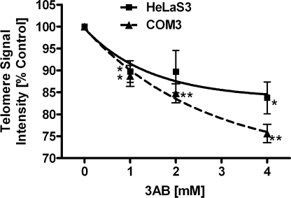
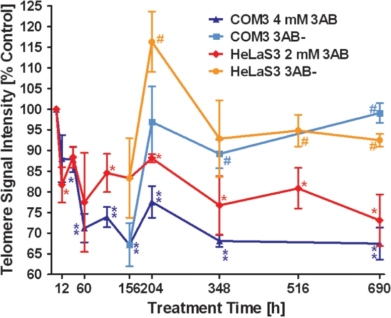
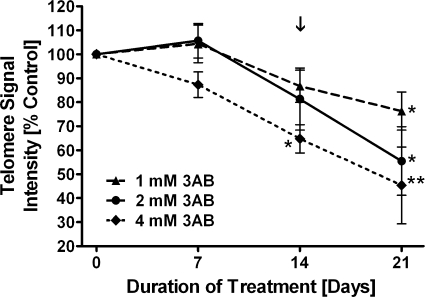
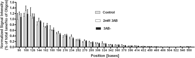
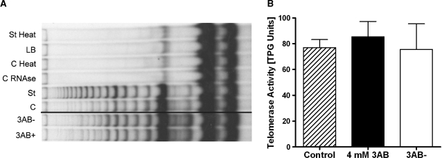
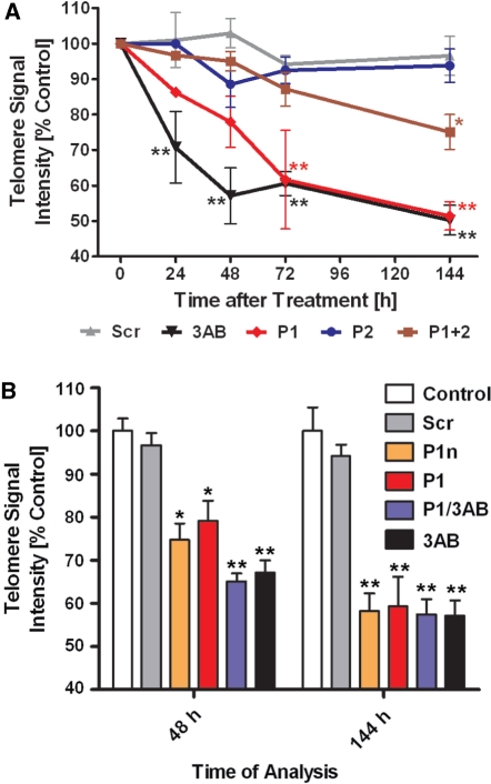
Shelterin/telosome is a multi-protein complex at mammalian telomeres, anchored to the double-stranded region by the telomeric-repeat binding factors-1 and -2. In vitro modification of these proteins by poly(ADP-ribosyl)ation through poly(ADP-ribose) polymerases-5 (tankyrases) and -1/-2, respectively, impairs binding. Thereafter, at least telomeric-repeat binding factor-1 is degraded by the proteasome. We show that pharmacological inhibition of poly(ADP-ribose) polymerase activity in cells from two different species leads to rapid decrease in median telomere length and stabilization at a lower setting. Specific knockdown of poly(ADP-ribose) polymerase-1 by RNA interference had the same effect. The length of the single-stranded telomeric overhang as well as telomerase activity were not affected. Release of inhibition led to a fast re-gain in telomere length to control levels in cells expressing active telomerase. We conclude that poly(ADP-ribose) polymerase-1 activity and probably its interplay with telomeric-repeat binding factor-2 is an important determinant in telomere regulation. Our findings reinforce the link between poly(ADP-ribosyl)ation and aging/longevity and also impact on the use of poly(ADP-ribose) polymerase inhibitors in tumor therapy.


Figures

Figure 1.
Figure 1.
Dose-dependent telomere shortening in mammalian cells exposed to increasing concentrations of 3AB. Telomere length was measured by Q-FISH and resulting values were expressed as percentage of the corresponding control. HeLaS3 and COM3 hamster cells were treated with concentrations of 3AB as indicated for 1 week. Both show a dose-dependent decrease in telomere signal intensity, with ∼85% and 75% of control level, respectively, at the highest concentration of the inhibitor. Squares, HeLaS3; triangles, COM3; *P < 0.05, **P < 0.01, compared to control.
Figure 2.
Figure 2.
Time course of telomere shortening during 3AB treatment. Telomere length was measured and plotted as in Figure 1. HeLaS3 and COM3 cells were treated with 2 mM and 4 mM 3AB, respectively. After 156 h incubation with the inhibitor, cultures were split and one part was released, whereas the other was further incubated with 3AB. 3AB− indicates cultures released from the inhibitor. After an initial drop, telomeres are stabilizing at a new lower level. Release leads to fast increase in median length, which stabilizes at control levels. Red diamonds, HeLaS3/3AB; dark blue triangles, COM3/3AB; orange circles, HeLaS3 released; light blue squares, COM3 released. *P < 0.05, **P < 0.01, compared to control. #P < 0.05 released compared to treated cultures; unpaired two-tailed t-test.
Figure 3.
Figure 3.
Irreversible telomere shortening in IMR90 fibroblasts exposed to the PARP inhibitor 3AB. IMR90 fibroblastswere incubated with 3AB for 14 days and released thereafter. After 14 days, significant telomere shortening is seen in IMR90 cells treated with 4 mM 3AB. After 21 days, all cultures show significant telomere shortening compared to telomeres from control cells. The arrow marks the time of release from 3AB. Triangles/dashed line, 1 mM 3AB; circles/closed line, 2 mM 3AB; diamonds/dotted line, 4 mM 3AB; *P < 0.05, **P < 0.01 compared to control.
Figure 4.
Figure 4.
3AB does not influence telomeric 3′-overhang. The length of the telomeric 3′-overhang was determined in HeLaS3 cells by using the T-OLA method at 204 h of 3AB treatment (greatest difference between treated and released cultures). Bars represent percentage of intensity of bands in one lane. Intensities were normalized to the number of ligated oligonucleotides. There is no significant difference between 3AB treated, released and control cells. Gray bars, controls; open bars, 2 mM 3AB; black bars, released.
Figure 5.
Figure 5.
3AB does not affect in vitro telomerase activity. After 4 weeks of incubation of cells, in vitro telomerase activity of COM3 cell extracts were measured by conventional PCR-based assay (TRAP). (A) Representative gel with TRAP products; 3AB+ and 3AB− lanes were from the same gel and only rearranged to the control lane. 3AB+, continuously 3AB treated; 3AB−, 3 weeks after 3AB release; C, control cells; St, standard reaction for normalization; C RNAse, control sample treated with RNaseA; C Heat, control sample treated with heat; LB, lysis buffer only; St Heat, standard reaction treated with heat. (B) Evaluation of three independent experiments. The activity is expressed as total product generated (TPG) units. No significant differences were observed. Hatched bar, control; black bar, 4 mM 3AB; open bar, released.
Figure 6.
Figure 6.
Knockdown of PARP1 shortens telomeres similar to 3AB. (A) HeLaS3 cells were transfected with different siRNAs or incubated with 3AB as telomere shortening control. Values were normalized to untreated controls. 3AB treated cells show the same effect as siRNA against PARP1, whereas Scr and PARP2 siRNA have no effect. Black upside down triangles, 3AB treated; red diamonds, PARP1 siRNA (P1); brown squares, PARP1 + PARP2 siRNAs (P1 + 2); blue dots, PARP2 siRNA (P2); gray triangles, scrambled siRNA (Scr); *P < 0.05, **P < 0.01 compared to control. (B) HeLaS3 cells were treated as in (A), but with a second unrelated PARP1 siRNA (P1n) and double treated with P1 siRNA and 3 mM 3AB (P1/3AB). Bar-code, empty, control; light gray, Scr; orange, P1n; red, P1; blue, P1/3AB; black, 3AB; *P < 0.01, **P < 0.001, compared to control.

References

    1. Celli GB, de Lange T. DNA processing is not required for ATM-mediated telomere damage response after TRF2 deletion. Nat. Cell Biol. 2005;7:712–718. - PubMed
    1. Liu D, O’Connor MS, Qin J, Songyang Z. Telosome, a mammalian telomere-associated complex formed by multiple telomeric proteins. J. Biol. Chem. 2004;279:51338–51342. - PubMed
    1. Wright WE, Tesmer VM, Huffman KE, Levene SD, Shay JW. Normal human chromosomes have long G-rich telomeric overhangs at one end. Genes Dev. 1997;11:2801–2809. - PMC - PubMed
    1. Griffith JD, Comeau L, Rosenfield S, Stansel RM, Bianchi A, Moss H, de Lange T. Mammalian telomeres end in a large duplex loop. Cell. 1999;97:503–514. - PubMed
    1. Smith S, Giriat I, Schmitt A, de Lange T. Tankyrase, a poly(ADP-ribose) polymerase at human telomeres. Science. 1998;282:1484–1487. - PubMed

Publication types