Case Reports
. 2009 Feb;46(2):86-93.
doi: 10.1136/jmg.2008.061580. Epub 2008 Oct 7.

Chromosome 15q11-13 duplication syndrome brain reveals epigenetic alterations in gene expression not predicted from copy number

Affiliations
Case Reports

Chromosome 15q11-13 duplication syndrome brain reveals epigenetic alterations in gene expression not predicted from copy number

A Hogart et al. J Med Genet. 2009 Feb.

Abstract

Background: Chromosome 15q11-13 contains a cluster of imprinted genes essential for normal mammalian neurodevelopment. Deficiencies in paternal or maternal 15q11-13 alleles result in Prader-Willi or Angelman syndromes, respectively, and maternal duplications lead to a distinct condition that often includes autism. Overexpression of maternally expressed imprinted genes is predicted to cause 15q11-13-associated autism, but a link between gene dosage and expression has not been experimentally determined in brain.

Methods: Postmortem brain tissue was obtained from a male with 15q11-13 hexasomy and a female with 15q11-13 tetrasomy. Quantitative reverse transcriptase-polymerase chain reaction (RT-PCR) was used to measure 10 15q11-13 transcripts in maternal 15q11-13 duplication, Prader-Willi syndrome, and control brain samples. Southern blot, bisulfite sequencing and fluorescence in situ hybridisation were used to investigate epigenetic mechanisms of gene regulation.

Results: Gene expression and DNA methylation correlated with parental gene dosage in the male 15q11-13 duplication sample with severe cognitive impairment and seizures. Strikingly, the female with autism and milder Prader-Willi-like characteristics demonstrated unexpected deficiencies in the paternally expressed transcripts SNRPN, NDN, HBII85, and HBII52 and unchanged levels of maternally expressed UBE3A compared to controls. Paternal expression abnormalities in the female duplication sample were consistent with elevated DNA methylation of the 15q11-13 imprinting control region (ICR). Expression of non-imprinted 15q11-13 GABA receptor subunit genes was significantly reduced specifically in the female 15q11-13 duplication brain without detectable GABRB3 methylation differences.

Conclusion: Our findings suggest that genetic copy number changes combined with additional genetic or environmental influences on epigenetic mechanisms impact outcome and clinical heterogeneity of 15q11-13 duplication syndromes.


Figures
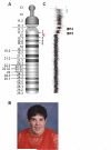
Figure 1
Figure 1
Ideograms and DNA fluorescence in situ hybridization (FISH) showing chromosome 15 duplications. A) Ideogram of the acrocentric normal chromosome 15 with relative positions of five common breakpoints (BP) indicated on the right. Position of the DNA FISH probe, located within the GABAAreceptor gene cluster, is indicated with a red line in between BP2 and BP3. B) Photo of Case 6856 showing epicanthal folds, exotropia, and moderate obesity. C) Array comparative genomic hybridization of Case 6856 genomic DNA to an oligonucleotide array with chromosome 15 probes shows tetrasomy for genomic sequences through BP4 and trisomy for genomic sequences between BP4 and BP5.
Figure 2
Figure 2
Fluorescence in situ hybridization (FISH) analysis of idic15 brain samples to confirm copy number and examine homologous pairing. A) Ideogram representing the tricentric derivative chromosome of Case 7014 with two sets of inverted 15q11-13 duplications with BP3 boundaries. DNA FISH signals (red spots) in neurons (DAPI nuclear stain) of Case 7014 confirm hexasomy in brain, with two closely spaced doublet observed for the derivative chromosome. B) Ideogram of the asymmetrical isodicentric chromosome 15 of Case 6856, including one BP4 and one BP5 boundary. DNA FISH of Case 6856 neurons confirm the partial tetrasomy for 15q11-13 in brain. C) Graphs for Case 7014 and D) Case 6856 reveal similar distributions of homologous pairing, with the derivative chromosomes (der) interacting nonselectively with the normal chromosome 15 alleles in equal proportions.
Figure 3
Figure 3
Gene expression analysis of 15q11-13 transcripts in post-mortem brain. A) Schematic representation of the genes analyzed in the critical region between BP2 and BP3 of 15q11-13. Arrows indicating the direction of transcription and the names of genes are shown to the left of the gray line, with the imprinting control region (ICR) shown as a filled circle at the 5′ end of SNRPN. B-K) Graphs summarizing quantitative RT-PCR measurements of 10 transcripts in the critical region normalized to GAPDH, with error bar representing +/- SEM. Fold changes relative to control expression are indicated above individual bars. Significant differences are indicated with * p < 0.01, ** p < 0.005, and *** p <0.0005. B and C are maternally expressed imprinted genes, D-G are paternally expressed imprinted genes, and H-K are nonimprinted biallelically expressed genes.
Figure 4
Figure 4
Methylation analysis of the imprinting control region (ICR) in the 5′ end of SNRPN A) Methylation-sensitive Southern blot analysis of Case 6856 reveals the ratio of methylated alleles (maternal, mat) to unmethylated alleles (paternal, pat) is higher than expected based on parental copy number, 4.35:1 observed versus 3:1 expected. B, C) Bisulfite sequencing of the ICR in Case 6856 (B) and Case 7014 (C). Circles represent the 33 CpG sites present in each clone, with filled circles representing methylated CpG sites, and unfilled circles representing unmethylated CpG sites. Each horizontal line represents the sequence of an individual clone. D) Graph of the percent methylation in individual clones from Prader-Willi syndrome samples (19 clones) compared to Case 6856 (21 clones) and Case 7014 (22 clones). All clones for Case 6856 and 7014 are either fully methylated (above 80%) or completely unmethylated.
Figure 5
Figure 5
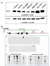
Analysis of GABRB3 protein and DNA methylation A) GABRB3 protein level in Case 6856 is reduced compared to controls, while Case 7014 has moderately elevated GABRB3. GAPDH was used as a loading control. B) Schematic representation of the proximal end of GABRB3 with the green line indicating the position of the CpG island. Numbered boxes represent exons and red lines with numbered circles represent the regions that were cloned during bisulfite sequencing of Case 6856. Bisulfite sequencing results, shown below the schematic, reveal hypomethylation of the GABRB3 CpG island. Partial methylation of Region 3 (formally Region 1) within GABRB3 intron 3 was compared to a matched control (1486) and determined to be within the normal range of methylation previously described [18].

References

    1. Robinson WP, Dutly F, Nicholls RD, et al. The mechanisms involved in formation of deletions and duplications of 15q11-q13. J Med Genet. 1998;35(2):130–6. - PMC - PubMed
    1. Bittel DC, Butler MG. Prader-Willi syndrome: clinical genetics, cytogenetics and molecular biology. Expert Rev Mol Med. 2005;7(14):1–20. - PMC - PubMed
    1. Clayton-Smith J, Laan L. Angelman syndrome: a review of the clinical and genetic aspects. J Med Genet. 2003;40(2):87–95. - PMC - PubMed
    1. Cook EH, Jr., Lindgren V, Leventhal BL, et al. Autism or atypical autism in maternally but not paternally derived proximal 15q duplication. Am J Hum Genet. 1997;60(4):928–34. - PMC - PubMed
    1. Schroer RJ, Phelan MC, Michaelis RC, et al. Autism and maternally derived aberrations of chromosome 15q. Am J Med Genet. 1998;76(4):327–36. - PubMed

Publication types

LinkOut - more resources