. 2009 Jan;58(1):104-15.
doi: 10.2337/db07-1344. Epub 2008 Oct 3.

Obesity-related upregulation of monocyte chemotactic factors in adipocytes: involvement of nuclear factor-kappaB and c-Jun NH2-terminal kinase pathways

Affiliations

Obesity-related upregulation of monocyte chemotactic factors in adipocytes: involvement of nuclear factor-kappaB and c-Jun NH2-terminal kinase pathways

Ping Jiao et al. Diabetes. 2009 Jan.

Abstract

Objective: We sought to evaluate the entire picture of all monocyte chemotactic factors that potentially contribute to adipose tissue macrophage accumulation in obesity.

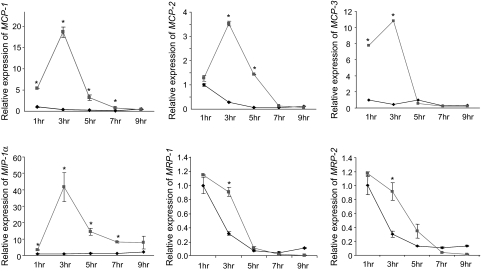
Research design and methods: Expression and regulation of members in the entire chemokine superfamily were evaluated in adipose tissue and isolated adipocytes of obese versus lean mice. Kinetics of adipose tissue macrophage infiltration was characterized by fluorescence-activated cell sorting. The effects of fatty acids on stimulation of chemokine expression in adipocytes and underlying mechanisms were investigated.

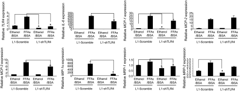
Results: Six monocyte chemotactic factors were found to be predominantly upregulated in isolated adipocytes versus stromal vascular cells in obese mice for the first time, although most of them were previously reported to be upregulated in whole adipose tissue. In diet-induced obese mice, adipose tissue enlargement, increase of adipocyte number, and elevation of multiple chemokine expression precede the initiation of macrophage infiltration. Free fatty acids (FFAs) are found to be inducers for upregulating these chemokines in 3T3-L1 adipocytes, and this effect can be partially blunted by reducing Toll-like receptor 4 expression. FFAs induce expression of monocyte chemotactic factors in adipocytes via both transcription-dependent and -independent mechanisms. In contrast to the reported role of JNK as the exclusive mediator of FFA-induced monocyte chemoattractant protein-1 (MCP-1) expression in macrophages, we show a novel role of inhibitor of kappaB kinase-beta (IKKbeta) in mediating FFA-induced upregulation of all six chemokines and a role of JNK in FFA-induced upregulation of MCP-1 and MCP-3.

Conclusions: Multiple chemokines derived from adipocytes might contribute to obesity-related WAT macrophage infiltration with FFAs as potential triggers and involvement of both IKKbeta and JNK pathways.


Figures

FIG. 1.
FIG. 1.
Obesity is associated with upregulation of multiple monocyte chemotactic factors in adipose tissue and adipocytes. A: Significantly upregulated monocyte chemotactic factors in epidydimal fat pads of ob/ob mice versus lean controls (n = 5, top left), in DIO mice versus lean controls (n = 5, top right), in isolated adipocytes of DIO mice versus lean controls (n = 4, bottom left), and in isolated stromal vascular cells in DIO mice versus controls (n = 4, bottom right). Expression of chemokines was measured by real-time PCR analysis. For comparison, the expression level of these genes in lean mice was arbitrarily set at 1. B: Protein levels of MCP-1, MCP-2, MCP-3, MIP-1α, MRP-1, and MRP-2 in adipose tissue of ob/ob and DIO mice. C: Plasma levels of MCP-1, MCP-2, MCP-3, MRP-1, and MRP-2 in ob/ob and DIO mice. D: Protein levels of MCP-1, MCP-2, MCP-3, MIP-1α, MRP-1, and MRP-2 in adipose tissue of lean mice on acute high-fat diet. Error bars represent ± SE. *P < 0.05, ob/ob (O) vs. lean (L) or high fat (HF) vs. low fat (LF). HFD, high-fat diet; WK, week.
FIG. 1.
FIG. 1.
Obesity is associated with upregulation of multiple monocyte chemotactic factors in adipose tissue and adipocytes. A: Significantly upregulated monocyte chemotactic factors in epidydimal fat pads of ob/ob mice versus lean controls (n = 5, top left), in DIO mice versus lean controls (n = 5, top right), in isolated adipocytes of DIO mice versus lean controls (n = 4, bottom left), and in isolated stromal vascular cells in DIO mice versus controls (n = 4, bottom right). Expression of chemokines was measured by real-time PCR analysis. For comparison, the expression level of these genes in lean mice was arbitrarily set at 1. B: Protein levels of MCP-1, MCP-2, MCP-3, MIP-1α, MRP-1, and MRP-2 in adipose tissue of ob/ob and DIO mice. C: Plasma levels of MCP-1, MCP-2, MCP-3, MRP-1, and MRP-2 in ob/ob and DIO mice. D: Protein levels of MCP-1, MCP-2, MCP-3, MIP-1α, MRP-1, and MRP-2 in adipose tissue of lean mice on acute high-fat diet. Error bars represent ± SE. *P < 0.05, ob/ob (O) vs. lean (L) or high fat (HF) vs. low fat (LF). HFD, high-fat diet; WK, week.
FIG. 2.
FIG. 2.
FFAs stimulate chemokine production in 3T3-L1 adipocytes. A: FFAs upregulate monocyte chemotactic factors at mRNA level in 3T3-L1 adipocytes. Left: FFAs, not glycerol, are capable of inducing MCP-1 expression in cultured 3T3-L1 adipocytes. RNAs were extracted from fully differentiated 3T3-L1 adipocytes treated with 0.5 mmol/l FFA/BSA, 0.5 mmol/l glycerol, 1 μg/ml insulin, or respective vehicles (Veh) for 3 h after overnight incubation with serum-free medium. Right: FFAs are capable of inducing expression of multiple chemokines in cultured 3T3-L1 adipocytes. For comparison, the expression level of these genes in vehicle-treated 3T3-L1 adipocytes was arbitrarily set at 1. B: FFAs upregulate monocyte chemotactic factors in 3T3-L1 adipocytes at protein level. Conditioned medium was collected from adipocytes treated with ethanol/BSA or FFA/BSA for 3 h after overnight incubation with serum-free medium and used for enzyme-linked immunosorbent assay (ELISA) analysis. Error bars represent ± SE. *P < 0.05, treated vs. vehicle. Results shown here are representative of three independent experiments.
FIG. 3.
FIG. 3.
Time course of FFA-induced upregulation of monocyte chemotactic factors in 3T3-L1 adipocytes. After overnight incubation with serum-free medium, 3T3-L1 adipocytes were treated with ethanol/BSA or FFA/BSA for 1, 3, 5, 7, or 9 h before RNA extraction. Expression of chemokines were evaluated by real-time PCR analysis. Error bars represent ±SE. *P < 0.05, treated vs. vehicle. Results shown here are representative of three independent experiments.
FIG. 4.
FIG. 4.
TLR4 is involved in FFA-induced upregulation of chemokine expression. 3T3-L1 preadipocytes stably expressing shRNA against TLR4 (L1-shTLR4) or scramble shRNA (L1-scramble) were differentiated into adipocytes, treated with ethanol/BSA, or 0.5 mmol/l FFAs/BSA for 3 h after overnight incubation with serum-free medium before RNA extraction. For comparison, the expression level of chemokines in vehicle-treated 3T3-L1 adipocytes expressing scramble shRNA was arbitrarily set at 1. *P < 0.05, L1-shTLR4 vs. L1-scramble. Results shown here are representative of three independent experiments.
FIG. 5.
FIG. 5.
Mechanism of FFA-induced upregulation of chemokines. A: FFA-induced upregulation of chemokine expression is both transcription dependent and independent. 3T3-L1 adipocytes were pretreated with 5 μg/ml actinomycin D for 30 min before stimulation by 0.5 mmol/l FFAs for 3 h. B: Protein synthesis is only required for mediating FFA-induced MIP1α expression. 3T3–L1 adipocytes were pretreated with 10 μg/ml cycloheximide for 30 min before stimulation by 0.5 mmol/l FFAs for 3 h. For comparison, the expression level of chemokines in vehicle-treated 3T3-L1 adipocytes was arbitrarily set at 1. Veh, vehicle; ActD, actinomycin D; CHX, cycloheximide. Error bars represent ± SE. *P < 0.05, treated vs. vehicle. Results shown here are representative of three independent experiments.
FIG. 6.
FIG. 6.
Inflammatory pathways involved in FFA-induced chemokine expression. A: Oxidative stress is not required for mediating FFA-induced chemokine expression in 3T3-L1 adipocytes. 3T3-L1 adipocytes were pretreated with 500 μmol/l apocynin (NADPH inhibitor) for 1 h before stimulation by 0.5 mmol/l FFAs for 3 h. B: NF-κB pathway is involved in FFA-mediated upregulation of chemokine expression in 3T3-L1 adipocytes. 3T3-L1 adipocytes were pretreated with 200 μmol/l NF-κB inhibitor for 1 h before stimulation by 0.5 mmol/l FFAs for 3 h. C: JNK is necessary for mediating FFA-induced expression of MCP-1 and MCP-3, but not other chemokines, in 3T3-L1 adipocytes. 3T3-L1 adipocytes were pretreated with 250 μmol/l JNK inhibitor for 1 h before stimulation by 0.5 mmol/l FFAs for 3 h. For comparison, the expression level of chemokines in vehicle-treated 3T3-L1 adipocytes was arbitrarily set at 1. *P < 0.05, treated vs. vehicle. NF-κB Inh, NF-κB inhibitor; SP600125, JNK inhibitor. Results shown here are representative of three independent experiments.
FIG. 7.
FIG. 7.
Effect of IKKβ and JNK knockdown on FFA-induced upregulation of chemokines. A: Top, reduced IKKβ expression in 3T3-L1 adipocytes by RNA interference. 3T3-L1 adipocytes were electroporated with either scrambled siRNA or siRNA against IKKβ. Twenty-four hours after electroporation, adipocytes were incubated in serum-free DMEM overnight. Forty-eight hours after electroporation, adipocytes were treated with 0.5 mmol/l FFA/BSA or ethanol/BSA for 2 h and then harvested for examination of IKKβ expression. Bottom, expression of chemokines in 3T3-L1 adipocytes with IKKβ knockdown. Adipocytes treated as described in A were harvested for examination of chemokine expression by real-time PCR analysis. *P < 0.05, scrambled vs. siIKKβ. B: Top, reduced JNK expression in 3T3-L1 adipocytes by RNA interference. Bottom, expression of chemokines in 3T3-L1 adipocytes with JNK knockdown. For comparison, the expression level of chemokines in scrambled siRNA–electroporated and ethanol/BSA-treated 3T3-L1 adipocytes was arbitrarily set at 1. Results shown here are representative of three independent experiments.
FIG. 8.
FIG. 8.
Effect of IKKβ overexpression on chemokine production. Wild-type, inactive, and constitutively active human IKKβ (named hIKK WT, hIKK KM, and hIKK SE, respectively) were overexpressed in 3T3-L1 CAR cells. A: Overexpression of hIKKβ in 3T3-L1 CAR cells at protein level. B: Expression of chemokine genes in 3T3-L1 CAR adipocytes overexpressing hIKKβ. Fully differentiated 3T3-L1 CAR adipocytes were infected with adenoviruses overexpressing green fluorescent protein (GFP), hIKKβ WT, hIKKβ KM, or hIKKβ SE at the dose of 1 × 10⁹ plaque-forming units/ml for 6 h. Cells were then incubated in fresh medium for 48 h. Forty-eight hours after infection, cells were incubated in serum-free medium overnight. Seventy-two hours after infection, cells were harvested for RNA extraction to examine expression of chemokine genes by real-time PCR analysis. C: Expression of chemokine protein in conditioned supernatant. Conditioned supernatant collected from the experiment described in B was used to measure secreted chemokine protein by ELISA. Error bars represent means ± SE. Methods for construction of adenovirus constructs are in supplementary materials available in the online appendix. *P < 0.05, IKKβ-overexpressing cells vs. GFP-overexpressing cells. Results shown here are representative of three independent experiments.

References

    1. Festa A, D'Agostino R Jr, Howard G, Mykkanen L, Tracy RP, Haffner SM: Chronic subclinical inflammation as part of the insulin resistance syndrome: the Insulin Resistance Atherosclerosis Study (IRAS). Circulation 102: 42–47, 2000 - PubMed
    1. Pickup JC, Mattock MB, Chusney GD, Burt D: NIDDM as a disease of the innate immune system: association of acute-phase reactants and interleukin-6 with metabolic syndrome X. Diabetologia 40: 1286–1292, 1997 - PubMed
    1. Ghanim H, Aljada A, Hofmeyer D, Syed T, Mohanty P, Dandona P: Circulating mononuclear cells in the obese are in a proinflammatory state. Circulation 110: 1564–1571, 2004 - PubMed
    1. Xu H, Barnes GT, Yang Q, Tan G, Yang D, Chou CJ, Sole J, Nichols A, Ross JS, Tartaglia LA, Chen H: Chronic inflammation in fat plays a crucial role in the development of obesity-related insulin resistance. J Clin Invest 112: 1821–1830, 2003 - PMC - PubMed
    1. Weisberg SP, McCann D, Desai M, Rosenbaum M, Leibel RL, Ferrante AW Jr: Obesity is associated with macrophage accumulation in adipose tissue. J Clin Invest 112: 1796–1808, 2003 - PMC - PubMed

Publication types

MeSH terms