. 2009 Jan;200(1):63-73.
doi: 10.1677/JOE-08-0383. Epub 2008 Oct 3.

Nuclear receptor co-regulator Krüppel-like factor 9 and prohibitin 2 expression in estrogen-induced epithelial cell proliferation in the mouse uterus

Affiliations

Nuclear receptor co-regulator Krüppel-like factor 9 and prohibitin 2 expression in estrogen-induced epithelial cell proliferation in the mouse uterus

J M P Pabona et al. J Endocrinol. 2009 Jan.

Abstract

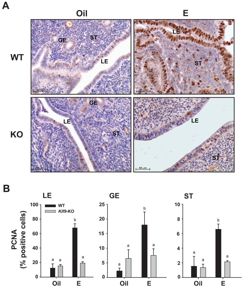
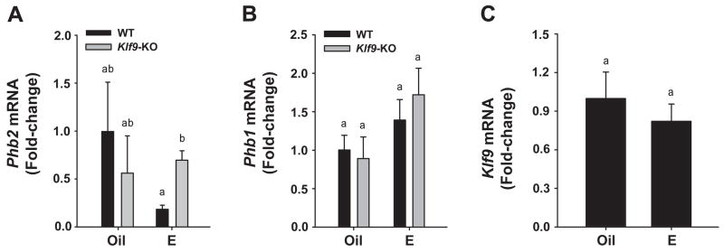
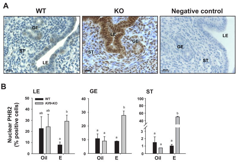
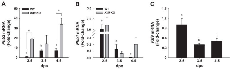
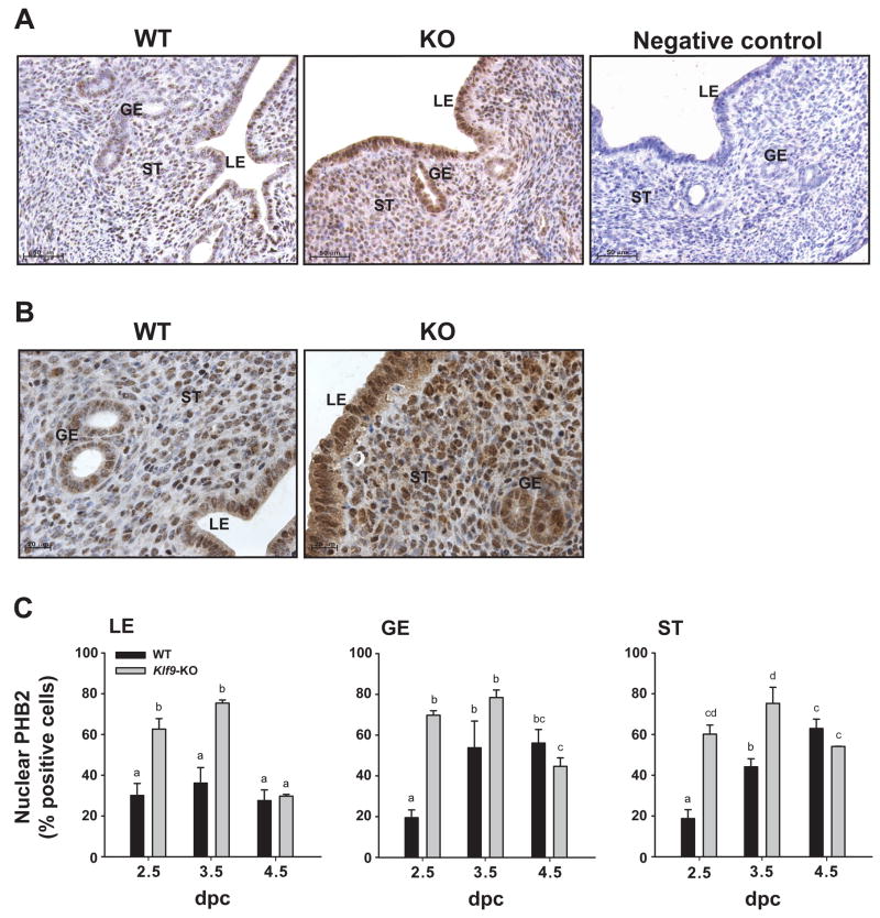
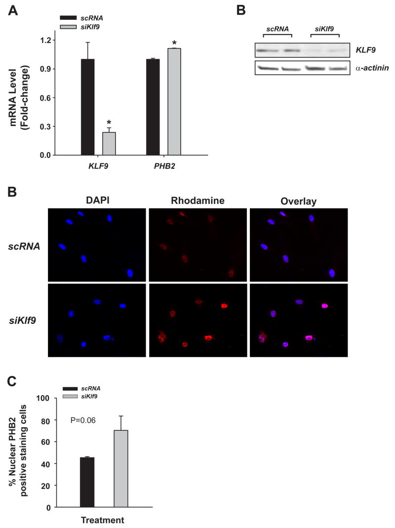
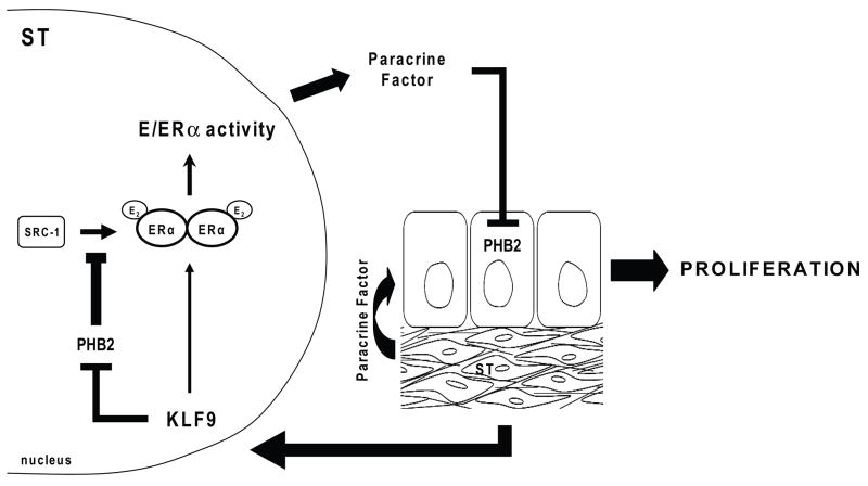
Estrogen, acting through its cognate receptor estrogen receptor-alpha (ESR1), is a critical regulator of uterine endometrial epithelial proliferation. Although the dynamic communication between endometrial stromal (ST) and epithelial cells is considered to be an important component in this process, key molecular players in particular compartments remain poorly defined. Here, we used mice null for Krüppel-like factor 9 (KLF9) to evaluate the contribution of this nuclear protein in ST-epithelial interactions underlying proliferative effects of estrogen. We found that in ovariectomized mice administered estradiol-17beta (E(2)) for 24 h, Klf9 null mutation resulted in lack of E(2)-induced proliferative response in all endometrial compartments. We demonstrated a negative association between Klf9 expression and nuclear levels of ESR1 transcriptional corepressor prohibitin (PHB) 2 in uterine ST and epithelial cells of E(2)-treated wild-type (WT) and Klf9 null mice. In early pregnancy uteri of WT mice, the temporal pattern of Klf9 transcript levels was inversely associated with that of Phb2. Deletion of Klf9 up-regulated uterine Phb2 expression and increased PHB2 nuclear localization in endometrial ST and epithelial cells, with no effects on the expression of the related Phb1. In the human endometrial ST cell line treated with E(2) for 24 h, Klf9 siRNA targeting augmented PHB2 transcript and increased nuclear PHB2 protein levels, albeit this effect was not to the extent seen in vivo with Klf9 null mutants. Our findings suggest a novel mechanism for control of estrogen-induced luminal epithelial proliferation involving ST KLF9 regulation of paracrine factor(s) to repress epithelial expression of corepressor PHB2.


Figures

Figure 1
Figure 1
Nuclear PCNA levels in uterine endometrial cells of Oil (vehicle) and E2-treated ovariectomized WT and Klf9 null mice. (A) Representative PCNA immunostaining of glandular epithelium (GE), luminal epithelium (LE), and stromal (ST) compartments are shown at 200X magnification. (B) The percentages of nuclear-staining cells are presented as mean ± SEM (n=3–5 mice per treatment per genotype). Significant differences were identified by two-way ANOVA, followed by Tukey’s test. Means with different superscripts differed at P<0.05.
Figure 2
Figure 2
Transcript levels of (A) Phb2, (B) Phb1, and (C) Klf9 in uteri of Oil (vehicle) and E2-treated ovariectomized WT and/or Klf9 null mice. mRNA expression was quantified by QPCR and normalized to that of the control gene Ppia. Transcript levels (mean ± SEM) are expressed as fold-change relative to WT oil treatment group (n=3–5 mice per treatment per genotype). Means with different superscripts differed at P<0.05.
Figure 3
Figure 3
PHB2 levels in uterine endometrial cells of Oil (vehicle) and E2-treated ovariectomized WT and Klf9 null mice. (A) Representative PHB2 immunostaining of GE, LE, and ST cells of E2-treated WT and Klf9 null (KO) mice are shown at 400X magnification to demonstrate nuclear localization of immunoreactive PHB2. Negative control was tissue from E2-treated WT mice processed similarly except for omission of primary antibody. (B) The percentages of PHB2 nuclear-staining cells are presented as mean ± SEM (n=3–5 mice per treatment per genotype). Significant differences were identified by two-way ANOVA, followed by Tukey’s test. Means with different superscripts differed at P<0.05.
Figure 4
Figure 4
Uterine transcript levels for Phb2 (A) and Phb1 (B) in WT and Klf9 null mice and for Klf9 in WT mice (C) during early pregnancy. mRNA expression was quantified by QPCR and normalized to that of the control gene Ppia. Transcript levels (mean ± SEM) were determined at dpc 2.5, 3.5 and 4.5 in WT (Phb2; Phb1; Klf9) and Klf9 KO (Phb2; Phb1) mice and expressed as fold-change relative to WT (dpc 2.5) group. Significant difference (*) due to genotype at P<0.05 was identified by Student’s t-test for dpc 2.5 and 4.5. One-way ANOVA was used to compare Phb2 and Phb1 mRNA levels of WT among pregnancy days. Means with different superscripts differed at P<0.05.
Figure 5
Figure 5
Nuclear PHB2 levels in uterine endometrial cells of WT and Klf9 null mice during early pregnancy. (A) Representative immunostaining for uterine PHB2 in pregnant WT and Klf9 null mice at dpc 2.5. GE, glandular epithelium; LE, luminal epithelium; ST, stroma. Panels are shown at 200X magnification. Negative control was tissue from Klf9 KO mice processed similarly except for omission of primary antibody. (B) Representative PHB2 immunostaining of GE, LE, and ST cells of pregnant WT and Klf9 null (KO) mice at dpc 2.5 are shown at 400X magnification to demonstrate nuclear localization of immunoreactive PHB2. (C) The percentages of nuclear-immunostained cells are presented as mean ± SEM (n=3-5 mice per treatment per genotype). Significant differences were identified by two-way ANOVA, followed by Tukey’s test. Means with different superscripts differed at P<0.05.
Figure 6
Figure 6
PHB2 levels in human endometrial stromal cells as a function of KLF9 expression status. HESC were incubated in phenol-red free DMEM/Ham’s F12 medium containing 10% charcoal-stripped serum and E2 (10 nM) in the presence of siRNA to scrambled (negative control) mRNA or siRNA to KLF9 (50 nM). (A) Harvested cells were analyzed for KLF9 and PHB2 transcripts by QPCR and normalized to control gene RPL7. Results (means ± SEM; relative to cells treated with scrambled siRNA) shown are representative of 2–3 independent experiments, with each experiment conducted in triplicates. (B) Western blots of whole cell lysates prepared from HESC cells as a function of Klf9 knockdown. Cells transfected with siRNA to scrambled sequence (negative control) or to Klf9 were subjected to Western blots using anti-KLF9 or anti-α-actinin antibodies. Each lane represents an independent experiment. (C) HESC treated with E2 for 24 h were immunostained for PHB2/REA and counterstained for DAPI. Immunopositive cells were visualized using fluorescent antibodies. Overlay of anti-PHB2 (red) and DAPI (blue)-staining cells showed nuclear localization of PHB2. (D) The percentages of nuclear PHB2-staining cells were expressed relative to the total number of cells counted. Results (mean ± SEM) were normalized to those of scrambled siRNA-treated cells and were from two independent experiments, with each experiment performed in triplicates. Differences between groups were determined by t-test (P=0.06)
Figure 7
Figure 7
Postulated model for E2 control of uterine epithelial cell proliferation involving stromal KLF9. By attenuating expression of stromal corepressor PHB2 which can inhibit the interaction of ERα coactivators such as SRC-1 with ERα, KLF9 may promote ligand-dependent ERα-mediated transactivation of paracrine factor(s) yet unknown to repress epithelial expression of PHB2, leading to cell proliferation.

References

    1. Cooke PS, Buchanan DL, Young P, Setiawan T, Brody J, Korach KS, Taylor J, Lubahn DB, Cunha GR. Stromal estrogen receptors mediate mitogenic effects of estradiol on uterine epithelium. Proc Natl Acad Sci USA. 1997;94:6535–6540. - PMC - PubMed
    1. Delage-Morroux R, Martini PG, Choi I, Kraichely DM, Hoeksema J, Katzenellenbogen BS. Analysis of estrogen receptor interaction with repressor of estrogen receptor activity (REA) and the regulation of estrogen receptor transcriptional activity by REA. J Biol Chem. 2000;275:35848–35856. - PubMed
    1. Dupont S, Krust A, Gansmuller A, Dierich A, Chambon P, Mark M. Effect of single and compound knockouts of estrogen receptors alpha (ERalpha) and beta (ERbeta) on mouse reproductive phenotypes. Development. 2000;127:4277–4291. - PubMed
    1. Dutertre M, Smith CL. Ligand-independent interactions of p160/steroid receptor coactivators and CREB-binding protein (CBP) with estrogen receptor α: regulation by phosphorylation sites in the A/B region depends on other receptor domains. Mol Endocrinol. 2003;17:1296–1314. - PubMed
    1. Font de Mora J, Brown M. AIB1 is a conduit for kinase-mediated growth factor signaling to the estrogen receptor. Mol Cell Biol. 2000;20:5041–5047. - PMC - PubMed

Publication types

MeSH terms

LinkOut - more resources