. 2008 Oct 14;105(41):16009-14.
doi: 10.1073/pnas.0806114105. Epub 2008 Oct 3.

Restoration of visual function in retinal degeneration mice by ectopic expression of melanopsin

Affiliations

Restoration of visual function in retinal degeneration mice by ectopic expression of melanopsin

Bin Lin et al. Proc Natl Acad Sci U S A. .

Abstract

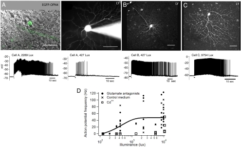
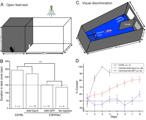
The rod and cone cells of the mammalian retina are the principal photoreceptors for image-forming vision. They transmit information by means of a chain of intermediate cells to the retinal ganglion cells, which in turn send signals from the retina to the brain. Loss of photoreceptor cells, as happens in a number of human diseases, leads to irreversible blindness. In a mouse model (rd/rd) of photoreceptor degeneration, we used a viral vector to express in a large number of retinal ganglion cells the light sensitive protein melanopsin, normally present in only a specialized subset of the cells. Whole-cell patch-clamp recording showed photoresponses in these cells even after degeneration of the photoreceptors and additional pharmacological or Cd(2+) block of synaptic function. Interestingly, similar responses were observed across a wide variety of diverse types of ganglion cell of the retina. The newly melanopsin-expressing ganglion cells provided an enhancement of visual function in rd/rd mice: the pupillary light reflex (PLR) returned almost to normal; the mice showed behavioral avoidance of light in an open-field test, and they could discriminate a light stimulus from a dark one in a two-choice visual discrimination alley. Recovery of the PLR was stable for at least 11 months. It has recently been shown that ectopic retinal expression of a light sensitive bacterial protein, channelrhodopsin-2, can restore neuronal responsiveness and simple visual abilities in rd/rd mice. For therapy in human photodegenerations, channelrhodopsin-2 and melanopsin have different advantages and disadvantages; both proteins (or modifications of them) should be candidates.


Conflict of interest statement

Conflict of interest statement: A patent application (U.S. no. 60/397,088; July 18, 2002) has been filed by R.H.M. and assigned to the Massachusetts General Hospital.

Figures

Fig. 1.
Fig. 1.
Ectopic expression of melanopsin protein in retinal ganglion cells of different morphological types in the rd/rd mouse. (A–C) Spatial distributions of native melanopsin-expressing ganglion cells in uninjected (A) and sham-injected (C) rd/rd mouse retinas, and of total melanopsin-expressing ganglion cells in an AAV-Opn4 injected rd/rd mouse retina (B). Orientation of the retina: T, temporal; N, nasal, D, dorsal; and V, ventral. (D–G) Different types of ganglion cells were targeted in the melanopsin-treated mouse retina. Two monostratified ganglion cells (D and E) and one bistratified ganglion cell (F) are indicated here. (I) Their dendritic arbors are denser and smaller than those of the native melanopsin cell (G). (H and I) Sections of mouse retina stained with DAPI. In the rd/rd mice there appeared to be total loss of rod photoreceptors. Rare cones, which lacked inner and outer segments, were present but limited to the retinal periphery. ONL, outer nuclear layer; INL, inner nuclear layer; GCL, ganglion cell layer. (Scale bars, 500 μm in A, B, and C; 100 μm in D–G; and 20 μm in H and I.) (J) Quantitation of melanopsin-expressing ganglion cells in rd/rd mice. For comparison, numbers of GFP-expressing ganglion cells in retinas injected with AAV-GFP are also indicated. Values are mean ± SD, with n indicating the number of retinas counted.
Fig. 2.
Fig. 2.
Long-lasting responses from retinal ganglion cells expressing Opn4 and EGFP. (A–C) Examples of EGFP- and Opn4-expressing retinal ganglion cells (Scale bars: 50 μm). (A Left) Combined DIC and fluorescence image of a cell that expresses EGFP (green) and Opn4. (A Right) After patch–clamp recording, the cell was filled with Lucifer yellow (0.1%) to visualize the morphology. Indicated in the traces below, the cell responded to light (480 nm peak; 2,289 lux; 1 s) with long-lasting trains of action potentials. A lower frequency and shorter duration train of action potentials were observed with a lower intensity of light (427 lux). (B and C) Heterogeneous types of ganglion cells (here, one very small and one large cell) expressed GFP and melanopsin, but they responded similarly to light. (D) Responses to light of the Opn4-transduced cells under normal or pharmacological conditions. The solid line is the average of all of the data in glutamate antagonists and control medium.
Fig. 3.
Fig. 3.
Restoration of light sensitivity in the eye of the AAV-Opn4 treated rd/rd mouse. (A and B) Representative infrared images of pupil area taken in dark (A) and light (B). White dots indicate the pupil areas. For a time series of images, see Fig. S2. Pupil area was measured from such images by using ImageJ. (C) Intensity-response curves for pupillary constriction. The stimulus was exposure to 20 s of white light. The threshold for response is dramatically reduced in melanopsin treated eyes (red curve) compared with sham-injected eyes (blue curve). Data from uninjected C57BL mice are shown for comparison. The data are fitted with a sigmoidal function. (D and E) Time course of pupil constriction over the first few seconds of dim (D) and bright (E) light exposure are shown for melanopsin-treated (red) and sham-injected (blue) rd/rd mice, and uninjected C57BL mice (black). The area of the pupil is depicted as a percentage of its size immediately preceding the onset of light. Values are mean ± SEM, with n indicating the number of eyes examined.
Fig. 4.
Fig. 4.
Enhancement of visual function in the AAV-Opn4 treated rd/rd mice. (A) The open-field test box consisted of a dark compartment (one third of the floor area) and a larger illuminated compartment (two thirds). A small opening located at floor level in the center of the dividing wall allowed mice to freely move between the lit and dark chambers. (B) Time spent in dark area by four groups of mice. The AAV-Opn4 treated rd/rd mice showed behavioral aversion to light: they spent significantly longer time in the dark chamber than their counterparts of either sham-injected or uninjected rd/rd mice (P < 0.01, t test). The exploratory behavior of uninjected C57BL mice is shown as comparison. Dotted line shows the behavior to be expected by chance. (C) Visual discrimination alley. Mice swam down a water-filled alley toward an illuminated or a dark target. The rewarded stimulus indicated the location of a submerged platform. (D) Melanopsin-treated (open circles) mice outperformed sham-injected (triangles) rd/rd mice in visual detection task over an 8-day trial (P < 0.001, two-way ANOVA test). Values represent mean ± SEM; n = number of mice.

References

    1. Berson DM, Dunn FA, Takao M. Phototransduction by retinal ganglion cells that set the circadian clock. Science. 2002;295:1070–1073. - PubMed
    1. Fu Y, et al. Intrinsically photosensitive retinal ganglion cells detect light with a vitamin A-based photopigment, melanopsin. Proc Natl Acad Sci USA. 2005;102:10339–10344. - PMC - PubMed
    1. Hattar S, Liao HW, Takao M, Berson DM, Yau KW. Melanopsin-containing retinal ganglion cells: architecture, projections, and intrinsic photosensitivity. Science. 2002;295:1065–1070. - PMC - PubMed
    1. Panda S, et al. Melanopsin is required for non-image-forming photic responses in blind mice. Science. 2003;301:525–527. - PubMed
    1. Provencio I, Jiang G, De Grip WJ, Hayes WP, Rollag MD. Melanopsin: An opsin in melanophores, brain, and eye. Proc Natl Acad Sci USA. 1998;95:340–345. - PMC - PubMed

Publication types