

doi: 10.1097/01.ASW.0000323563.59885.1c.
Using gene transcription patterns (bar coding scans) to guide wound debridement and healing
Affiliations
- PMID: 18836328
- PMCID: PMC2948232
- DOI: 10.1097/01.ASW.0000323563.59885.1c
Item in Clipboard
Using gene transcription patterns (bar coding scans) to guide wound debridement and healing
Adv Skin Wound Care.
2008 Oct.
Abstract
Purpose: To acquaint wound care practitioners with new information related to debridement of chronic wounds.
Target audience: This continuing education activity is intended for physicians and nurses with an interest in wound care.
Objectives: After reading this article and taking this test, the reader should be able to: 1. Explain the role of keratinocytes in wound healing. 2. Discuss new research findings on the physiological differences between healing and nonhealing wounds.3. Identify implications of the new research for debridement of chronic wounds.
Figures
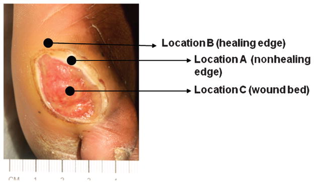
A typical diabetic ulcer is shown. Arrows indicate specific wound locations: Wound bed = location C; nonhealing edge = location A; healing edge = location B.

Histopathology of nonhealing edge (left) reveals thick cornified layer (stratum corneum) and thick, hyperproliferative epidermis. Section of normal skin (right) is shown for comparison.
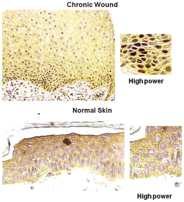
C-myc is a pathogenic marker of chronic wounds. Immunohistochemistry is showing positive staining with c-myc–specific antibody of the nuclei of the biopsy from a patient with chronic wound (top), whereas there is no c-myc staining in biopsy of normal skin (bottom).
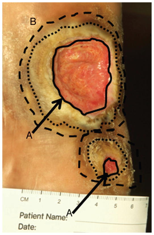
Digital photograph of a neuropathic plantar foot ulcer in a patient with diabetes. The solid black lines indicate nonhealing edge (location A, indicated by black arrows). Two outer circles (broken lines) indicate 2 possible margins of debridement, the presumed location B or healing-edge of the wound. At this edge, keratinocytes have the ability to migrate and participate in the wound healing process.

Histology and gene expression profiles obtained from biopsies from a patient with venous ulcer are shown for 3 locations: nonhealing edge (location A), healing edge (location B), and normal skin. Specific gene expression pattern is detected from each of the locations, indicating that it can be used to determine the subpopulation of cells within the wound. Colors indicate the degree of regulation of specific genes.
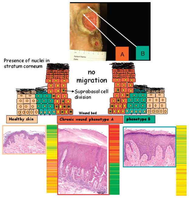
Debridement aims at removal of cells from location A, thus exposing cells from location B exposed to wound healing stimuli.
References
-
- Center for Medicare & Medicaid Services. Expert Advisory Panel on the Usual Care of Chronic Wounds [cited 10/28/2006] [Last accessed August 1, 2008]. Available at: http://www.cms.hhs.gov/mcd/viewmcac.asp?where=index&mid=28.
-
- Brem H, Jacobs T, Vileikyte L, et al. Wound-healing protocols for diabetic foot and pressure ulcers. Surg Technol Int. 2003;11:85–92. - PubMed
-
- Whitney J, Phillips L, Aslam R, et al. Guidelines for the treatment of pressure ulcers. Wound Repair Regen. 2006;14:663–79. - PubMed
-
- Steed DL. Clinical evaluation of recombinant human platelet-derived growth factor for the treatment of lower extremity ulcers. Plast Reconstr Surg. 2006;117(7 Suppl):143S–149S. discussion 150S–151S. - PubMed
-
- Robson MC, Cooper DM, Aslam R, et al. Guidelines for the treatment of venous ulcers. Wound Repair Regen. 2006;14:649–62. - PubMed
Publication types
MeSH terms
Grants and funding
LinkOut - more resources
Full Text Sources
Other Literature Sources
Medical
