. 2008 Nov;11(11):1302-10.
doi: 10.1038/nn.2204. Epub 2008 Oct 5.

Palmitoylation-dependent neurodevelopmental deficits in a mouse model of 22q11 microdeletion

Affiliations

Palmitoylation-dependent neurodevelopmental deficits in a mouse model of 22q11 microdeletion

Jun Mukai et al. Nat Neurosci. 2008 Nov.

Abstract

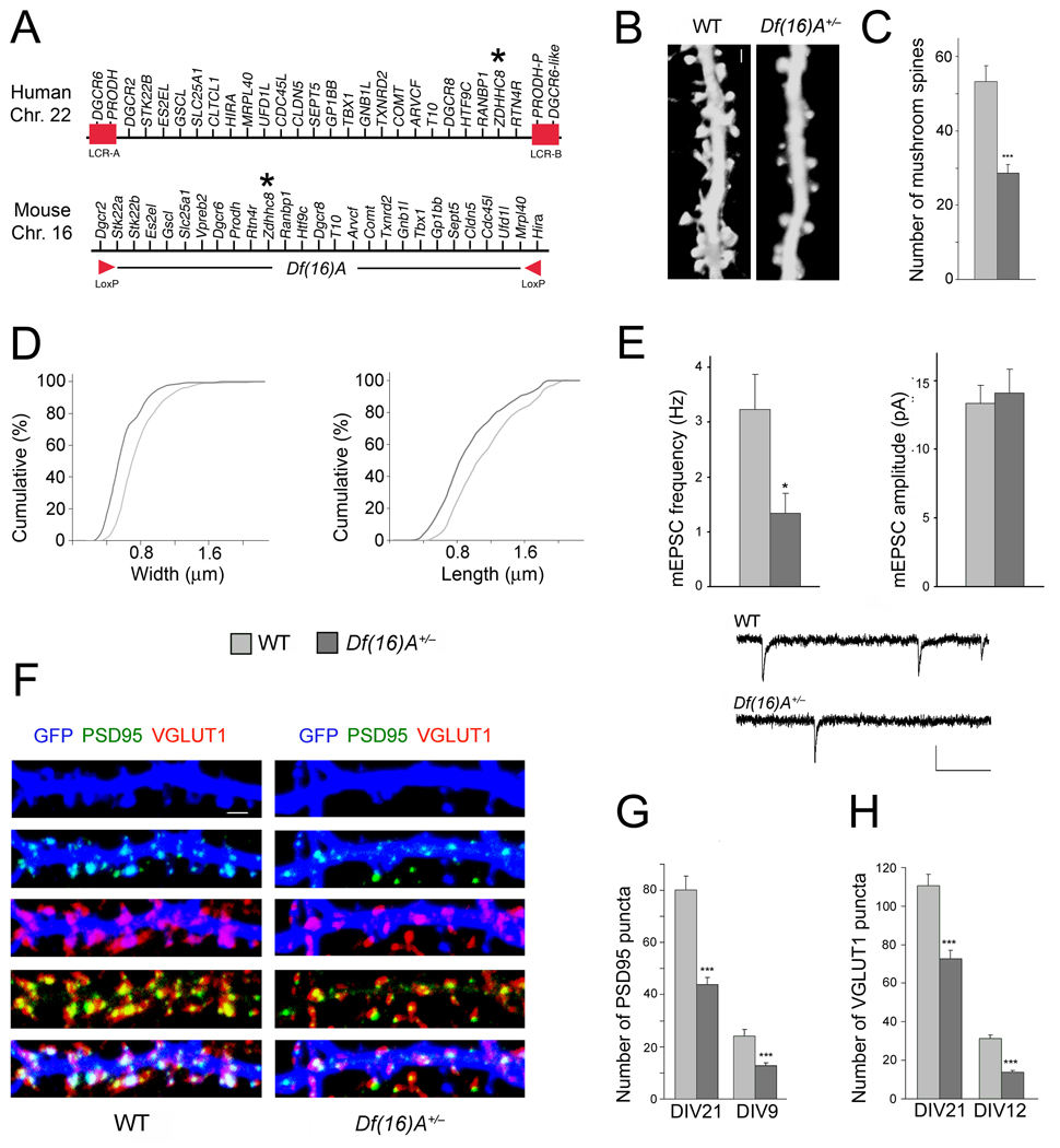
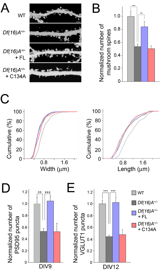
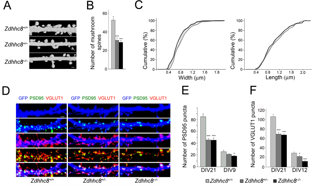
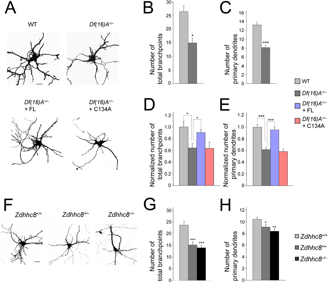
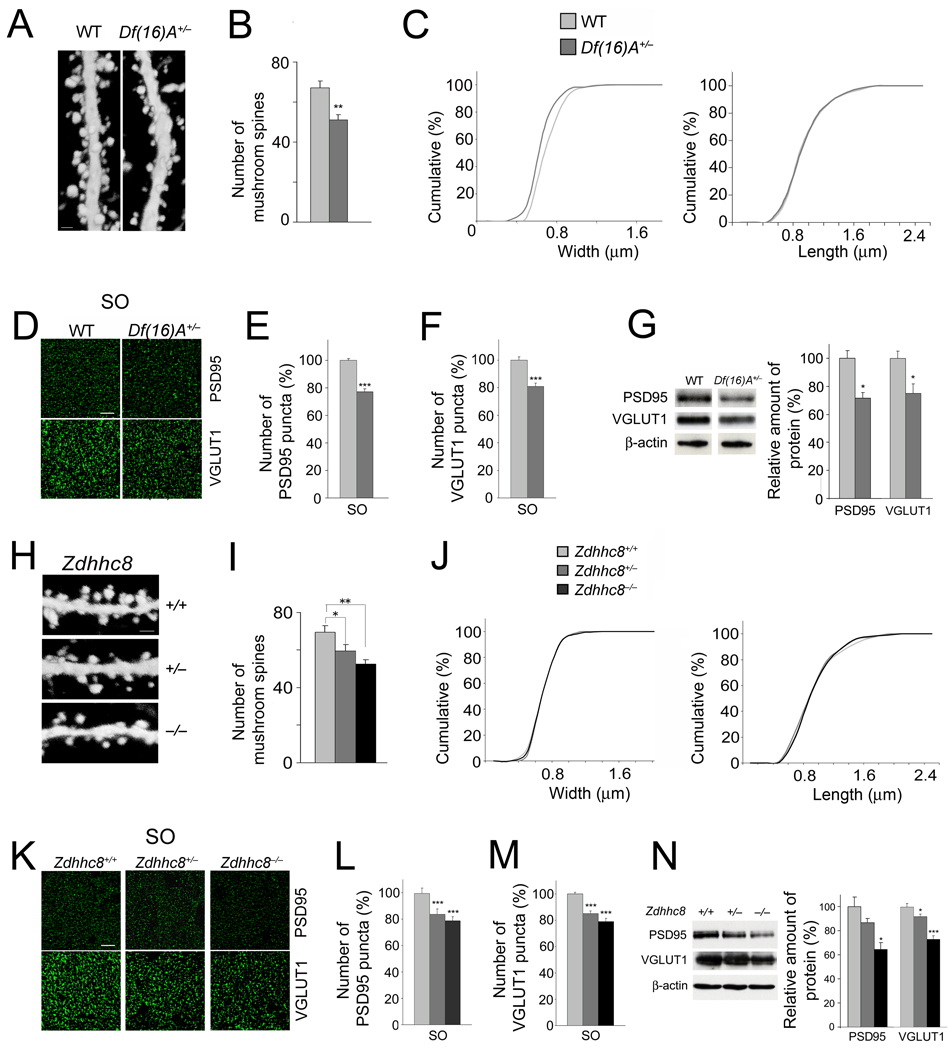
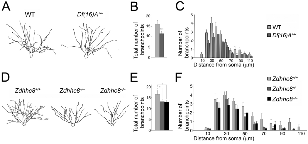
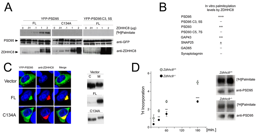
Individuals with 22q11.2 microdeletions have cognitive deficits and a high risk of developing schizophrenia. Here we provide evidence that primary hippocampal neurons from a mouse model of 22q11.2 deletion (Df(16)A(+/-) mice) have decreased density of dendritic spines and glutamatergic synapses, as well as impaired dendritic growth. These deficits were prevented by introduction of the enzymatically active ZDHHC8 palmitoyltransferase encoded by a gene in the 22q11.2 locus, and they were also observed in primary cultures from Zdhhc8-deficient mice. Many of these deficits were also present in the hippocampi of adult Df(16)A(+/-) and Zdhhc8-deficient mice. Finally, we provide evidence that PSD95 is one of the substrates of ZDHHC8. Our analysis reveals that 22q11.2 microdeletion results in deficits in neuronal development and suggests that impaired neuronal protein palmitoylation contributes to many of these deficits.


Figures

Figure 1
Figure 1. Df(16)A+/− neurons show reduced density of spines and glutamatergic synapses
(a) Schematic showing the human 22q11.2 region and the syntenic mouse region. The 1.5-Mb deletion found in patients is mediated by low copy repeat sequences LCR-A and LCR-B (red boxes). Dgcr2 and Hira, the two endpoints of the targeted deletion where loxP sites were inserted, are indicated by red arrowheads. The location of the human and mouse ZDHHC8 gene is indicated by an asterisk. (b) Representative images of spines in Df(16)A+/− neurons (DIV21) transfected with a EGFP, which fills and clearly defines dendritic protrusions. Scale bar, 1 µm. (c) Reductions in the number of mushroom spines observed at DIV21 on Df(16)A+/− neurons (estimated over 75 µm of dendritic length). (d) Reductions in the width (left) and length (right) of mushroom spines on Df(16)A+/− neurons. P < 0.05, K-S test. (e) Properties of mEPSC recorded from autaptic individual pyramidal neurons grown on astrocytic microislands. Top left: the frequency at which events occurred was lower in Df(16)A+/− neurons (n = 10) than in WT controls (n = 11). Top right: The average amplitude of mEPSCs was not different between genotypes. Bottom: Traces of recordings from a WT and a Df(16)A+/− neuron. Scale bars, vertical: 20 pA, horizontal: 100 msec. (f) Representative images of PSD95 puncta (green) and VGLUT1 puncta (red) and their co-localization in GFP (blue)-labeled hippocampal neurons at DIV21. Scale bar, 1 µm. (g) Reduction in the number of PSD95 puncta in Df(16)A+/− neurons at DIV21 and DIV9. (h) Reduction in the number of VGLUT1 puncta in Df(16)A+/− neurons at DIV21 and DIV12. Data are shown as mean ± S.E.M. * P < 0.05, *** P < 0.0001.
Figure 2
Figure 2. Enzymatically active ZDHHC8 protein prevents deficits
(a) Representative images of spines in Df(16)A+/− neurons (DIV21) transfected with a plasmid expressing EGFP. Scale bar, 2 µm. (b) Transfection of ZDHHC8-FL (but not of -C134A) restores spine number density to WT levels in Df(16)A+/− neurons. (c) Transfection of active ZDHHC8-FL or -C134A does not influence the length or width of mushroom spines of Df(16)A+/− neurons. P > 0.05, K-S test. (d,e) The reduction in the puncta number of PSD95 at DIV9 (d), VGLUT1 at DIV12 (e), in Df(16)A+/− neurons is reversed upon introduction of ZDHHC8-FL, but not -C134A. Bar graphs shows puncta density normalized to WT (first bar). Data are shown as mean ± S.E.M. ** P < 0.001, *** P < 0.0001.
Figure 3
Figure 3. Zdhhc8-deficiency affects the density of spines and glutamatergic synapses
(a) Representative images of spines in DIV21 Zdhhc8-deficient neurons transfected with a plasmid expressing EGFP. Scale bar, 1 µm. (b) Reduction in the number of mushroom spines (estimated over 75 µm of dendritic length) in Zdhhc8+/− and Zdhhc8−/− neurons. (c) No differences are observed in the width (left) or length (right) of mushroom spines in Zdhhc8+/− or Zdhhc8−/− neurons relative to WT neurons. P > 0.05, K-S test. (d) PSD95 (green) and VGLUT1 puncta (red) and their co-localization in GFP (blue)-labeled WT or Zdhhc8-deficient neurons at DIV21. Scale bar, 1 µm. (e) Reduced PSD95 puncta number (per 50 µm of dendritic length) at DIV21 and DIV9 in Zdhhc8-deficient neurons. (f) Reduced VGLUT1 puncta number at DIV21 and DIV12 in Zdhhc8-deficient neurons. Data are shown as mean ± S.E.M. * P < 0.05, *** P < 0.0001.
Figure 4
Figure 4. ZDHHC8-dependent reduction in dendritic complexity in Df(16)A+/− neurons
(a) Df(16)A+/− neurons transfected with EGFP at DIV9. Scale bar, 20 µm. (b,c) Reduction in the number of branchpoints (b) and number of primary dendrites emanating from the soma (c) in Df(16)A+/− neurons. (d,e) Reduction in the number of branchpoints (d) and number of primary dendrites (e) in Df(16)A+/− neurons is prevented by transfection of ZDHHC8-FL (but not of -C134A) into Df(16)A+/− neurons. Bar graphs shows puncta density normalized to WT (first bar). (f) Representative images of Zdhhc8-deficient neurons at DIV9, transfected with a plasmid expressing EGFP. Scale bar, 20 µm. (g,h) Reduction in the number of branchpoints (g) and number of primary dendrites emanating from the soma (h) in Zdhhc8+/− and Zdhhc8−/− neurons. Data are shown as mean ± S.E.M. * P < 0.05, ** P < 0.001, *** P < 0.0001.
Figure 5
Figure 5. Alterations in spines/excitatory synapses in the HPC of mutant mice
(a) High magnification representative images of EGFP-expressing neurons in the CA1 region of the HPC of Thy1-GFP+/− (left) and Df(16)A+/−;Thy1-GFP+/− (right) mice. Dendritic spines located at basal dendrites are shown. Scale bar, 1 µm. (b) The density of mushroom spines (estimated over 75 µm of dendritic length) is reduced in Df(16)A+/−;Thy1-GFP+/− neurons. (c) Reduction in the width (left: P < 0.05, K-S test), but not length (right: P > 0.05, K-S test), of mushroom spines of Df(16)A+/−;Thy1-GFP+/− CA1 hippocampal neurons relative to WT Thy1-GFP+/− CA1 neurons. (d) Representative images of the immunohistochemical analysis for PSD95 (top) and VGLUT1 (bottom) puncta performed on hippocampal slices from adult Df(16)A+/− mice and WT littermates. Scale bar, 10 µm. (e,f) Quantification of PSD95 (e) and VGLUT1 (f) puncta number in the SO layer sampled along the dorsal/ventral axis of the HPC in the Df(16)A+/− mice. (g) Total protein levels of PSD95 and VGLUT1 in HPC lysates from adult Df(16)A+/− mice and WT littermates. Representative western blot images are shown on the left. (h) Representative images of CA1 EGFP-expressing neurons from the HPC of Zdhhc8+/+;Thy1-GFP+/− (top), Zdhhc8+/−;Thy1-GFP+/− (center) and Zdhhc8−/−;Thy1-GFP+/− (bottom) mice. Spines located on basal dendrites are shown. Scale bar, 1 µm. (i) The density of mushroom spines (estimated over 75 µm of dendritic length) is reduced in Zdhhc8+/−;Thy1-GFP+/− and Zdhhc8−/−;Thy1-GFP+/− neurons. (j) No differences are observed in the width (left: P > 0.05, K-S test) or length (right: P > 0.05, K-S test) of mushroom spines in Zdhhc8+/− or Zdhhc8−/− neurons. (k) Representative images of the immunohistochemical analysis for PSD95 (top) and VGLUT1 (bottom) puncta performed on hippocampal slices from adult Zdhhc8+/− and Zdhhc8−/− mice and WT littermates. Scale bar, 10 µm. (l,m) PSD95 (l) and VGLUT1 (m) puncta numbers in the SO layer, sampled along the dorsal/ventral axis of the HPC of adult Zdhhc8+/− or Zdhhc8−/− mice and WT littermates. (n) Western blot analysis of total protein levels of PSD95 and VGLUT-1 in HPC lysates from adult Zdhhc8+/− and Zdhhc8−/− mice and WT littermates. Representative images are shown on the left. Data are shown as mean ± S.E.M. * P < 0.05, ** P < 0.001, *** P < 0.0001.
Figure 6
Figure 6. Alterations in dendritic complexity in the HPC of mutant mice
(a) Representative tracings of the basal dendritic tree of pyramidal neurons from the CA1 region of the HPC of Df(16)A+/−;Thy1-GFP+/− and WT Thy1-GFP+/− mice. (b) Reduction in the total number of branchpoints of basal dendrites in Df(16)A+/−;Thy1-GFP+/− CA1 hippocampal neurons relative to WT Thy1-GFP+/− CA1 neurons. (c) Sholl analysis of dendritic complexity using 10 µm concentric circles around the soma. A repeated measures ANOVA examining the interaction between branchpoints and distance from the soma shows an overall genotype effect indicating a significant reduction in branching prevalent throughout the dendritic tree (n = 18, P < 0.008). (d) Representative tracings of the basal dendritic tree of pyramidal neurons from the CA1 region of the HPC of Zdhhc8+/+;Thy1-GFP+/–, Zdhhc8+/–;Thy1-GFP+/− and Zdhhc8−/− ;Thy1-GFP+/− mice. (e) Reduction in the total number of branchpoints of basal dendrites in Zdhhc8+/−;Thy1-GFP+/− and Zdhhc8−/−;Thy1-GFP+/− CA1 hippocampal neurons. (f) Sholl analysis of dendritic complexity. Analysis of the interaction between branchpoints and distance from the soma shows an overall genotype effect (n = 36, P < 0.01). Data are shown as mean ± S.E.M. * P < 0.05, ** P < 0.001, *** P < 0.0001.
Figure 7
Figure 7. ZDHHC8 shows PAT activity toward PSD95
(a) ZDHHC8 activity increases the amount of palmitate incorporated into PSD95 in HEK293 cells. HEK293 cells were transiently transfected with YFP-PSD95 or YFPPSD95 C3,5S and ZDHHC8-FL or -C134A. YFP-PSD95 (but not YFP-PSD95 C3,5S) was palmitoylated by ZDHHC8-FL (but not -C134A) in a dose dependent manner. (b) In addition to PSD95, ZDHHC8 increased incorporation of palmitate in PSD93, as well as GAP43. There was very little, if any, ZDHHC8-dependent incorporation of palmitate in SNAP25, GAD65, or Synaptotagmin-1 in HEK293 cells. (c) Left: The effect of ZDHHC8 on PSD95 distribution in HEK293 cells. YFP-PSD95 was co-expressed with ZDHHC8-FL into HEK293 cells. ZDHHC8-FL but not -C134A specifically increases the accumulation of WT PSD95 at the perinuclear region. Right: Sub-fractionation of PSD95 in the presence or absence of ZDHHC8 in HEK293 cells. The distribution of PSD95 into Triton X-100-soluble (C) and Triton X-100-insoluble (M) fractions was analyzed by western blotting with a PSD95 antibody. (d) Palmitate incorporation into endogenous PSD95 is compromised in Zdhhc8-deficient neurons at DIV21. Left: Graph indicating levels of palmitoylated PSD95 in Zdhhc8−/− cultures compared to WT cultures (t = 10, 30, 60, 180 minutes: n = 3, 6, 6, 11, respectively, at each time point). Right: Representative fluorographs and immunoblots of PSD95 at 180 min following incubation with [3H] palmitate.

References

    1. McDonald-McGinn DM, et al. Phenotype of the 22q11.2 deletion in individuals identified through an affected relative: cast a wide FISHing net! Genetics in Medicine. 2001;3:23–29. - PubMed
    1. Woodin M, et al. Neuropsychological profile of children and adolescents with the 22q11.2 microdeletion. Genetics in Medicine. 2001;3:34–39. - PubMed
    1. Bearden CE, et al. The neurocognitive phenotype of the 22q11.2 deletion syndrome: selective deficit in visual-spatial memory. J. Clin. & Experim. Neuropsychol. 2001;23:447–464. - PubMed
    1. Sobin C, et al. Neuropsychological characteristics of children with the 22q11 Deletion Syndrome: a descriptive analysis. Child Neuropsychol. 2005;11:39–53. - PMC - PubMed
    1. Chow EW, Watson M, Young DA, Bassett AS. Neurocognitive profile in 22q11 deletion syndrome and schizophrenia. Schizophr. Res. 2006;87:270–278. - PMC - PubMed

Publication types

MeSH terms