. 2008 Oct;14(10):1088-96.
doi: 10.1038/nm.1874. Epub 2008 Oct 5.

Molecular mimicry in pauci-immune focal necrotizing glomerulonephritis

Affiliations

Molecular mimicry in pauci-immune focal necrotizing glomerulonephritis

Renate Kain et al. Nat Med. 2008 Oct.

Abstract

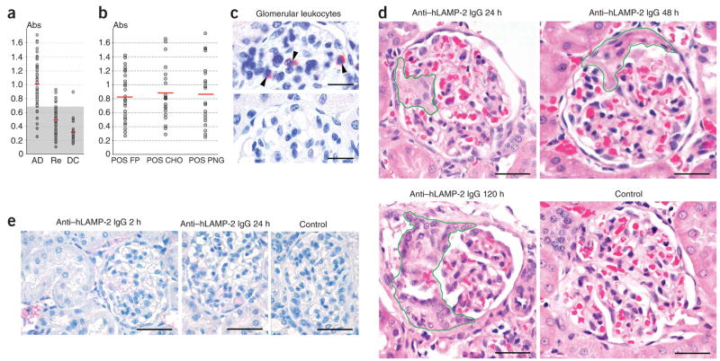
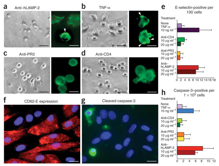
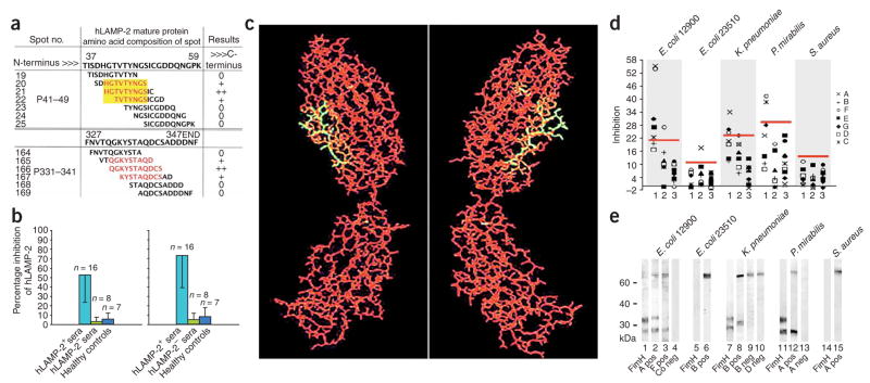
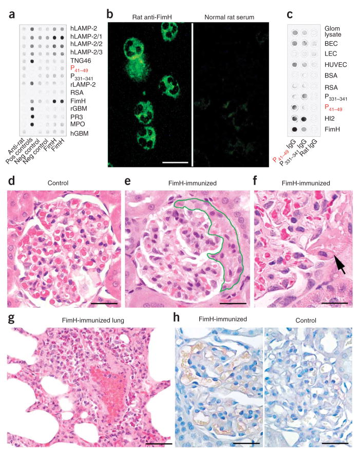
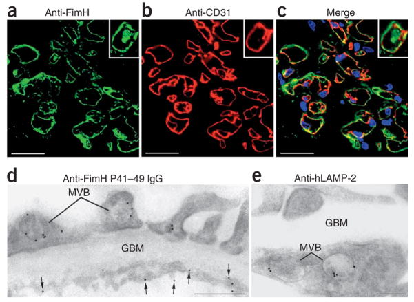
Pauci-immune focal necrotizing glomerulonephritis (FNGN) is a severe inflammatory disease associated with autoantibodies to neutrophil cytoplasmic antigens (ANCA). Here we characterize autoantibodies to lysosomal membrane protein-2 (LAMP-2) and show that they are a new ANCA subtype present in almost all individuals with FNGN. Consequently, its prevalence is nearly twice that of the classical ANCAs that recognize myeloperoxidase or proteinase-3. Furthermore, antibodies to LAMP-2 cause pauci-immune FNGN when injected into rats, and a monoclonal antibody to human LAMP-2 (H4B4) induces apoptosis of human microvascular endothelium in vitro. The autoantibodies in individuals with pauci-immune FNGN commonly recognize a human LAMP-2 epitope (designated P(41-49)) with 100% homology to the bacterial adhesin FimH, with which they cross-react. Rats immunized with FimH develop pauci-immune FNGN and also develop antibodies to rat and human LAMP-2. Finally, we show that infections with fimbriated pathogens are common before the onset of FNGN. Thus, FimH-triggered autoimmunity to LAMP-2 provides a previously undescribed clinically relevant molecular mechanism for the development of pauci-immune FNGN.


Figures

Figure 1
Figure 1
Antibodies to human LAMP-2 (hLAMP-2) in pauci-immune FNGN. (a) Absorbance values of a specific ELISA to assay autoantibodies to LAMP-2 measured in sera from individuals with pauci-immune FNGN and active disease (AD), from the same individuals after treatment when in remission (Re) or from ANCA-negative disease controls (DC) with other types of glomerulonephritis. The shaded area indicates the 99.5% confidence interval for the normal range. (b) Absorbance values of sera from 32 individuals with autoantibodies to LAMP-2 assayed in three different ELISAs with the following substrates: purified unglycosylated, E. coli–expressed human LAMP-2 fusion protein (POS FP); glycosylated mammalian expressed human LAMP-2 (POS CHO); and PNGaseF-treated mammalian-expressed human LAMP-2 (POS PNG). The mean value for each ELISA is indicated as is the upper limit of normal. (c) Glomerular CD45 positive leukocytes (arrowheads) in WKY rats injected 24 h earlier with 10 mg of rabbit antibodies to LAMP-2 (anti-hLAMP2 IgG, top) or 10 mg normal rabbit IgG (bottom). Scale bar, 25 μm. (d) Morphology of representative glomeruli from WKY rats injected intravenously with IgG to rat LAMP-2. Segmental necrosis is present after 24 h, cellular crescents after 48 h and segmental scarring with fibrocellular crescents and adhesions after 120 h. Scale bars, 25 μm. (e) Direct immunoperoxidase staining of representative glomeruli from WKY rats injected with rabbit IgG to LAMP-2. Small quantities of deposited IgG is seen in blood vessels and occasional circulating leukocytes after 2 h but not at later time points. Scale bar, 50 μm.
Figure 2
Figure 2
Antibodies to hLAMP-2 activate neutrophils and kill human microvascular endothelium. (ad) Purified human neutrophils were incubated with 10 μg ml−1 H4B4, a monoclonal antibody to human LAMP-2 (a); 2 ng ml−1 TNF-α (b), 10 μg ml−1 1F11, a monoclonal antibody to proteinase-3 (c) or 10 μg ml−1 of monoclonal antibody to CD4 (d). H4B4 and TNF induced significantly more shape change than the other two treatments, and the insets confirm that the shape change is associated with actin condensation (insets in ad; scale bars, 50 μm). When the same antibodies were incubated with purified human dermal microvascular blood endothelial cells (BECs), H4B4 significantly increased CD62E (E-selectin) expression (P < 0.01) whereas 1F11 and CD4 had no effect (e, f). H4B4 also significantly increased the number of cells expressing activated caspase-3 (P < 0.05), a marker of apoptosis (g, h). Scale bars, 50 μm. Values in e and h represent means ± s.d.
Figure 3
Figure 3
Autoantibodies to hLAMP-2 cross-react with the bacterial adhesin FimH. (a) Two epitopes, P41–49 (HGTVTYNGS) and P331–341 (QGKYSTAQDCS) recognized by autoantibodies to hLAMP-2 in active FNGN are shown in red. The yellow box illustrates the homologous sequence shared by human LAMP-2 and FimH. Depicted are examples of overlapping peptides synthesized onto a SPOTs membrane aligned to the human LAMP-2 sequence. (b) Inhibition ELISA used to determine whether synthetic peptides corresponding to P41–49 (left) and P331–341 (right) prevented autoantibodies in active pauci-immune FNGN from binding to human LAMP-2. Both peptides significantly inhibited binding (P < 0.001). Values represent means ± s.d. (c) Three-dimensional projection of the structure of the FimH mannose binding pocket. Amino acids 72–80 of FimH, homologous to P41–49 (yellow), are exposed on the surface. (d) ELISA was used to determine whether bacterial lysates from FimH-expressing (E. coli 12900, K. pneumoniae., P. mirabilis) or non-expressing (E. coli 23510, S. aureus) organisms inhibit binding of antibodies to LAMP-2 from subjects (A–G) with active FNGN (lane 1), FNGN in remission (lane 2) or healthy controls (lane 3). Red bars, 99.5% confidence interval for inhibition of binding. (e) Western blot showing binding of antibodies from subjects with FNGN to lysates of bacteria that do (lanes 1–4, 7–10 and 11–13) or do not (lanes 5–6 and 14–15) express FimH. Two sera with specificity for hLAMP-2 (A pos, B pos and F pos) recognize the same bands as an antibody to FimH (fimH), whereas negative sera (A neg, B neg and D neg) and controls (Co neg) do not.
Figure 4
Figure 4
Immunization with FimH induces antibodies to LAMP-2 and FNGN in rats. (a) Sera from FimH-immunized rats and controls were probed for FimH, full-length and N-terminal hLAMP-2 and 100–amino acid (hLAMP-2/1) fusion proteins. The specificity of rat IgG for human or rat glomerular basement membrane (rGBM and hGBM), proteinase-3 (PR3) or myeloperoxidase (MPO), trans-Golgi network protein 46 (TGN46) or rat serum albumin (RSA) was tested with purified proteins. Specific antibodies to these proteins, where available, were used as positive controls, and anti–rat IgG alone as negative control. (b) Sera from FimH-immunized WKY rats bound neutrophil granules, resulting in a cytoplasmic staining pattern similar to that observed with sera from subjects with FNGN (left). Sera from control rats remained negative. Scale bar, 25 μm. (c, d) IgG specific for P41–49 or P331–341, purified from rat serum after immunization with FimH was probed on purified FimH and hLAMP-2 fusion proteins, peptides P41–49 and P331–341, and proteins from isolated glomeruli, microvascular blood endothelial cells (BECs), HUVECs and lymphatic endothelial cells (LECs) (c). Purified normal rat IgG was used as control. Glomerular histology from un-immunized control animals appears normal (d). WKY rats immunized with FimH show glomerular crescent formation (e), segmental necrosis of capillaries (f) and marked neutrophilic capillaritis of the lung (g). Direct immunoperoxidase staining shows no glomerular IgG deposition in glomeruli from FimH immunized nephritic rats (h; left) nor from adjuvant treated controls (h; right). Scale bars in dh, 50 μm.
Figure 5
Figure 5
Antibodies induced by FimH immunization bind human glomerular endothelium. (a, b) Double labeling of frozen sections of normal human glomeruli with purified rabbit IgG specific for the FimH mannose binding pocket (green, a) and with an antibody to CD31 (red, b). (c) Overlay of a and b (yellow; scale bars, 25 μm). (d, e) IgG with specificity for peptide P41–49, purified from rat sera after immunization with FimH, was used for indirect immunolabeling of ultrathin frozen sections of human glomeruli. The staining pattern on endothelial cell membrane (arrows) as well as within multivesicular bodies (MVB) of endothelial and epithelial (E) is compared to that of a monoclonal antibody to hLAMP-2 (e). GBM, glomerular basement membrane. Scale bar in d, 500 nm; scale bar in e, 200 nm.

Comment in

References

    1. Jennette JC, Falk RJ. Small-vessel vasculitis. N Engl J Med. 1997;337:1512–1523. - PubMed
    1. Morgan MD, Harper L, Williams J, Savage C. Anti-neutrophil cytoplasm associated glomerulonephritis. J Am Soc Nephrol. 2006;17:1224–1234. - PubMed
    1. Davies DJ, Moran JE, Niall JF, Ryan GB. Segmental necrotizing glomerulo-nephritis with antineutrophil antibody: possible arbovirus aetiology? . Br Med J (Clin Res Ed) 1982;285:606. - PMC - PubMed
    1. van der Woude FJ, et al. Autoantibodies against neutrophils and monocytes: tool for diagnosis and marker of disease activity in Wegener’s granulomatosis. Lancet. 1985;1:425–429. - PubMed
    1. Jennette JC, Xiao H, Falk RJ. Pathogenesis of vascular inflammation by anti-neutrophil cytoplasmic antibodies. J Am Soc Nephrol. 2006;17:1235–1242. - PubMed

Publication types

MeSH terms