. 2008 Oct;14(10):1123-7.
doi: 10.1038/nm.1754. Epub 2008 Sep 28.

Chemical control of protein stability and function in living mice

Affiliations

Chemical control of protein stability and function in living mice

Laura A Banaszynski et al. Nat Med. 2008 Oct.

Abstract

Conditional control of protein function in vivo offers great potential for deconvoluting the roles of individual proteins in complicated systems. We recently developed a method in which a small protein domain, termed a destabilizing domain, confers instability to fusion protein partners in cultured cells. Instability is reversed when a cell-permeable small molecule binds this domain. Here we describe the use of this system to regulate protein function in living mammals. We show regulation of secreted proteins and their biological activity with conditional secretion of an immunomodulatory cytokine, resulting in tumor burden reduction in mouse models. Additionally, we use this approach to control the function of a specific protein after systemic delivery of the gene that encodes it to a tumor, suggesting uses for enhancing the specificity and efficacy of targeted gene-based therapies. This method represents a new strategy to regulate protein function in living organisms with a high level of control.


Figures

Figure 1
Figure 1
Conditional regulation of protein stability in vivo. (a) SCID mice bearing HCT116 L106P-tsLuc xenografts (50-100 mm3) were either untreated (top) or treated ip with Shield-1 (10 mg/kg, bottom) and bioluminescent signals were imaged over time. (b) Quantification of tumor signals from panel A. (c) Mice as in a were treated ip with Shield-1 at 3 mg/kg (diamonds), 6 mg/kg (inverted triangles), or 10 mg/kg (triangles) and imaged over time. (d) Mice bearing HCT116-tsLuc xenografts were either untreated (squares) or treated with Shield-1 (10 mg/kg, triangles) every 48 hr and imaged over time. Data for panels b-d are presented as the average bioluminescence detected within regions of interest drawn around the tumors ± SEM (n=4 to 10).
Figure 2
Figure 2
Conditional regulation of a secreted immunomodulatory protein leads to a reduction of tumor burden in vivo. (a) HCT116 L-L106P-IL-2 cells were treated with various concentrations of Shield-1 and culture media was assayed for the presence of IL-2. Data are represented as the average IL-2 concentration ± SEM (n=3). (b) CD1 nu-/nu- mice bearing subcutaneous HCT116 L-L106P-IL-2 tumors were either untreated (diamonds) or treated ip with Shield-1 at 5 mg/kg (triangles) or 10 mg/kg (inverted triangles) every 48 hr beginning 5 days post-transplantation (arrow). Alternatively, mice received HCT116 L-L106P-IL-2 cells that had been pre-treated with 1 μM Shield-1 for 24 hr, and were then treated with Shield-1 (10 mg/kg) every 48 hr beginning on day 0 (squares). Tumor volume was determined by caliper measurement and monitored over time. Data are represented as the average tumor volume ± SEM (n=5). At Day 16, all Shield-1 treated groups displayed significantly reduced tumor burden relative to controls (p=0.0019 for 10 mg/kg; p=0.0002 for 5 mg/kg and p=0.0046 for pre-treat group). (c) Tumors from mice treated with Shield-1 post-transplantation were collected 48 hr after the start of Shield-1 treatment. Tumors were weighed ex vivo and then homogenized, and the concentration of IL-2 per gram tumor tissue was determined by ELISA (n=4). Tumors from mice treated with Shield-1 at 10 mg/kg contained significantly higher levels of IL-2 than tumors from mice treated at 5 mg/kg (p=0.032), which in turn produced more IL-2 than tumors from untreated mice (p=0.0028). (d) Mice treated with Shield-1 post-transplantation were bled 48 hr after the start of Shield-1 treatment, and the concentration of IL-2 in the serum was determined by ELISA. Shield-1 treatment at 10 mg/kg (n=7) produced significantly higher levels of serum IL-2 relative to control mice (n=6, p=0.0004), whereas treatment at 5 mg/kg (n=6) did not produce any increase in serum IL-2 levels.
Figure 3
Figure 3
Systemic, targeted-delivery of a conditionally stabilized protein. (a) HCT116 cells were infected with vvDD L106P-tsLuc and then mock treated (squares) or treated with Shield-1 at 1 μM (triangles), 100 nM (inverted triangles), or 10 nM (diamonds). Data are represented as the average luminescence ± SEM (n=3). (b) SCID mice bearing subcutaneous HCT116 xenografts (50-100 mm3) received a single tail vein injection of vvDD L106P-tsLuc (1×10⁸ PFU/mouse). After 72 hr, mice were either untreated (C, control) or treated with Shield-1 (10 mg/kg, #1-4). Bioluminescent signals were imaged over time. (c) Quantification of bioluminescent signal produced from regions of interest drawn around tumors as in panel b. Data presented are the average bioluminescence ± SEM (n=4).
Figure 4
Figure 4
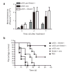
Antitumor benefit of conditional regulation of a targeted gene therapy vector. CD1 nu-/nu- mice bearing subcutaneous HCT116 tumors (150-250 mm3) were treated via a single tail vein injection with either PBS (circles) or the vaccinia strain vvDD expressing both luciferase as well as the L-L106P-TNF-α fusion protein (1 × 10⁸ PFU/mouse). Treated mice also received Shield-1 (10 mg/kg) every 48 hr by three different protocols, starting (i) 24 hr prior to vvDD therapy (diamonds); (ii) 72 hr post-vvDD therapy (squares) or (iii) not at all (triangles) (n=8 mice/group). Tumor-bearing mice that did not receive vvDD therapy were also treated with Shield-1 (10 mg/kg) as a negative control (inverted triangles, n=5). (a) Viral load within the tumor was assayed by measuring constitutive viral gene expression (bioluminescence) for each group at the indicated time points after vvDD treatment. Shield-1 treatment starting 72 hr after vvDD (vvDD + Shield-1) resulted in significantly greater levels of viral gene expression in the tumor than when Shield-1 treatment was started prior to vvDD therapy (vvDD Pre-Shield-1; i.e., constitutive TNF-α expression) (p=0.035 at 2 days; p=0.035 at 4 days and p=0.002 at 7 days). (b) Tumor volume was determined by caliper measurement and mice were sacrificed once tumor volume reached 1.44 cm3. Kaplan-Meier survival graphs are shown, and all surviving mice at 50 days had no detectable tumor. Mice treated with Shield-1 prior to vvDD therapy (i.e., constitutive TNF-α expression) produced significantly enhanced survival relative to mice treated with vvDD alone (p=0.017). Addition of Shield-1 72 hr post-vvDD (vvDD + Shield-1) produced a further significant increase in survival relative to mice treated with Shield-1 prior to vvDD (vvDD pre-Shield-1) (p=0.031).

References

    1. Bockamp E, et al. Of mice and models: improved animal models for biomedical research. Physiol Genomics. 2002;11:115–132. - PubMed
    1. Bradley A, Evans M, Kaufman MH, Robertson E. Formation of germ-line chimeras from embryo-derived teratocarcinoma cell lines. Nature. 1984;309:255–256. - PubMed
    1. Thomas KR, Capecchi MR. Site-directed mutagenesis by gene targeting in mouse embryo-derived stem cells. Cell. 1987;51:503–512. - PubMed
    1. Doetschman T, et al. Targetted correction of a mutant HPRT gene in mouse embryonic stem cells. Nature. 1987;330:576–578. - PubMed
    1. Yamada G, et al. Targeted mutation of the murine goosecoid gene results in craniofacial defects and neonatal death. Development. 1995;121:2917–2922. - PubMed

Publication types