Review
. 2008 Oct 6;27(45):5904-12.
doi: 10.1038/onc.2008.271.

The tumor microenvironment and its role in promoting tumor growth

Affiliations
Review

The tumor microenvironment and its role in promoting tumor growth

T L Whiteside. Oncogene. .

Abstract

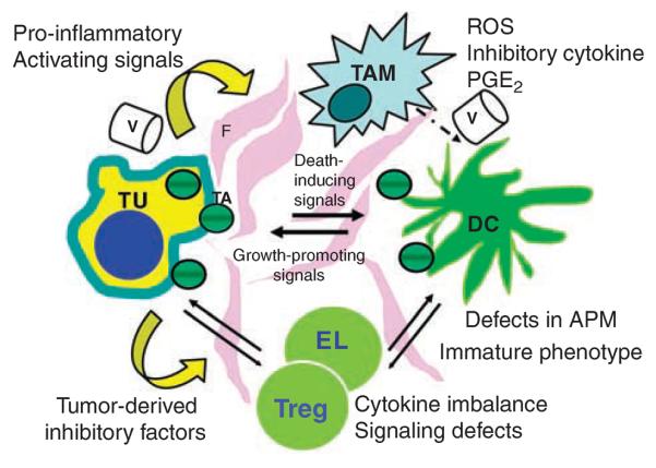
The tumor microenvironment is created by the tumor and dominated by tumor-induced interactions. Although various immune effector cells are recruited to the tumor site, their anti-tumor functions are downregulated, largely in response to tumor-derived signals. Infiltrates of inflammatory cells present in human tumors are chronic in nature and are enriched in regulatory T cells (T(reg)) as well as myeloid suppressor cells (MSC). Immune cells in the tumor microenvironment not only fail to exercise antitumor effector functions, but they are co-opted to promote tumor growth. Sustained activation of the NF-kappaB pathway in the tumor milieu represents one mechanism that appears to favor tumor survival and drive abortive activation of immune cells. The result is tumor escape from the host immune system. Tumor escape is accomplished through the activation of one or several molecular mechanisms that lead to inhibition of immune cell functions or to apoptosis of anti-tumor effector cells. The ability to block tumor escape depends on a better understanding of cellular and molecular pathways operating in the tumor microenvironment. Novel therapeutic strategies that emerge are designed to change the pro-tumor microenvironment to one favoring acute responses and potent anti-tumor activity.


Figures

Figure 1
Figure 1
A diagram depicting the tumor microenvironment. Interactions of various cells with each other, fibroblasts (F) in the tumor stroma and blood vessels (V) are indicated by arrows. Although the tumor (TU) generates signals inducing dysfunction and death of immune cells, the latter are a source of signals promoting tumor growth. TAM, tumor-associated macrophages; DC, dendritic cells; EL, effector lymphocytes; Treg, regulatory T cells; TA, tumor-derived antigens; ROS, reactive oxygen species; PGE2, prostaglandin E2.
Figure 2
Figure 2
Accumulation and expansion of Treg in the tumor microenvironment may be a result of the cross talk between the tumor and DC. The tumor coopts DC differentiation, and in the presence of tumor-derived factors, immature DC develop abnormalities in APM, have decreased expression levels of MHC molecules, upregulate B7–H1 (PD-L1) and produce excess of IL-10 and TGF-β1. Cross-presentation of TA by these DC to T cells (as in the lower part of figure) lead to emergence of Treg and their expansion. Once generated, Treg interfere with functions of anti-tumor effector cells (CTL). Even if cross-presentation of TA by DC to naive CD8 + T cells is successful (as in the upper part of figure), Treg proceed to block CTL functions. Reproduced with changes from Ferrone and Whiteside, 2007.
Figure 3
Figure 3
Tumors recruit MSC from the bone marrow by means of tumor-derived soluble factors. Immature myeloid cells migrate to lymph nodes, where DC cross-prime T cells, and interfere with this process. They also migrate to the tumor site and become tumor-associated MSC, which are adept in blocking T cell functions through the production of arginase I and activation of iNOs.
Figure 4
Figure 4
Mechanisms responsible for `immunoediting' of tumor cells in the tumor microenvironment. The various mechanisms listed collaborate in immunoediting of the tumor cells. The tumor, stromal cells and infiltrating leukocytes all contribute to pro-inflammatory milieu. Infiltrating immune cells and stromal elements are re-programmed by the tumor to the pro-inflammatory mode favoring its survival. In this milieu, the tumor evolves a phenotype allowing it to escape and to counterattack immune cells. Tumor stem cells are resistant to anti-tumor therapies and thus represent yet another means of escape.

References

    1. Albers AE, Kim G, Ferris RL, Chikamatsu K, DeLeo AB, Whiteside TL, et al. Immune responses to p53 in patients with cancer: enrichment in tetramer+p53 peptide-specific T cells and regulatory CD4+CD25+ cells at tumor sites. Cancer Immunol Immunother. 2002;62:670–679. - PMC - PubMed
    1. Aller MA, Arias JL, Nava MP, Arias J. Posttraumatic inflammation is a complex response based on the pathological expression of the nervous, immune and endocrine function systems. Exp Biol Med. 2004;229:170–181. - PubMed
    1. Almand B, Clark JI, Nikitina E, van Beynen J, English NR, Knight SC, et al. Increased production of immature myeloid cells in cancer patients: a mechanism of immunosuppression in cancer. J Immunol. 2001;166:678–689. - PubMed
    1. Almand B, Resser JR, Lindman B, Nadaf S, Clark JI, Kwon ED, et al. Clinical significance of defective dendritic cell differentiation in cancer. Clin Cancer Res. 2000;6:1755–1766. - PubMed
    1. Bacchetta R, Gambineri E, Roncarolo MG. Role of regulatory T cells and FOXP3 in human diseases. J Allergy Clin Immunol. 2007;120:227–235. - PubMed

Publication types