. 2008 Oct 6;3(10):e3331.
doi: 10.1371/journal.pone.0003331.

The IL-1-like cytokine IL-33 is constitutively expressed in the nucleus of endothelial cells and epithelial cells in vivo: a novel 'alarmin'?

Affiliations

The IL-1-like cytokine IL-33 is constitutively expressed in the nucleus of endothelial cells and epithelial cells in vivo: a novel 'alarmin'?

Christine Moussion et al. PLoS One. .

Abstract

Background: Interleukin-33 (IL-33) is an IL-1-like cytokine ligand for the IL-1 receptor-related protein ST2, that activates mast cells and Th2 lymphocytes, and induces production of Th2-associated cytokines in vivo. We initially discovered IL-33 as a nuclear factor (NF-HEV) abundantly expressed in high endothelial venules from lymphoid organs, that associates with chromatin and exhibits transcriptional regulatory properties. This suggested that, similarly to IL-1alpha and chromatin-associated cytokine HMGB1, IL-33 may act as both a cytokine and a nuclear factor. Although the activity of recombinant IL-33 has been well characterized, little is known yet about the expression pattern of endogenous IL-33 in vivo.

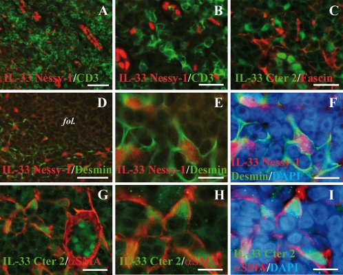
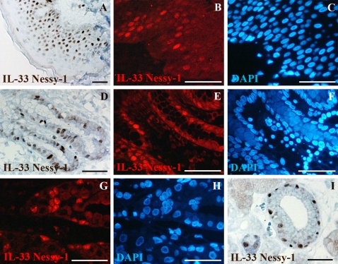
Methodology/principal findings: Here, we show that IL-33 is constitutively and abundantly expressed in normal human tissues. Using a combination of human tissue microarrays and IL-33 monoclonal and polyclonal antibodies, we found that IL-33 is a novel nuclear marker of the endothelium widely expressed along the vascular tree. We observed abundant nuclear expression of IL-33 in endothelial cells from both large and small blood vessels in most normal human tissues, as well as in human tumors. In addition to endothelium, we also found constitutive nuclear expression of IL-33 in fibroblastic reticular cells of lymphoid tissues, and epithelial cells of tissues exposed to the environment, including skin keratinocytes and epithelial cells of the stomach, tonsillar crypts and salivary glands.

Conclusions/significance: Together, our results indicate that, unlike inducible cytokines, IL-33 is constitutively expressed in normal human tissues. In addition, they reveal that endothelial cells and epithelial cells constitute major sources of IL-33 in vivo. Based on these findings, we speculate that IL-33 may function, similarly to the prototype 'alarmin' HMGB1, as an endogenous 'danger' signal to alert the immune system after endothelial or epithelial cell damage during trauma or infection.


Conflict of interest statement

Competing Interests: The authors have declared that no competing interests exist.

Figures

Figure 1
Figure 1. IL-33 is a chromatin-associated nuclear factor constitutively expressed in human secondary lymphoid tissues by HEVs and isolated cells in the interfollicular T cells areas.
A: Immunohistochemical staining of a human tonsil section with IL-33 mAb Nessy-1. B–D: Double staining of human tonsils sections with HEV-specific mAb MECA79 (green) and IL-33 mAb Nessy-1 (B, red) or two distinct IL-33 polyclonal antisera, Cter1 (C, red) or Cter2 (D, red). The follicles (fol) are indicated. E and F: Nuclear staining of HEVs and isolated cells in the T cell areas was abrogated by pre-incubating the IL-33 monoclonal (E) and polyclonal (F) antibodies with IL-33 peptides but not control peptides. G and H: Nuclear accumulation of IL-33 in HEV blood vessels and isolated cells was also observed in lymph node (G, IL-33 mAb, red; CD31, green) and appendix (H, IL-33 mAb, red; vWF, green). I and J: Higher magnification of a human tonsil section double-stained with IL-33 and MECA-79 antibodies and counterstained with the DNA-binding dye DAPI. In the nucleus of both HEVs (arrow, upper panel) and isolated cells (arrowhead, lower panel), IL-33 accumulates in nuclear domains that colocalize with dense regions of DAPI staining (J), indicating association with chromatin. Magnification bars: A, I 10 µm; B, G, H 20 µm; J 5 µm; C, D, E, F 60 µm.
Figure 2
Figure 2. IL-33 is a nuclear marker of FRCs in the interfollicular T cells areas.
A and B: Double staining of human tonsils sections with IL-33 mAb Nessy-1 (red) and anti-CD3 polyclonal antibody (green). C: Double staining of a human tonsil section with IL-33 polyclonal antibody Cter2 (green) and anti-fascin mAb (red). D–F: Double staining of a human tonsil section with IL-33 mAb Nessy-1 (red) and anti-desmin polyclonal antibody (green). A follicle is indicated (fol). Higher magnification (E) and DAPI counterstaining (F) are shown to reveal nuclear accumulation of IL-33 in desmin+ cells. G–I: Double staining of a human tonsil section with IL-33 polyclonal antibody Cter2 (green) and anti-α-SMA mAb (red). α-SMA is detected in the basal lamina of an HEV blood vessel (G) but is not expressed by the HEV endothelial cells. Higher magnification (H) and DAPI counterstaining (I) reveals nuclear accumulation of IL-33 in α-SMA+ cells. Magnification bars: A, D 60 µm; B, C, G 25 µm; E, F, H, I 10 µm.
Figure 3
Figure 3. IL-33 is constitutively expressed in the nucleus of endothelial cells from large blood vessels in normal human tissues.
Expression of IL-33 in human tissues was analyzed using both immunohistochemistry (lefts panels) and immunofluorescence staining (right panels). Double staining was performed with IL-33 mAb Nessy-1 (red) and anti-CD31 or anti-vWF polyclonal antibodies (green). DNA was counter-stained with DAPI. Arrowheads label the nuclei of smooth muscle cells in arterioles that are not stained with IL-33 antibodies. Magnification bars: 35 µm.
Figure 4
Figure 4. IL-33 is constitutively expressed in the nucleus of endothelial cells from small blood vessels in normal human tissues.
Expression of IL-33 in the microvasculature was analyzed using immunofluorescence staining. Double staining was performed with IL-33 mAb Nessy-1 (red) and anti-CD31 or anti-vWF polyclonal antibodies (green). DNA was counter-stained with DAPI. Magnification bars: 20 µm.
Figure 5
Figure 5. IL-33 is abundantly expressed in the nucleus of endothelial cells in human tumor tissues.
Expression of IL-33 in the indicated human tumor tissues was analyzed using immunofluorescence staining. Double staining was performed with IL-33 mAb Nessy-1 (red) and anti-CD31 or anti-vWF polyclonal antibodies (green). DNA was counter-stained with DAPI. Magnification bars: A,B,G,H 50 µm; C,D,E,F 20 µm.
Figure 6
Figure 6. IL-33 is constitutively expressed in the nucleus of epithelial cells in tissues exposed to the environment.
A–C: Skin keratinocytes; D–F: Epithelial cells of the mucosal surface in the stomach; G–H: Epithelial cells of the gastric glands in the stomach; I: Epithelial cells of the salivary glands. Immunohistochemical and immunofluorescence staining were performed with IL-33 mAb Nessy-1. For immunofluorescence, IL-33 expression was detected in red and DNA was counter-stained with DAPI (blue). Magnification bars: 70 µm.

References

    1. Baekkevold ES, Roussigne M, Yamanaka T, Johansen FE, Jahnsen FL, et al. Molecular characterization of NF-HEV, a nuclear factor preferentially expressed in human high endothelial venules. American Journal of Pathology. 2003;163:69–79. - PMC - PubMed
    1. Schmitz J, Owyang A, Oldham E, Song Y, Murphy E, et al. IL-33, an interleukin-1-like cytokine that signals via the IL-1 receptor-related protein ST2 and induces T helper type 2-associated cytokines. Immunity. 2005;23:479–490. - PubMed
    1. Carriere V, Roussel L, Ortega N, Lacorre DA, Americh L, et al. IL-33, the IL-1-like cytokine ligand for ST2 receptor, is a chromatin-associated nuclear factor in vivo. Proc Natl Acad Sci U S A. 2007;104:282–287. - PMC - PubMed
    1. Sanada S, Hakuno D, Higgins LJ, Schreiter ER, McKenzie AN, et al. IL-33 and ST2 comprise a critical biomechanically induced and cardioprotective signaling system. J Clin Invest. 2007;117:1538–1549. - PMC - PubMed
    1. Allakhverdi Z, Smith DE, Comeau MR, Delespesse G. Cutting edge: The ST2 ligand IL-33 potently activates and drives maturation of human mast cells. J Immunol. 2007;179:2051–2054. - PubMed

Publication types