. 2009 May;58(5):709-18.
doi: 10.1007/s00262-008-0593-3. Epub 2008 Oct 3.

Characterization of MHC class-I restricted TCRalphabeta+ CD4- CD8- double negative T cells recognizing the gp100 antigen from a melanoma patient after gp100 vaccination

Affiliations

Characterization of MHC class-I restricted TCRalphabeta+ CD4- CD8- double negative T cells recognizing the gp100 antigen from a melanoma patient after gp100 vaccination

Simon Voelkl et al. Cancer Immunol Immunother. 2009 May.

Abstract

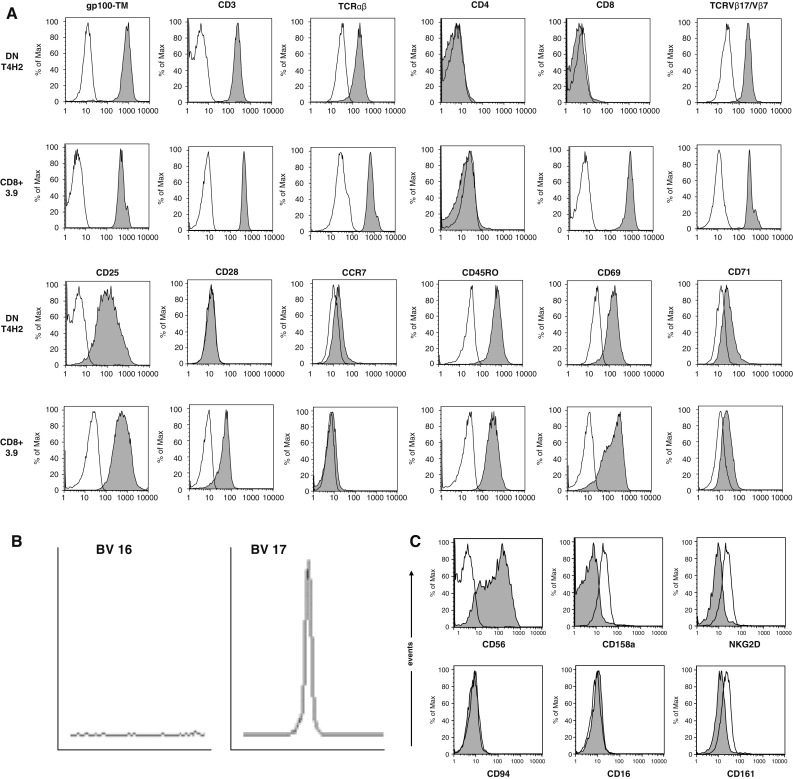
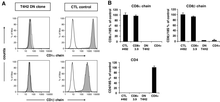
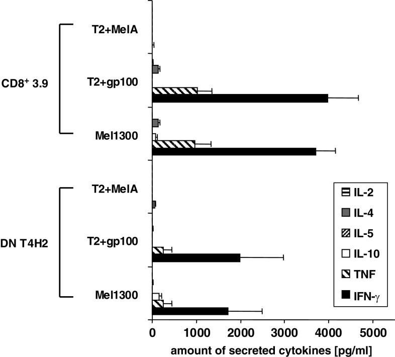
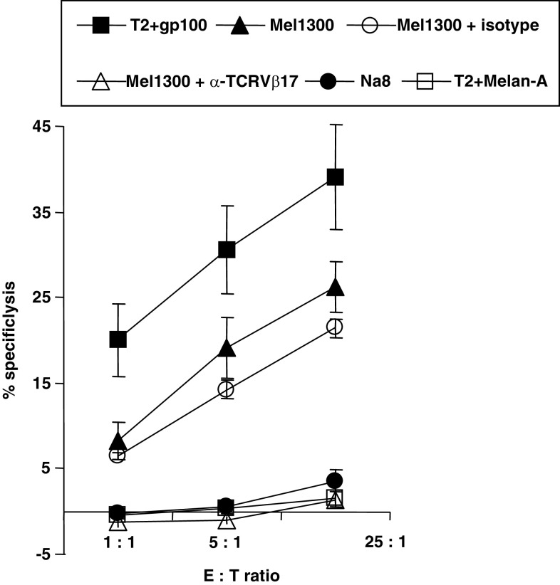
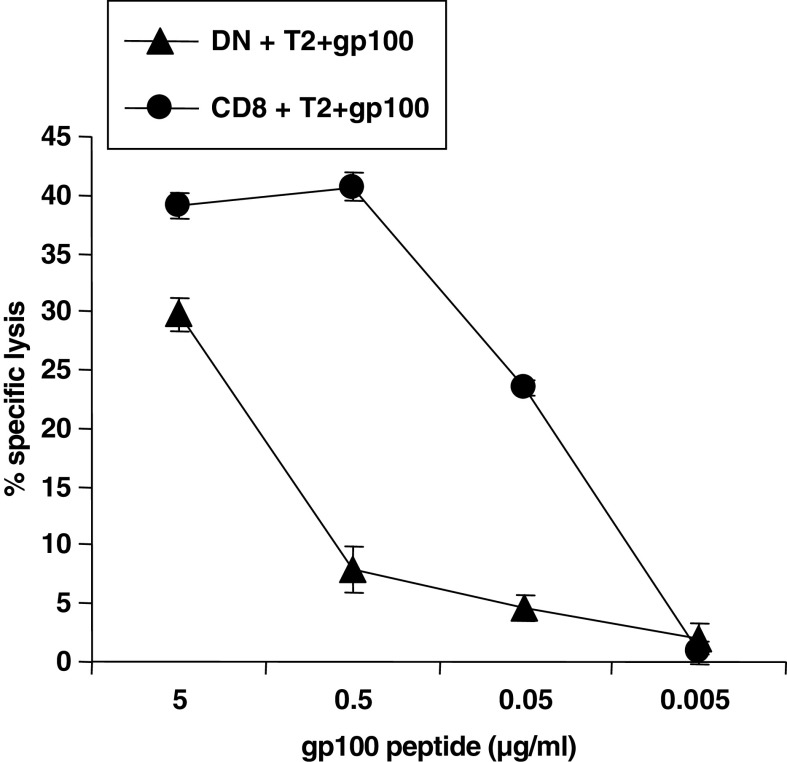
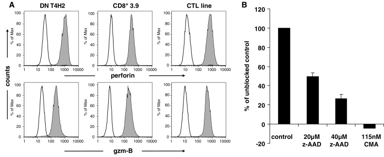
The immune attack against malignant tumors require the concerted action of CD8+ cytotoxic T lymphocytes (CTL) as well as CD4+ T helper cells. The contribution of T cell receptor (TCR) alphabeta+ CD4- CD8- double-negative (DN) T cells to anti-tumor immune responses is widely unknown. In previous studies, we have demonstrated that DN T cells with a broad TCR repertoire are present in humans in the peripheral blood and the lymph nodes of healthy individuals. Here, we characterize a human DN T cell clone (T4H2) recognizing an HLA-A2-restricted melanoma-associated antigenic gp100-peptide isolated from the peripheral blood of a melanoma patient. Antigen recognition by the T4H2 DN clone resulted in specific secretion of IFN-gamma and TNF. Although lacking the CD8 molecule the gp100-specific DN T cell clone was able to confer antigen-specific cytotoxicity against gp100-loaded target cells as well as HLA-A2+ gp100 expressing melanoma cells. The cytotoxic capacity was found to be perforin/granzymeB-dependent. Together, these data indicate that functionally active antigen-specific DN T cells recognizing MHC class I-restricted tumor-associated antigen (TAA) may contribute to anti-tumor immunity in vivo.


Figures

Fig. 1
Fig. 1
Phenotype of the gp100-reactive DN T cell clone T4H2. T4H2 cells were isolated from a melanoma patient after gp100-peptide vaccination and expanded over several months by repetitive stimulation with allogeneic feeder cells. A classical gp100-reactive CD8+ CTL clone (3.9), isolated from an HLA-A2+ healthy donor, served as a control. a T4H2 and 3.9 cells were harvested and stained with a panel of mAbs recognizing CD3, CD4, CD8, TCRαβ, TCRVβ17 (for T4H2) or TCRVβ7 (for 3.9), CD28, CD45RO, CCR7, CD69, CD25, CD71 and the HLA-A2/gp100-tetramer and analyzed by flow cytometry. Histograms are shown on gated lymphocytes (by forward and side scatter) to exclude residual feeder cells and fluorescence intensity is presented by the gray line. Open histograms show the respective isotype controls. b Analysis of the TCRVβ family distribution in T4H2 DN T cells via the TCR CDR3 spectratyping method. A clonal T cell population is represented by a distinct peak in the appropriate TCRVβ17 receptor family. One negative (TCRVβ16) and the TCRVβ17 positive run off results are shown. c Analysis of NK cell marker expression on T4H2 cells. T4H2 cells were stained with mAbs against NK cell markers CD56, CD158a, NKG2D, CD94, CD16, CD161, and analyzed by flow cytometry
Fig. 2
Fig. 2
Lack of CD8 coreceptor expression on the T4H2 DN T cell clone. a Intracellular expression of the CD8α chain and CD8β chain on the DN clone T4H2. For intracellular mAb staining cells were fixed with 0.25% paraformaldehyde, permeabilized with 10% saponine and stained with the respective anti-CD8 mAbs and analyzed by flow cytometry. A CD8+ Melan-A-specific CTL line served as a positive control. Histograms are shown on gated lymphocytes (by forward and side scatter) and fluorescence intensity is presented by the gray line. Open histograms show the isotype controls. b Quantitative light cycler PCR was performed to detect CD8α-chain, CD8β-chain and CD4 mRNA in T4H2 cells. Values were normalized to the amount of CD8 transcripts either in Melan-A specific CD8+ CTL lines (CTL#492) or CD4 transcripts in freshly isolated CD4+ T cells. Mean values ± SEM from triplicates are shown
Fig. 3
Fig. 3
CD8 gene demethylation in T4H2 DN T cells. Schematic overview of human CD8α promoter and positions of CpG motifs (upper panel). Genomic DNA from DN T cell clone T4H2, CD8+ T cell clone 3.9 and a CD4+ T cell clone was analyzed by bisulfite sequencing. In the matrix, each row represents one sequenced insert, each box shows methylation status of a single CpG. Filled boxes indicate methylation (lower panel)
Fig. 4
Fig. 4
gp100-reactive DN T cells secrete cytokines upon recognition of the target cells. Cytokine profile of the T4H2 DN clone and the CD8+ T cell clone 3.9 after a 4 h coculture with either gp100- or Melan-A (MelA)-pulsed T2 cells or HLA-A2+, gp100+ expressing melanoma cells (Mel1300). Melan-A loaded T2 cells were used as control stimulator cells. The amount of cytokines in the supernatant was determined by the Cytometric Bead Array (CBA). Data show mean ± SEM of three independent experiments
Fig. 5
Fig. 5
Cytotoxic activity of T4H2 DN T cells against endogenously presented gp100 antigen on melanoma cells. The cytotoxic capacity of the T4H2 DN T cell clone was assessed by a standard 4 h 51Cr-release assay. T2 cells loaded exogenously with gp100 peptide (filled squares) as well as the gp100-expressing melanoma cell line Mel1300 (filled triangles) served as target cells. As negative controls Melan-A-loaded T2 (open squares) and the melanoma cell line Na8 (HLA-A2+, gp100, filled circles) were used. For TCR blocking studies, effector cells were preincubated for 30 min with a neutralizing anti-TCRVβ17 mAb (open triangles) or the isotype control (open circles), washed and coincubated with Mel1300 cells. Data represent means ± SEM of triplicates
Fig. 6
Fig. 6
Cytotoxic activity of the gp100-reactive DN (T4H2) and CD8+ (3.9) T cell clone was examined at an effector-to-target (E/T) ratio of 5:1 using indicated concentrations of gp100 peptide loaded onto T2 cells. The cytotoxic capacity of the T cell clones was assessed by a standard 4 h 51Cr-release assay. Shown are mean values of triplicates ±SEM
Fig. 7
Fig. 7
Death pathway of T4H2 clone mediated cytotoxicity. a T4H2 cells were harvested, stained intracellularly with mAbs recognizing human effector molecules perforin and granzyme-B (gzm-B) (gray histograms) or with an isotypic control (open histograms), and analyzed by flow cytometry. The gp100-specific CD8+ T cell clone 3.9 as well as a Melan-A specific CD8+ CTL line (CTL line) served as a positive control. b Inhibition of the T4H2 cell-mediated target cell killing by concanamycin-A (CMA, inhibitor of perforin-mediated killing) and z-AAD (granzyme-B inhibitor). T4H2 cells were cultured with CMA or z-AAD. Cytotoxic activity was examined at an E/T ratio of 5:1 using the gp100 expressing melanoma cell line Mel1300. Data are shown as means of triplicates in percentage of unblocked control ±SEM

Similar articles

Cited by

References

    1. Fischer K, Voelkl S, Heymann J, Przybylski GK, Mondal K, Laumer M, Kunz-Schughart L, Schmidt CA, Andreesen R, Mackensen A. Isolation and characterization of human antigen-specific TCR alpha beta+ CD4–CD8- double-negative regulatory T cells. Blood. 2005;105:2828–2835. doi: 10.1182/blood-2004-07-2583. - DOI - PubMed
    1. Zinkernagel RM, Doherty PC. The discovery of MHC restriction. Immunol Today. 1997;18:14–17. doi: 10.1016/S0167-5699(97)80008-4. - DOI - PubMed
    1. Sprent J, Schaefer M, Lo D, Korngold R. Functions of purified L3T4+ and Lyt-2+ cells in vitro and in vivo. Immunol Rev. 1986;91:195–218. doi: 10.1111/j.1600-065X.1986.tb01489.x. - DOI - PubMed
    1. Nishimura MI, Avichezer D, Custer MC, Lee CS, Chen C, Parkhurst MR, Diamond RA, Robbins PF, Schwartzentruber DJ, Rosenberg SA. MHC class I-restricted recognition of a melanoma antigen by a human CD4+ tumor infiltrating lymphocyte. Cancer Res. 1999;59:6230–6238. - PubMed
    1. Takahama Y, Kosugi A, Singer A. Phenotype, ontogeny, and repertoire of CD4–CD8- T cell receptor alpha beta+ thymocytes. Variable influence of self-antigens on T cell receptor V beta usage. J Immunol. 1991;146:1134–1141. - PubMed

Publication types

MeSH terms