. 2008 Oct;9(10):835-41.
doi: 10.1631/jzus.B0860005.

Fungal diversity on fallen leaves of Ficus in northern Thailand

Affiliations

Fungal diversity on fallen leaves of Ficus in northern Thailand

Hong-Kai Wang et al. J Zhejiang Univ Sci B. 2008 Oct.

Abstract

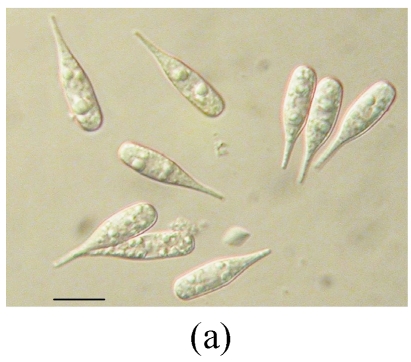
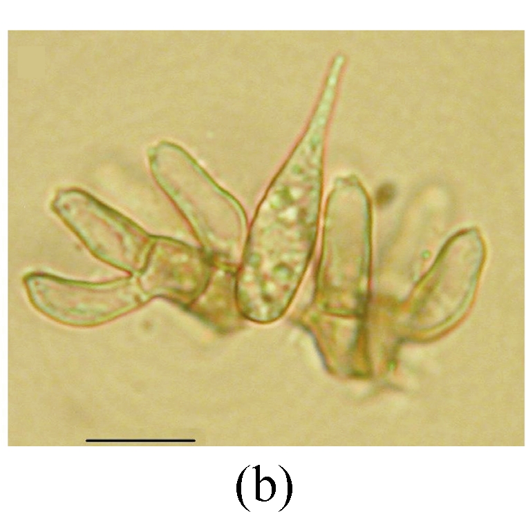
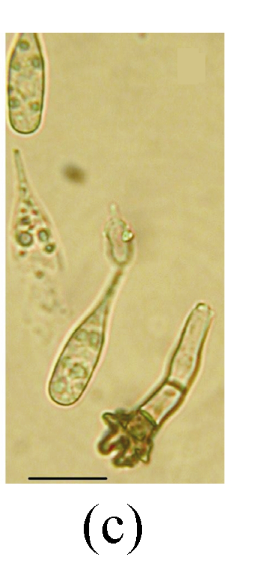
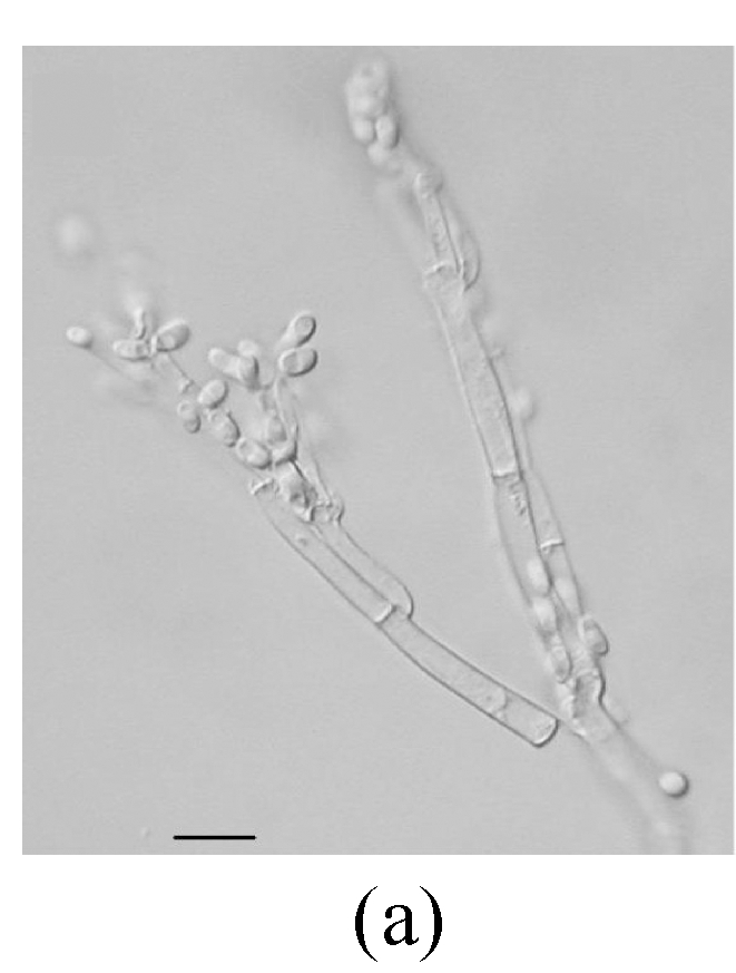
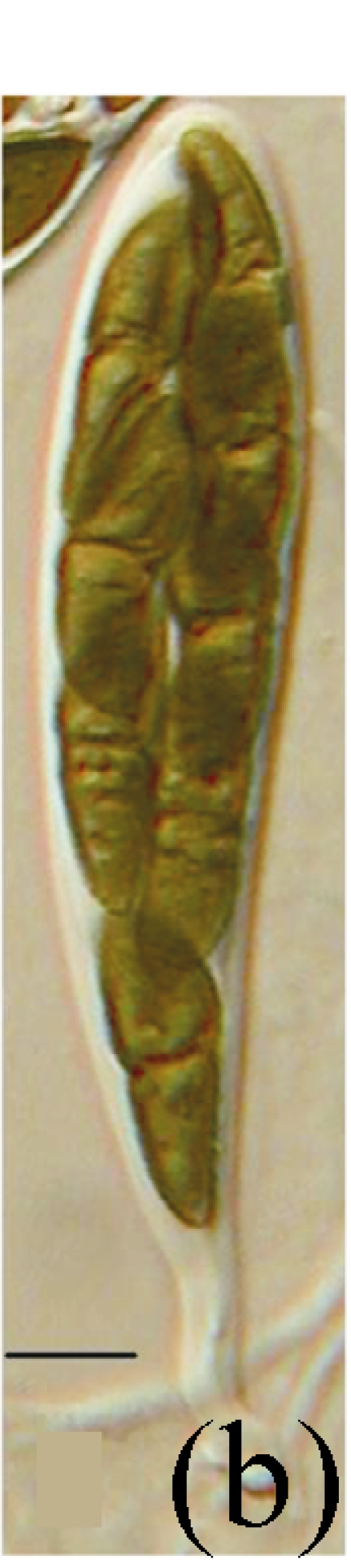
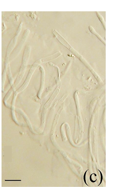
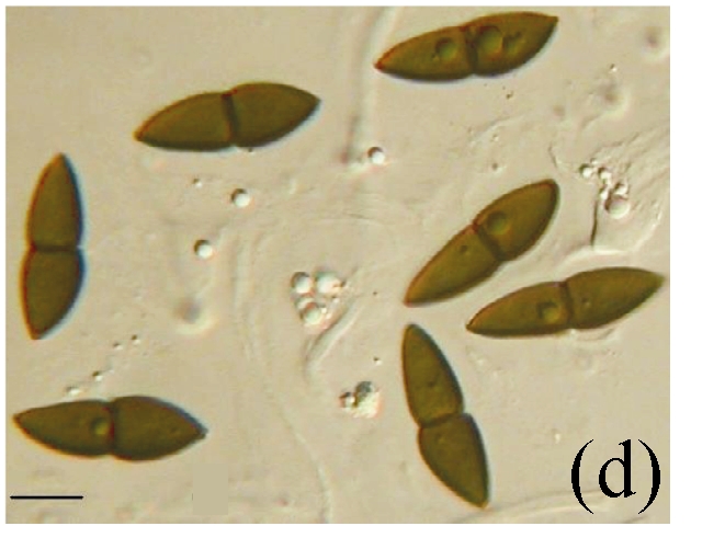
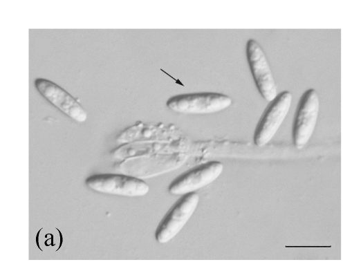
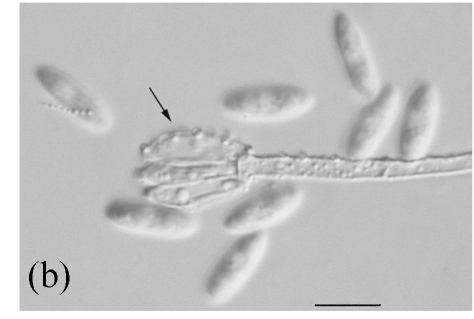
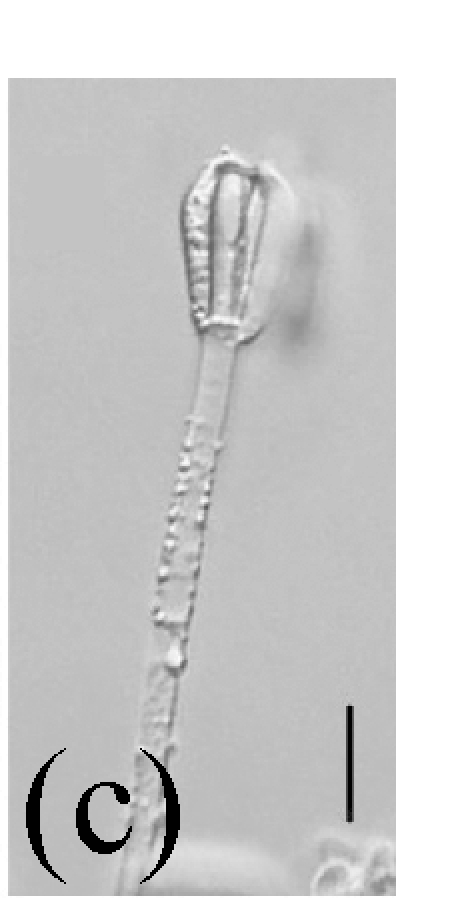
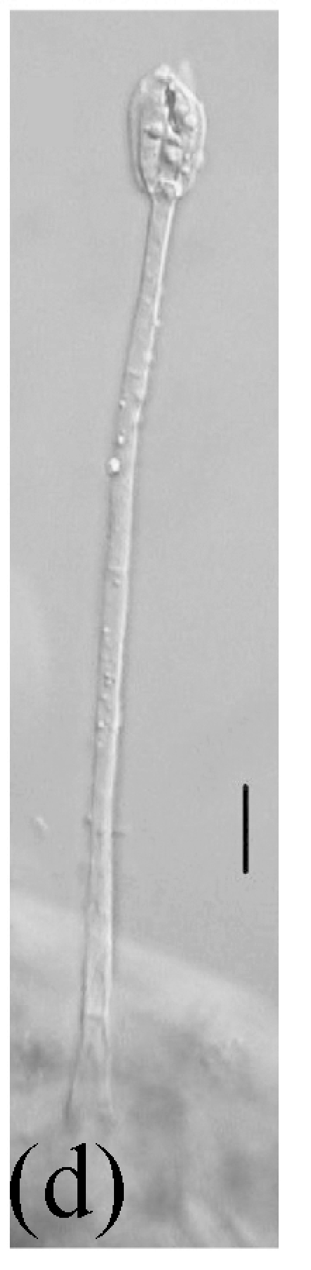
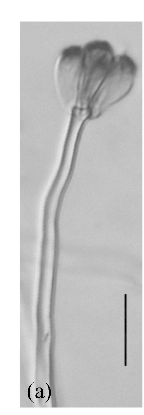
Fallen leaves of Ficus altissima, F. virens, F. benjamina, F. fistulosa and F. semicordata, were collected in Chiang Mai Province in northern Thailand and examined for fungi. Eighty taxa were identified, comprising 56 anamorphic taxa, 23 ascomycetes and 1 basidiomycete. Common fungal species occurring on five host species with high frequency of occurrence were Beltraniella nilgirica, Lasiodiplodia theobromae, Ophioceras leptosporum, Periconia byssoides and Septonema harknessi. Colletotrichum and Stachybotrys were also common genera. The leaves of different Ficus species supported diverse fungal taxa, and the fungal assemblages on the different hosts showed varying overlap. The fungal diversity of saprobes at the host species level is discussed.


Figures

Fig. 1
Fig. 1
Ellisiopsis gallesiae on fallen leaves of Ficus altissima. (a) Morphology of conidia; (b) and (c) Morphology of conidiophores. Bar=10 µm
Fig. 1
Fig. 1
Ellisiopsis gallesiae on fallen leaves of Ficus altissima. (a) Morphology of conidia; (b) and (c) Morphology of conidiophores. Bar=10 µm
Fig. 1
Fig. 1
Ellisiopsis gallesiae on fallen leaves of Ficus altissima. (a) Morphology of conidia; (b) and (c) Morphology of conidiophores. Bar=10 µm
Fig. 2
Fig. 2
Morphology of Sagenomella striatispora on fallen leaves of Ficus fistulosa. (a) Characters of conidiophore; (b) Characters of conidia and conidiophore; (c) and (d) developing conidia from conidiophores. Arrow indicates the young conidium. Bar=10 µm
Fig. 2
Fig. 2
Morphology of Sagenomella striatispora on fallen leaves of Ficus fistulosa. (a) Characters of conidiophore; (b) Characters of conidia and conidiophore; (c) and (d) developing conidia from conidiophores. Arrow indicates the young conidium. Bar=10 µm
Fig. 2
Fig. 2
Morphology of Sagenomella striatispora on fallen leaves of Ficus fistulosa. (a) Characters of conidiophore; (b) Characters of conidia and conidiophore; (c) and (d) developing conidia from conidiophores. Arrow indicates the young conidium. Bar=10 µm
Fig. 2
Fig. 2
Morphology of Sagenomella striatispora on fallen leaves of Ficus fistulosa. (a) Characters of conidiophore; (b) Characters of conidia and conidiophore; (c) and (d) developing conidia from conidiophores. Arrow indicates the young conidium. Bar=10 µm
Fig. 3
Fig. 3
Morphology of Kirschsteiniothelia spp. on fallen leaves of Ficus semicordata. (a) Characters of asci. Arrows indicate the outer layer of the ascus wall; (b) Characters of ascus; (c) Characters of pseudoparaphyses; (d) Characters of ascospores. (a) Bar=20 µm; (b)~(d) Bar=10 µm
Fig. 3
Fig. 3
Morphology of Kirschsteiniothelia spp. on fallen leaves of Ficus semicordata. (a) Characters of asci. Arrows indicate the outer layer of the ascus wall; (b) Characters of ascus; (c) Characters of pseudoparaphyses; (d) Characters of ascospores. (a) Bar=20 µm; (b)~(d) Bar=10 µm
Fig. 3
Fig. 3
Morphology of Kirschsteiniothelia spp. on fallen leaves of Ficus semicordata. (a) Characters of asci. Arrows indicate the outer layer of the ascus wall; (b) Characters of ascus; (c) Characters of pseudoparaphyses; (d) Characters of ascospores. (a) Bar=20 µm; (b)~(d) Bar=10 µm
Fig. 3
Fig. 3
Morphology of Kirschsteiniothelia spp. on fallen leaves of Ficus semicordata. (a) Characters of asci. Arrows indicate the outer layer of the ascus wall; (b) Characters of ascus; (c) Characters of pseudoparaphyses; (d) Characters of ascospores. (a) Bar=20 µm; (b)~(d) Bar=10 µm
Fig. 4
Fig. 4
Morphology of Stachybotrys bisbyi on fallen leaves of Ficus benjamina. (a) Characters conidia. Arrow indicates the smooth wall of the conidium; (b) Characters of conidiophore. Arrow indicates the rough wall of conidiophore cell; (c) and (d) Characters of conidiophores. Bar=10 µm
Fig. 4
Fig. 4
Morphology of Stachybotrys bisbyi on fallen leaves of Ficus benjamina. (a) Characters conidia. Arrow indicates the smooth wall of the conidium; (b) Characters of conidiophore. Arrow indicates the rough wall of conidiophore cell; (c) and (d) Characters of conidiophores. Bar=10 µm
Fig. 4
Fig. 4
Morphology of Stachybotrys bisbyi on fallen leaves of Ficus benjamina. (a) Characters conidia. Arrow indicates the smooth wall of the conidium; (b) Characters of conidiophore. Arrow indicates the rough wall of conidiophore cell; (c) and (d) Characters of conidiophores. Bar=10 µm
Fig. 4
Fig. 4
Morphology of Stachybotrys bisbyi on fallen leaves of Ficus benjamina. (a) Characters conidia. Arrow indicates the smooth wall of the conidium; (b) Characters of conidiophore. Arrow indicates the rough wall of conidiophore cell; (c) and (d) Characters of conidiophores. Bar=10 µm
Fig. 5
Fig. 5
Morphology of Stachybotrys renispora on fallen leaves of Ficus virens. (a) Characters of conidiophore; (b) and (c) Characters of conidia and conidiophores. Arrow indicates the succession of conidium; (d) and (e) Characters of conidia. Bar=10 µm
Fig. 5
Fig. 5
Morphology of Stachybotrys renispora on fallen leaves of Ficus virens. (a) Characters of conidiophore; (b) and (c) Characters of conidia and conidiophores. Arrow indicates the succession of conidium; (d) and (e) Characters of conidia. Bar=10 µm
Fig. 5
Fig. 5
Morphology of Stachybotrys renispora on fallen leaves of Ficus virens. (a) Characters of conidiophore; (b) and (c) Characters of conidia and conidiophores. Arrow indicates the succession of conidium; (d) and (e) Characters of conidia. Bar=10 µm
Fig. 5
Fig. 5
Morphology of Stachybotrys renispora on fallen leaves of Ficus virens. (a) Characters of conidiophore; (b) and (c) Characters of conidia and conidiophores. Arrow indicates the succession of conidium; (d) and (e) Characters of conidia. Bar=10 µm
Fig. 5
Fig. 5
Morphology of Stachybotrys renispora on fallen leaves of Ficus virens. (a) Characters of conidiophore; (b) and (c) Characters of conidia and conidiophores. Arrow indicates the succession of conidium; (d) and (e) Characters of conidia. Bar=10 µm

References

    1. Arnold AE, Zuleyka MZ, Gilbert GS. Fungal endophytes in dicotyledonous neotropical trees: patterns of abundance and diversity. Mycol Res. 2001;105(12):1502–1507. doi: 10.1017/S0953756201004956. - DOI
    1. Begon M, Harper JL, Townsend CR. Ecology: Individuals, Population and Communities. 3rd Ed. Boston: Blackwell Science; 1993.
    1. Bucher VVC, Pointing SB, Hyde KD, Reddy CA. Production of wood decay enzymes, loss of mass, and lignin solubilization in wood by diverse tropical freshwater fungi. Microbial Ecology. 2004;48(3):331–337. doi: 10.1007/s00248-003-0132-x. - DOI - PubMed
    1. Duarte S, Pascoal C, Cássio F, Bärlocher F. Aquatic hyphomycete diversity and identity affect leaf litter decomposition in microcosms. Oecologia. 2006;147(4):658–666. doi: 10.1007/s00442-005-0300-4. - DOI - PubMed
    1. Dulymamode R, Cannon PF, Peerally A. Fungi on endemic plants of Mauritius. Mycol Res. 2001;105(12):1472–1479. doi: 10.1017/S0953756201004701. - DOI

Publication types

MeSH terms