Review
. 2008:24:597-620.
doi: 10.1146/annurev.cellbio.24.110707.175250.

Dscam-mediated cell recognition regulates neural circuit formation

Affiliations
Review

Dscam-mediated cell recognition regulates neural circuit formation

Daisuke Hattori et al. Annu Rev Cell Dev Biol. 2008.

Abstract

The Dscam family of immunoglobulin cell surface proteins mediates recognition events between neurons that play an essential role in the establishment of neural circuits. The Drosophila Dscam1 locus encodes tens of thousands of cell surface proteins via alternative splicing. These isoforms exhibit exquisite isoform-specific binding in vitro that mediates homophilic repulsion in vivo. These properties provide the molecular basis for self-avoidance, an essential developmental mechanism that allows axonal and dendritic processes to uniformly cover their synaptic fields. In a mechanistically similar fashion, homophilic repulsion mediated by Drosophila Dscam2 prevents processes from the same class of cells from occupying overlapping synaptic fields through a process called tiling. Genetic studies in the mouse visual system support the view that vertebrate DSCAM also promotes both self-avoidance and tiling. By contrast, DSCAM and DSCAM-L promote layer-specific targeting in the chick visual system, presumably through promoting homophilic adhesion. The fly and mouse studies underscore the importance of homophilic repulsion in regulating neural circuit assembly, whereas the chick studies suggest that DSCAM proteins may mediate a variety of different recognition events during wiring in a context-dependent fashion.


Figures
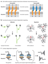
Figure 1
Figure 1
(a) Cell surface proteins mediate interactions between neurons. Interactions between neuronal processes are mediated by direct contact between cell surface molecules. Two interacting neurites (i.e., neurite A and neurite B) are shown. Binding between the extracellular regions of cell surface proteins is translated to changes in the cytoplasmic domain and elicits intracellular signaling events (black arrows), which lead to directed changes in neurite motility through cytoskeletal rearrangements. Binding between identical proteins is referred to as homophilic, whereas binding between different proteins is referred to as heterophilic. (b) Cell surface protein interactions have different signaling outputs. Contact-dependent interactions between cell surface proteins elicit signal transduction cascades within the cytoplasm that lead to two different outputs: adhesion (also called attraction) and repulsion. (Left) The neurites of two different neurons expressing adhesive molecules (green) encounter one another. Adhesion can lead to various responses, two of which, synapse formation and fasciculation, are depicted. (Right) Neurites of the same neuron (left) or two different neurons (right) expressing repulsive molecules (red) encounter one another. Repulsion causes the neurites to grow away from one another and mediates patterning events such as self-avoidance and tiling. (c) Discrete steps underlying contact-dependent repulsion. First, recognition cell surface molecules expressed on opposing neurites bind to each other. Next, intracellular signaling promotes downregulation of receptor binding and activation of cytoskeletal rearrangements that promote repulsion. Two mechanisms have been described to mediate dissociation: proteolytic cleavage of the interacting molecules (shown) and endocytosis (not shown).
Figure 2
Figure 2
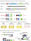
(a) Drosophila Dscam1 encodes a vast repertoire of cell surface recognition proteins. The Drosophila Dscam1 gene encodes a large family of single-pass transmembrane proteins of the immunoglobulin (Ig) superfamily. Dscam1 contains four blocks of alternative exons that encode 12 different variants for the N-terminal half of Ig2 (red), 48 different variants for the N-terminal half of Ig3 (blue), 33 different variants for Ig7 (green), and two different variants for the transmembrane domain (TM) (yellow). Splicing leads to the incorporation of one alternative exon from each block, and as such, Dscam1 encodes 19,008 (i.e., 12 × 48 × 33) different ectodomains linked to one of two different transmembrane domains. (b) Dscam1 proteins exhibit isoform-specific homophilic binding. Each isoform binds to itself but rarely, if at all, to other isoforms. The three variable Ig domains mediate homophilic binding specificity. Variants of each domain engage in self-binding but do not bind to other variants (with rare exceptions). Therefore, homophilic binding occurs between identical isoforms that match at all three variable Ig domains. Isoform pairs that contain only two matches and differ at the third variable domain do not bind to one another. The quaternary structure of the first four Ig domains constrains the Ig2 and Ig3 domains, literally tethering them to one another. As such, if opposing Ig2 domains do not match, Ig3 self-binding is sterically inhibited, even if the Ig3 domains match, and vice versa. Additional intramolecular interactions between constant domains in the linker region between Ig3 and Ig7 are formed when the three variable domains match; such interactions play a crucial role in stabilizing the homophilic dimer (see panel d). An asterisk indicates that Ig2 difference is shown. (c) Electrostatic and shape complementarity underlies self-binding of each variant. Complementarity is illustrated by the Ig2 interface as an example. The Ig2 self-binding interface occurs between identical segments in opposing Ig2 domains. These segments are oriented in an antiparallel fashion. The interface comprises a symmetry center (SC) residue and flanking left and right networks. The interface segments of two different Ig2 variants (i.e., A and B) are shown in red and pink, respectively. Each self-binding interface exhibits electrostatic and shape complementarity at the SC and the left and right networks (complementarity is illustrated by the yellow boxes). The heterophilic interface formed between these two Ig2 variants does not exhibit complementarity at the SC, the left network (note the three negatively charged residues), or the right network (note the three positively charged residues), and thus these different Ig2 variants do not bind to one another. (d) A conformational change occurs upon homophilic binding. (Right) The Dscam11–8 crystal structure reveals a dimer of two S-shaped monomers with direct contacts between opposing Ig2, Ig3, and Ig7 variable domains. Electron micrographs of Dscam11–8 demonstrated that, whereas the first four Ig domains form a compact horseshoe structure, the remainder of the domains are highly flexible. (Left) These differences in structure suggest that the bottom half of the S shape observed in the crystal structure forms upon homophilic binding as opposing Ig2, Ig3, and Ig7 domains interact. Stabilizing intramolecular contacts are formed between regions within constant domains Ig5 and Ig6. This large conformational change that occurs within the Dscam ectodomain upon homophilic binding may provide a molecular mechanism for transducing the signal of homophilic binding to the cytoplasmic domain, where subsequent signaling events occur.
Figure 3
Figure 3
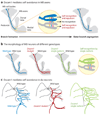
Dscam1 regulates self-avoidance. (a) Dscam1 mediates self-avoidance in mushroom body (MB) neurons. The MB is a central brain structure comprising thousands of neurons. Each MB extends a single axon within a nerve bundle called the peduncle. At the base of the peduncle MB axons bifurcate and extend one branch medially and the other dorsally. Each MB neuron expresses a unique combination of isoforms. As a consequence, sister branches recognize each other through Dscam1 matching. This signals repulsion and subsequent segregation of axons to separate pathways. (b) Axon branching patterns of MB neurons of different genotypes. (Left) Wild-type MB axon branches segregate with high fidelity. (Middle) The branches of a single Dscam mutant neuron in a wild-type background frequently do not segregate appropriately. (Right) Expression of a single arbitrarily chosen isoform promotes branch segregation in a single Dscam1 null mutant cell. These and other experiments demonstrated that, although it is unimportant which isoform a single MB neuron expresses for appropriate branch segregation, it is crucial that each MB neuron express isoforms different from its neighbors. (c) Dscam1 mediates self-avoidance in dendritic arborization (da) neurons. Different classes of da neurons elaborate overlapping dendritic fields in the body wall of Drosophila larva. (Left) Two wild-type neurons are shown. (Middle) The dendrites of a Dscam1 null mutant cell form fascicles, supporting the notion that Dscam1 binding promotes repulsion. (Right) Overexpression of the same Dscam1 isoform in both neurons leads to nonoverlapping receptive fields, consistent with homophilic binding inducing repulsion.
Figure 4
Figure 4
Dscam proteins participate in several aspects of neuronal wiring. (a) Drosophila Dscam2 is a tiling receptor. (Left panel) Schematic of Dscam2 phenotypes in lamina 1 (L1) axons in the fly medulla region of the visual system. L1 axons (pink and green) elaborate processes in two distinct layers. Interactions between these processes mediated by Dscam2 restrict the formation of connections to a single column, as indicated in the far left and right columns. Mutant (pink) L1 axons are not restricted to a single column. Similarly, the processes of wild-type (WT) (green) axons extend axons into columns with a mutant L1. Columns are delineated with dashed lines. (Right panel) Schematic of L1 column development. Dscam2 homophilic binding (blue bars) occurs between wild-type L1 neurites during pupal development. This induces a repulsive signal that results in the retraction of neurites back to their column of origin and the formation of columnar boundaries. Mutant neurites (pink) cannot interact with wild-type L1 neurites (green) because the former lack Dscam2. Without Dscam2 homophilic binding, neither mutant nor wild-type L1 neurites are restricted to their column of origin, and both can form connections in neighboring columns. (b) Mouse DSCAM mediates both self-avoidance and tiling. (Left panel) DSCAM-positive amacrine cells exhibit both self-avoidance and tiling properties. (Right panel) In the absence of DSCAM, both self-avoidance and tiling are lost. Branches from individual amacrine cells fasciculate with one another and with other cells of the same type. (c) Chick DSCAM contributes to layer-specific targeting. (Left panel) Ganglion cells (bottom) and amacrine cells (top) in the chick retina, which express the same DSCAM protein or a related Ig superfamily protein in the Sidekick (Sdk) family, target to the same layer. (Right panel) When either DSCAM or Sdk2 is knocked down in ganglion cells that normally express these proteins, dendritic targeting is less precise. Similarly, when cells that do not normally express DSCAM are engineered to misexpress it, they target to the DSCAM-positive S5 layer.

References

    1. Agarwala KL, Ganesh S, Tsutsumi Y, Suzuki T, Amano K, Yamakawa K. Cloning and functional characterization of DSCAML1, a novel DSCAM-like cell adhesion molecule that mediates homophilic intercellular adhesion. Biochem. Biophys. Res. Commun. 2001;285:760–772. - PubMed
    1. Baker MW, Macagno ER. The role of a LAR-like receptor tyrosine phosphatase in growth cone collapse and mutual-avoidance by sibling processes. J. Neurobiol. 2000;4:194–203. - PubMed
    1. Chen BE, Kondo M, Garnier A, Watson FL, Puettmann-Holgado R, et al. The molecular diversity of Dscam is functionally required for neuronal wiring specificity in Drosophila. Cell. 2006;125:607–620. Demonstrated branching defects in Dscam1 mutants lacking specific exon 4 variants, suggesting a role for specific isoforms in patterning connections.

    1. Cheng HJ, Flanagan JG. Identification and cloning of ELF-1, a developmentally expressed ligand for the Mek4 and Sek receptor tyrosine kinases. Cell. 1994;79:157–168. - PubMed
    1. Drescher U, Kremoser C, Handwerker C, Loschinger J, Noda M, Bonhoeffer F. In vitro guidance of retinal ganglion cell axons by RAGS, a 25 kDa tectal protein related to ligands for Eph receptor tyrosine kinases. Cell. 1995;82:359–370. - PubMed

Publication types

MeSH terms

LinkOut - more resources