Review
. 2008 Oct:225:212-25.
doi: 10.1111/j.1600-065X.2008.00676.x.

The structural immunology of antibody protection against West Nile virus

Affiliations
Review

The structural immunology of antibody protection against West Nile virus

Michael S Diamond et al. Immunol Rev. 2008 Oct.

Abstract

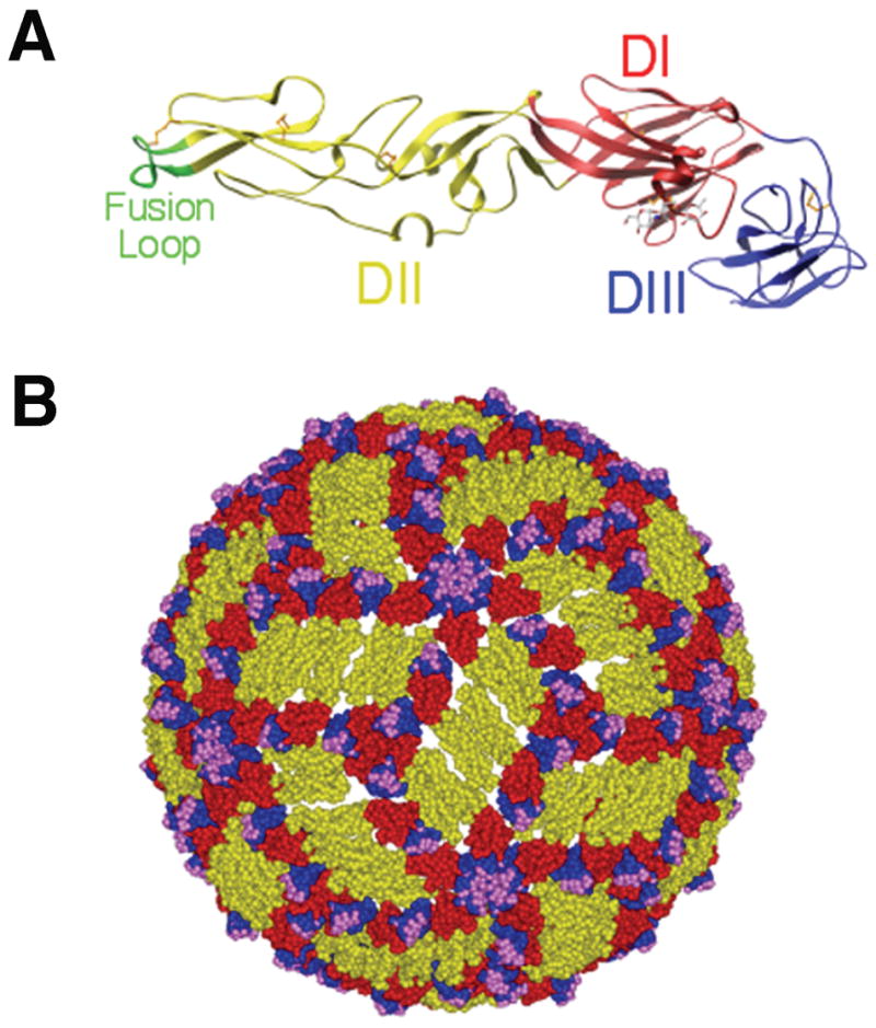
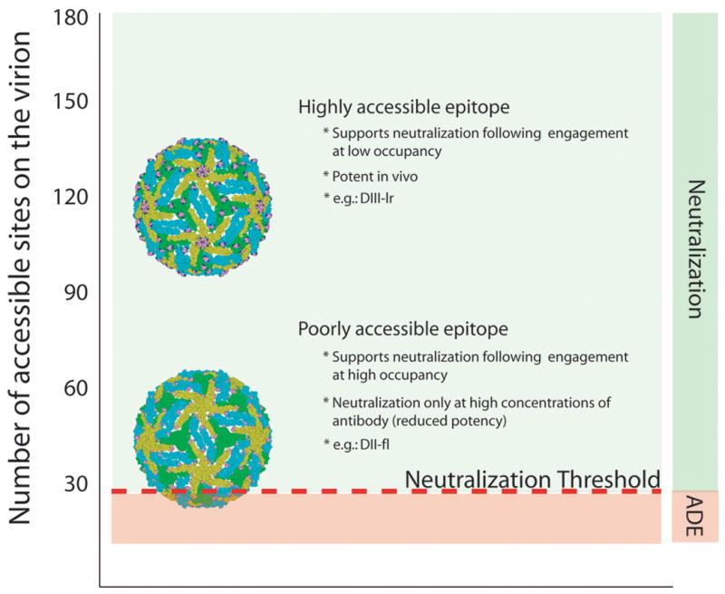
Recent investigations of the interaction between the West Nile virus (WNV) envelope protein (E) and monoclonal antibodies (mAbs) have elucidated fundamental insights into the molecular mechanisms of neutralization. Structural studies have defined an epitope on the lateral ridge of domain III (DIII-lr) of the WNV E protein that is recognized by antibodies with the strongest neutralizing activity in vitro and in vivo. Antibodies that bind this epitope are highly potent because they efficiently block at a post-entry step of viral infection with relatively low virion occupancy requirements. In this review, we discuss the structural, molecular, and immunologic basis for antibody-mediated protection against WNV, and its implications for novel therapeutic or vaccine strategies.


Figures

Fig. 1
Fig. 1. E protein and mature WNV virion structure
(A). Ribbon diagram of the WNV E protein crystal structure. Domains are labeled and the fusion loop is shown in green. (B). Pseudoatomic model of the mature WNV virion based on cryo-electron microscopy studies. E protein domains I, II, and III are indicated in red, yellow and blue, respectively. Residues critical for binding of E16, a DIII-lr mAb, are shown in magenta. Adapted from (21, 68, 117).
Fig. 2
Fig. 2. Strongly neutralizing DIII-lr specific antibodies define a single consensus epitope that is divergent in other flaviviruses
(A). Sequence of the four segments of WNV DIII contacted by E16 aligned with the analogous residues of other flaviviruses. The DIII contact residues as determined by epitope mapping are highlighted in magenta, while those identified only structurally are highlighted in blue. Deletions are indicated with a hash symbol. (B). Structure of the WNV dominant neutralizing epitope as defined by the E16-DIII complex. Adapted from (68).
Fig. 3
Fig. 3. . Epitopes of several different anti-WNV neutralizing mAbs as determined by yeast surface display screening of E protein mutants
The backbone colors red, yellow, blue, and green indicate domains I, II, III, and the fusion loop respectively. Mutations that resulted in ≥ 50% reduction of mAb binding were mapped (shown in magenta and circled) onto the WNV E protein crystal structure. Epitopes are labeled using the nomenclature defined in Oliphant et al. (21).
Fig. 4
Fig. 4. Relationship between epitope accessibility and the occupancy requirements for neutralization
The accessibility of epitopes recognized by two different mAbs on the mature WNV virion is illustrated using molecular modeling: residues that comprise each determinant are illustrated as solid spheres. E proteins are colored according to their proximity to the 2-, 3-, or 5-fold symmetry axes (blue, green, and yellow, respectively). The number of accessible binding sites for each antibody is indicated on the left, whereas the ‘threshold’ for neutralization is indicated as a red line (modeled in this instance as 30 mAbs based on studies using the mAb E16). To exceed the threshold requirements for neutralization, only a fraction of highly accessible determinants must be simultaneously occupied by antibody (a low occupancy requirement). For cryptic epitopes (fewer accessible sites), a significantly greater percentage of accessible epitopes must be bound to achieve the same number of antibodies docked on the average virion (a high occupancy requirement). Adapted from (112, 139).
Fig. 5
Fig. 5. C1q modulates mAb enhancement of WNV infection
(A). Serial dilutions of E16 (mouse IgG2b) were mixed with PBS, 5% fresh or heat-inactivated mouse serum, incubated with WNV RVP, and added to FcγRIIa+ K562 cells. Forty-eight hours later, cells were analyzed by flow cytometry for GFP expression. The data are expressed as the fold enhancement of infection compared to no antibody. (B). Experiments were performed as in panel (A) except that fresh C1q−/− or C3−/− serum or purified C1q was mixed with the mouse E16 mAb. (C, D). Experiments were performed as in panel (B) except the epitope matched DIII-specific (C) E24 (mouse IgG2a) or (D) E34 (mouse IgG1) mAbs were used. Adapted from (126).
Fig. 6
Fig. 6. Mechanism of protection against WNV by anti-NS1 antibodies
(A). Schematic diagram of NS1 fragments. (Top) A secondary structure prediction model, putative disulfide bonding patterns, and N-linked glycosylated sites are depicted. The secondary structure model is a consensus prediction based on NS1 from several different flaviviruses. (Bottom) Diagram of expression constructs used for yeast surface display and bacterial expression of NS1 fragments. NS1(125–352)B and NS1(158–352)Y indicate E. coli and yeast expressed FR-II-III, respectively. Adapted from (27). (B). Flow cytometry histograms showing immunoreactivity of NS1 on the surface of yeast, within permeabilized Raji-WNV cells, and on the surface of Raji-WNV-cells with individual anti-NS1 mAbs. Representative histograms are shown for four NS1 mAbs (8NS1, 15NS1, 10NS1, and 17NS1). The E1 mAb against WNV E protein was used as a negative control. Green and red arrows indicate the absence or presence of binding of mAbs to cell surface-associated NS1, respectively. The fragment localization of NS1 antibodies is indicated to the right. (C). Model of antibody and NS1-dependent clearance of WNV-infected cells by Fc-γ receptor I and IV-expressing phagocytes. WNV-infected cells express cell-associated forms of NS1 on their surface, which can be bound by subsets (fragment II and III-specific) antibodies. NS1-specific antibodies of a given IgG subclass (e.g. mouse IgG2a) are recognized by activating Fc-γ receptors (mouse Fc-γR I or IV) resulting in phagocytosis and clearance of infected cells.

References

    1. Hayes EB, Komar N, Nasci RS, Montgomery SP, O’Leary DR, Campbell GL. Epidemiology and transmission dynamics of West Nile virus disease. Emerg Infect Dis. 2005;11:1167–1173. - PMC - PubMed
    1. Sejvar JJ, et al. Neurologic manifestations and outcome of West Nile virus infection. JAMA. 2003;290:511–515. - PubMed
    1. Lanciotti RS, et al. Complete genome sequences and phylogenetic analysis of West Nile virus strains isolated from the United States, Europe, and the Middle East. Virology. 2002;298:96–105. - PubMed
    1. Beasley DW, Li L, Suderman MT, Barrett AD. Mouse neuroinvasive phenotype of West Nile virus strains varies depending upon virus genotype. Virology. 2002;296:17–23. - PubMed
    1. Beasley DW, et al. Genome sequence and attenuating mutations in West Nile virus isolate from Mexico. Emerg Infect Dis. 2004;10:2221–2224. - PMC - PubMed

Publication types

MeSH terms