Review
. 2009 Jan;30(1):154-63.
doi: 10.1016/j.peptides.2008.08.022. Epub 2008 Sep 16.

Kisspeptin and seasonality in sheep

Affiliations
Review

Kisspeptin and seasonality in sheep

Iain J Clarke et al. Peptides. 2009 Jan.

Abstract

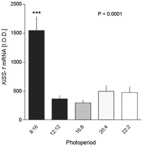
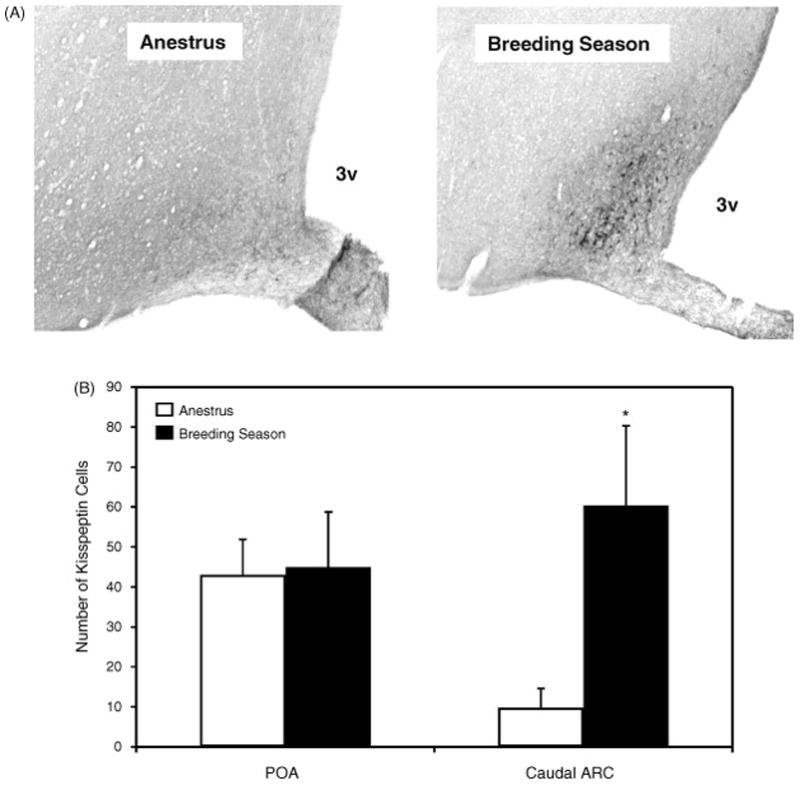
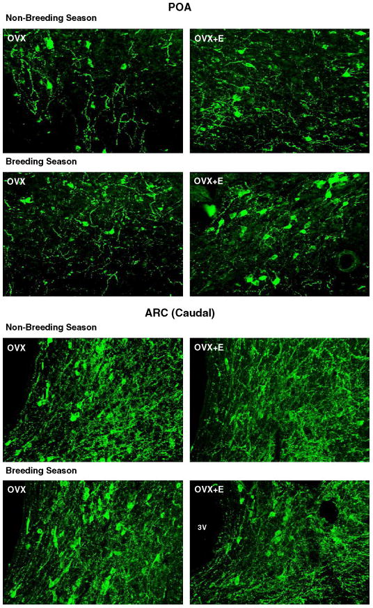
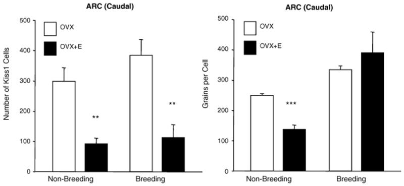
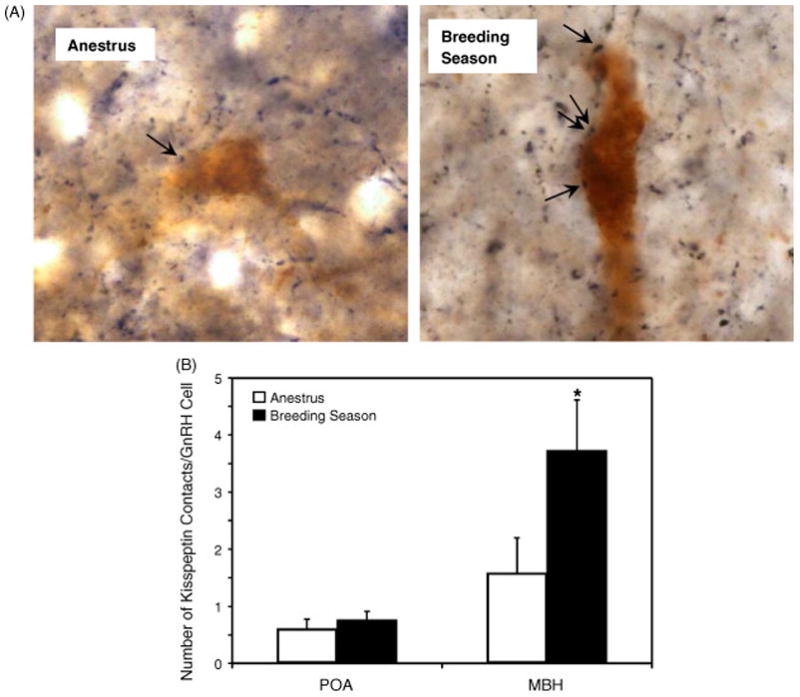
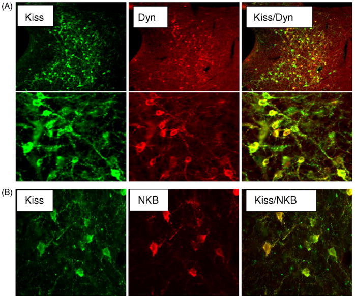
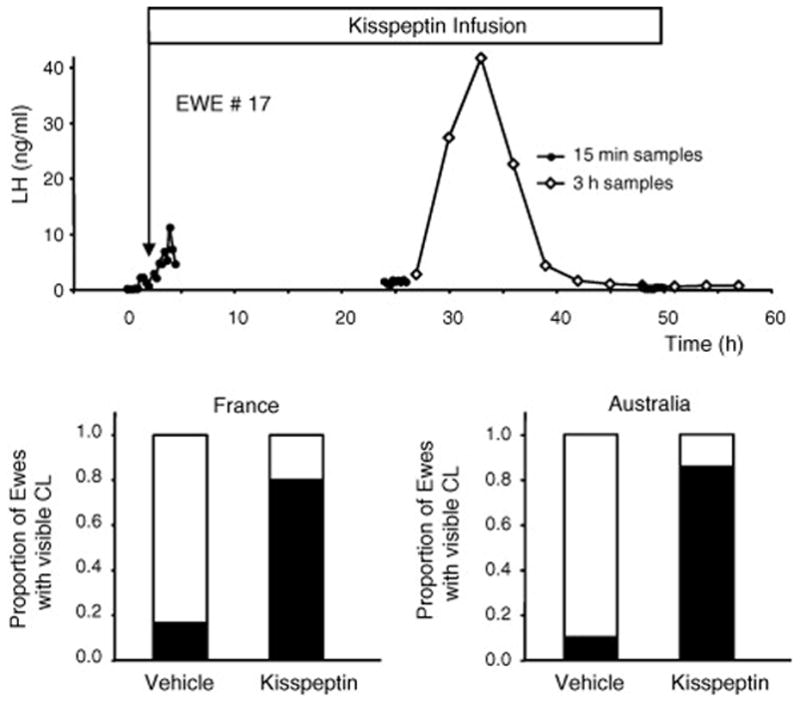
Sheep are seasonal breeders, experiencing a period of reproductive quiescence during spring and early summer. During the non-breeding period, kisspeptin expression in the arcuate nucleus is markedly reduced. This strongly suggests that the mechanisms that control seasonal changes in reproductive function involve kisspeptin neurons. Kisspeptin cells appear to regulate GnRH neurons and transmit sex-steroid feedback to the reproductive axis. Since the non-breeding season is characterized by increased negative feedback of estrogen on GnRH secretion, the kisspeptin neurons seem to be fundamentally involved in the determination of breeding state. The reduction in kisspeptin neuronal function during the non-breeding season can be corrected by infusion of kisspeptin, which causes ovulation in seasonally acyclic females.


Figures

Figure 1
Figure 1
Kiss1 expression in the arcuate nucleus (ARC) of the Soay ewe brain in different photoperiods. The animals were held in artificial photoperiods of 8h light (L), 16h dark (D), 12L:12D, 16L:8D, 20L:24D or 22L:2D. Integrated optical density measurements of Kiss1 expression at the level of the ARC are shown and data are mean ± SEM (n ≤ 8). ***P<0.0001 compared to other groups. Adapted from Reference [39] with permission.
Figure 2
Figure 2
Photomicrographs of immunohistochemistry showing kisspeptin-ir cells in the ARC (A) of the ewe brain. 3V, third ventricle. The number of kisspeptin-ir cells in the POA and the caudal ARC.of the ovariectomised ewes bearing estrogen implants during the non-breeding and breeding seasons ewes are shown in panel B. Values are means (± SEM). *, P < 0.05 non-breeding v breeding season. Adapted from Reference [36], with permission.
Figure 3
Figure 3
Photomicrographs showing kisspeptin-ir cells in sections of the POA and caudal ARC of ovariectomised (OVX) with or without estrogen (E) treatment during the non-breeding and breeding seasons. 3V, third ventricle. Adapted from Reference [36], with permission.
Figure 4
Figure 4
Expression of KiSS1 expression in the caudal ARC of ovariectomised (OVX) with or without estrogen (E) treatment during the non-breeding and breeding seasons. Left panels show the number of Kiss1 mRNA-expressing cells and right panels show the number of silver grains/Kiss1 cell (expression per cell). Values are means (± SEM). **, P < 0.01 ***, P < 0.001 non-breeding v breeding season. Adapted from Reference [36], with permission.
Figure 5
Figure 5
Photomicrographs of GnRH and kisspeptin double-label immunohistochemistry in the MBH of non-breeding and breeding season ewes showing kisspeptin-ir terminal (black) contacts with GnRH-ir cell bodies (brown). Arrows indicate points of contact. Panel B shows mean (± SEM) number of kisspeptin contacts per GnRH cell. *P < 0.05 non-breeding v breeding season. Adapted from Reference [36], with permission.
Figure 6
Figure 6
Confocal microscopic images of sections through the caudal ARC, showing kisspeptin cells that co-stain for either dynorphin (Dyn) (A) or neurokinin B (NKB) (B). The left panels show kisspeptin (green fluorescence) and the centre panels show either dynorphin (A) or NKB (B) (red fluorescence). Panels on right are computer-generated overlays of the left and middle panels. The upper panels in A are at low power and the lower panels are at a higher power to exemplify co-localisation of the two peptides. There is a high degree of co-localization of kisspeptin and dynorphin (yellow fluorescence). Adapted from Reference [16] with permission.
Figure 7
Figure 7
LH secretion and ensuing ovulation in ewes treated during the non-breeding season with an intravenous infusion of kisspeptin. The upper panel shows an example of plasma LH levels in a kisspeptin-infused ewe, indicating that the treatment led to increased secretion but the preovulatory surge did not occur until 25h later. This strongly suggests that the infusion initiated an artificial follicular phase and that the increased estrogen resulting from this activated a positive feedback response. The experiment was repeated in France and in Australia with similar results. Approximately 80% of the ewes treated with kisspeptin ovulated, as evidenced by resulting corpora lutea on the ovaries. Adapted from Reference [4] with permission.

References

    1. Adams VL, Goodman RL, Salm AK, Coolen LM, Karsch FJ, Lehman MN. Morphological plasticity in the neural circuitry responsible for seasonal breeding in the ewe. Endocrinology. 2006;147:4843–4851. - PubMed
    1. Bentley GE, Perfito N, Ukena K, Tsutsui K, Wingfield JC. Gonadotropin-inhibitory peptide in song sparrows (Melospiza melodia) in different reproductive conditions, and in house sparrows (Passer domesticus) relative to chicken-gonadotropin-releasing hormone. J Neuroendocrinol. 2003;15:794–802. - PubMed
    1. Caraty A, Fabre-Nys C, Delaleu B, Locatelli A, Bruneau G, Karsch FJ, Herbison A. Evidence that the mediobasal hypothalamus is the primary site of action of estradiol in inducing the preovulatory gonadotropin releasing hormone surge in the ewe. Endocrinology. 1998;139:1752–1760. - PubMed
    1. Caraty A, Smith JT, Lomet D, Ben Said S, Morrissey A, Cognie J, Doughton B, Baril G, Briant C, Clarke IJ. Kisspeptin synchronizes preovulatory surges in cyclical ewes and causes ovulation in seasonally acyclic ewes. Endocrinology. 2007;148:5258–5267. - PubMed
    1. Clarke IJ. Effector mechanisms of the hypothalamus that regulate the anterior pituitary gland. In: Unsicker K, editor. The Autonomic Nervous System. Harwood; London: 1996. pp. 45–88.