

Selective hyposmia in Parkinson disease: association with hippocampal dopamine activity
- PMID: 18838108
- PMCID: PMC2634293
- DOI: 10.1016/j.neulet.2008.09.070
Selective hyposmia in Parkinson disease: association with hippocampal dopamine activity
Abstract
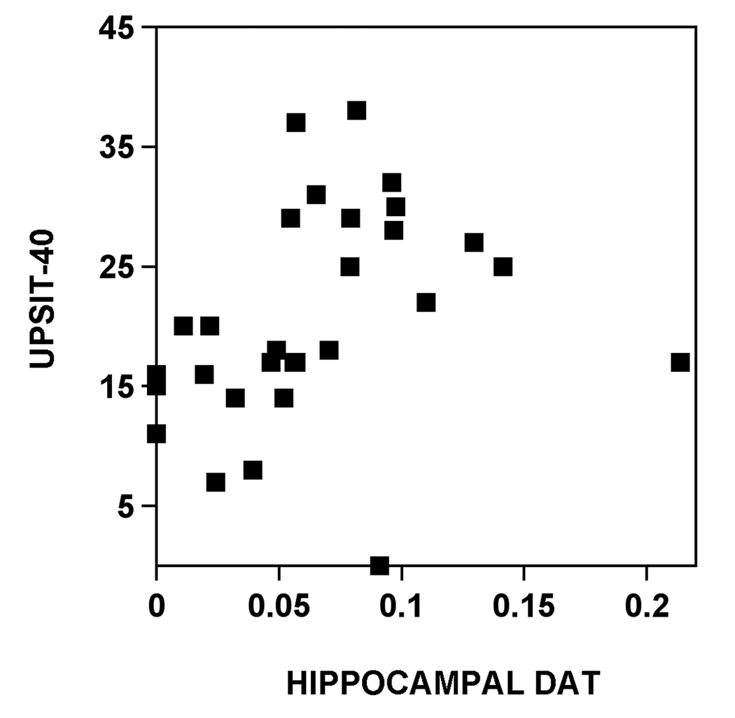
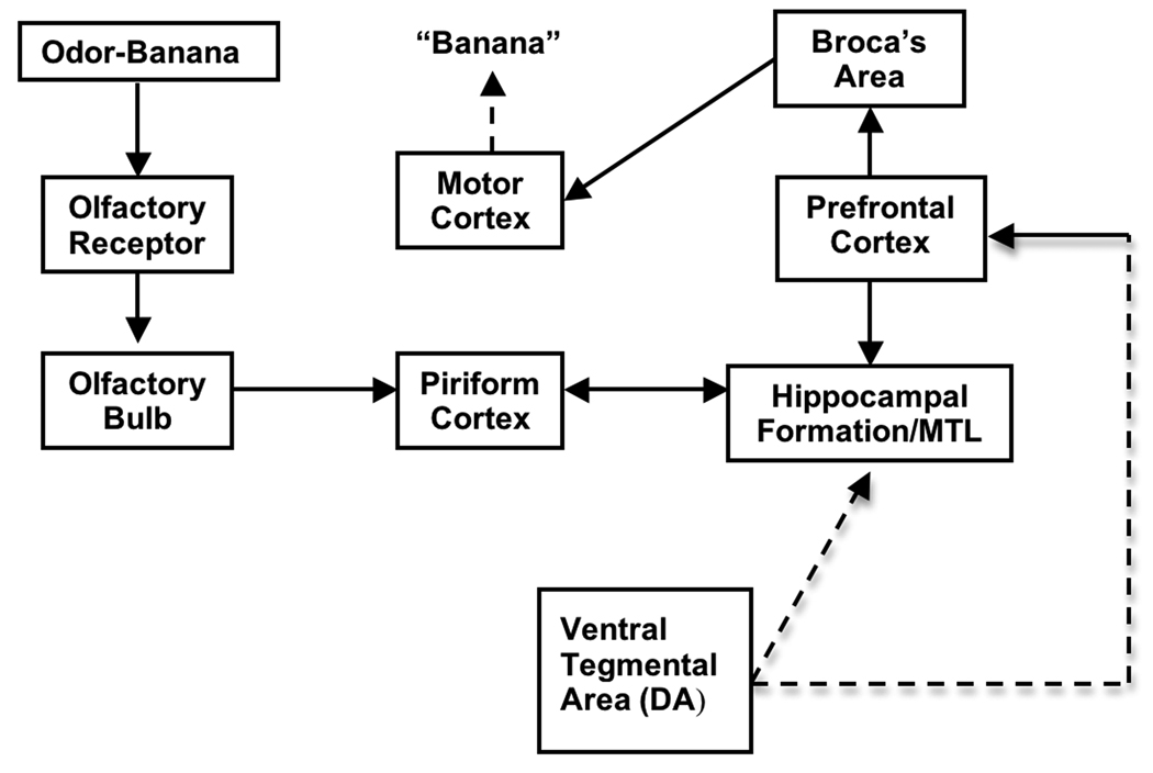
Olfactory dysfunction is common in patients with Parkinson disease (PD) and has been attributed to early pathological deposition of Lewy bodies and Lewy neurites in primary olfactory centers. However, olfactory deficits do not always worsen over time despite progression of disease raising the possibility of additional pathobiological mechanisms contributing to olfactory functions in PD, such as changes in olfactory neurotransmitter functions. Neurotransmitter changes, such as altered dopaminergic status, may also better explain the selective nature of odor identification deficits in PD. Proper odor identification depends on higher order structures, such as the hippocampus, for olfactory cognitive or memory processing. Using the University of Pennsylvania Smell Identification Test (UPSIT), we previously identified three odors (banana, licorice, dill pickle, labeled as UPSIT-3) that PD subjects most frequently failed to recognize compared to age- and gender-matched controls. We also identified six odors that were equally successfully identified by controls and PD subjects (NPD-Olf6). A ratio of UPSIT-3 divided by NPD-Olf6 scores provides another descriptor of selective hyposmia in PD ("olfactory ratio"). In this study we investigated the pathophysiology of hyposmia in PD using dopamine transporter (DAT) PET. Twenty-nine PD patients (Hoehn and Yahr stages I-III; 7f/22m; age 60.2+/-10.8) underwent olfactory testing using the UPSIT and [(11)C]beta-CFT DAT PET. DAT binding potentials (BP) were assessed in the hippocampus, amygdala, ventral and dorsal striatum. We found that correlation coefficients between total UPSIT scores and regional brain DAT BP were highest for the hippocampus (Rs=0.54, P=0.002) and lower for the amygdala (Rs=0.44, P=0.02), ventral (Rs=0.48, P=0.008) and dorsal striatum (Rs=0.39, P=0.03). Correlations were most significant for the selective hyposmia measures and hippocampal DAT: UPSIT-3 (Rs=0.65, P=0.0001) and the olfactory ratio (Rs=0.74, P<0.0001). We conclude that selective hyposmia in PD is more robustly correlated with hippocampal rather than amygdala, ventral or dorsal striatal dopamine innervation as shown by DAT binding. These findings indicate that mesolimbic dopamine innervation of the hippocampus may be a determinant of selective hyposmia in PD.
Figures
References
-
- Albin RL, Koeppe RA, Bohnen NI, Wernette K, Kilbourn MA, Frey KA. Spared caudal brainstem SERT binding in early Parkinson's disease. J Cereb Blood Flow Metab. 2008;28:441–444. - PubMed
-
- Antonini A, Schwarz J, WH O, Pogarell O, Leenders KL. Long-term changes of striatal dopamine D2 receptors in patients with Parkinson's disease: a study with positron emission tomography and [11C]raclopride. Mov Disord. 1997;12:33–38. - PubMed
-
- Bjorklund A, Dunnett SB. Dopamine neuron systems in the brain: an update. Trends Neurosci. 2007;30:194–202. - PubMed
-
- Bohnen NI, Gedela S, Kuwabara H, Constantine GM, Mathis CA, Studenski SA, Moore RY. Selective hyposmia and nigrostriatal dopaminergic denervation in Parkinson's disease. J Neurol. 2007;254:84–90. - PubMed
-
- Braak H, Del Tredici K, Rub U, de Vos RA, Jansen Steur EN, Braak E. Staging of brain pathology related to sporadic Parkinson's disease. Neurobiol Aging. 2003;24:197–211. - PubMed
Publication types
MeSH terms
Substances
Grants and funding
LinkOut - more resources
Full Text Sources
Medical
