Review
. 2008 Dec;29(12):616-25.
doi: 10.1016/j.tips.2008.08.006. Epub 2008 Oct 4.

Conformational changes involved in G-protein-coupled-receptor activation

Affiliations
Review

Conformational changes involved in G-protein-coupled-receptor activation

Jürgen Wess et al. Trends Pharmacol Sci. 2008 Dec.

Abstract

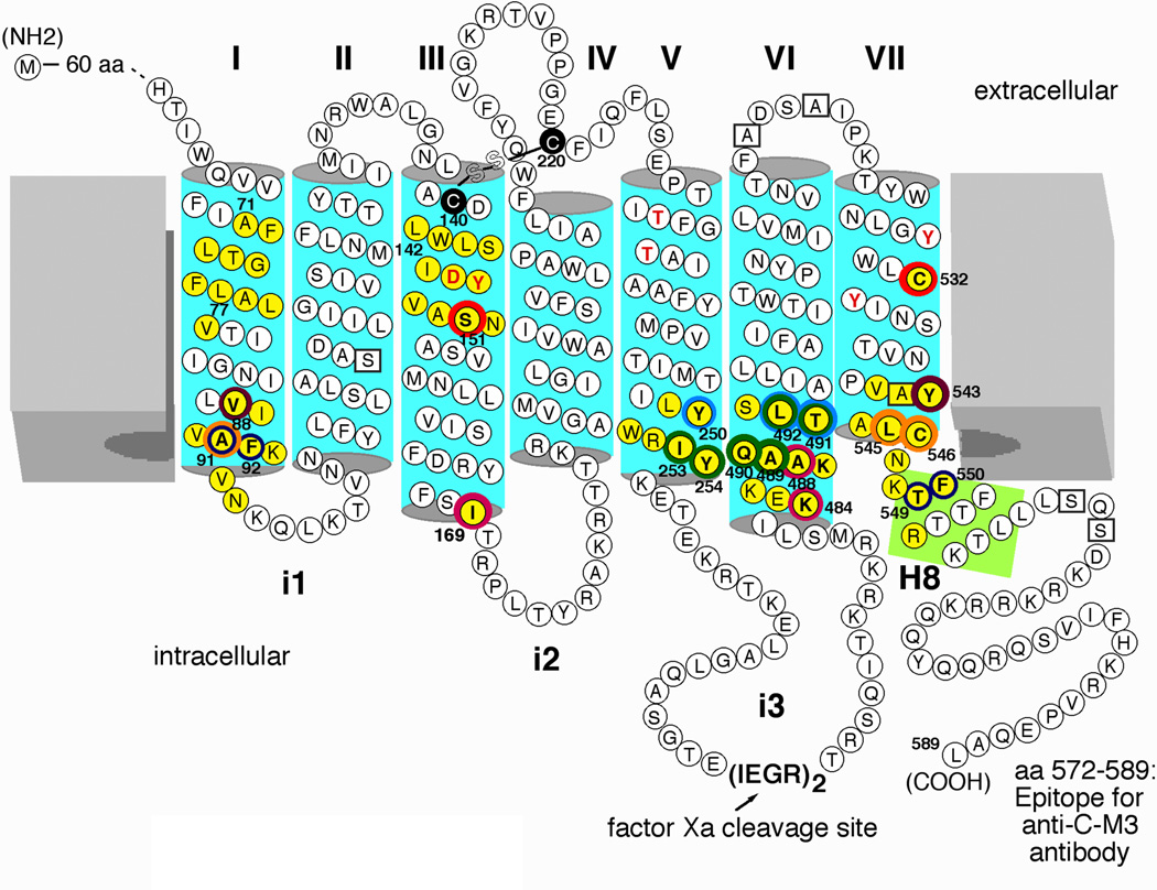
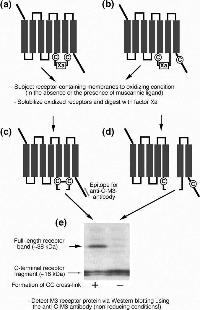
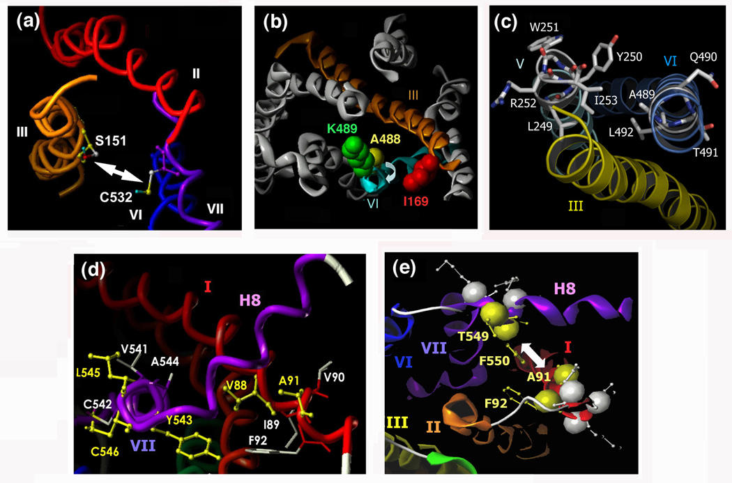
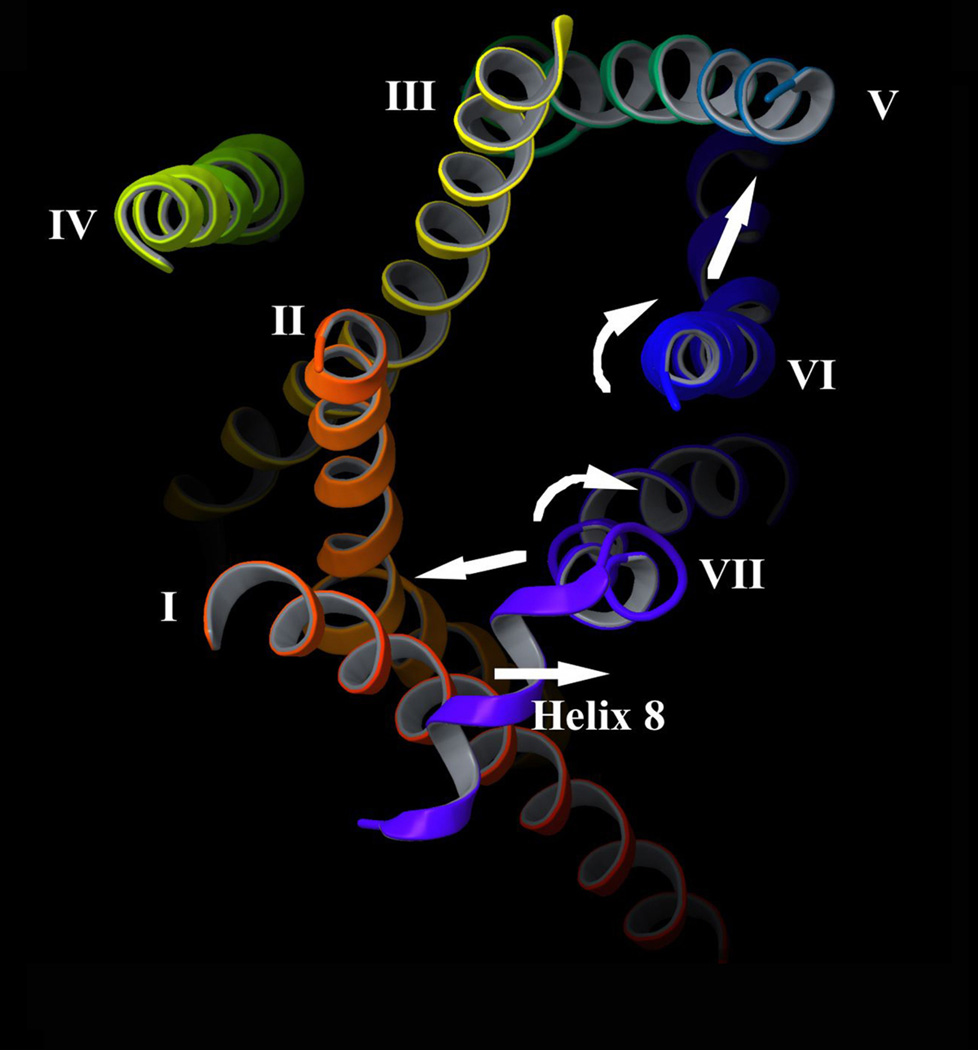
Little is known about the nature of the conformational changes that convert G-protein-coupled receptors (GPCRs), which bind diffusible ligands, from their resting into their active states. To gain structural insight into this process, various laboratories have used disulfide cross-linking strategies involving cysteine-substituted mutant GPCRs. Several recent disulfide cross-linking studies using the M(3) muscarinic acetylcholine receptor as a model system have led to novel insights into the conformational changes associated with the activation of this prototypical class I GPCR. These structural changes are predicted to involve multiple receptor regions, primarily distinct segments of transmembrane helices III, VI and VII and helix 8. Given the high degree of structural homology found among most GPCRs, it is likely that these findings will be of considerable general relevance. A better understanding of the molecular mechanisms underlying GPCR activation might lead to novel strategies aimed at modulating GPCR function for therapeutic purposes.


Figures

Figure 1
Figure 1
Agonist-modulated disulfide cross-linking in double Cys mutant M3 mAChRs. All Cys substitutions were introduced into the M3’(3C)-Xa receptor background, a modified version of the rat M3 mAChR [18]. In this construct, most endogenous Cys residues were replaced with either serine or alanine (boxed residues). The M3'(3C)-Xa construct contains only three remaining native Cys residues, C140, C220, and C5327.42 (note that C140 and 220 are engaged in a disulfide bond). In addition, the central portion of the i3 loop (A274-K469) was replaced with two adjacent factor Xa cleavage sites. The five potential N-glycosylation sites present in the N-terminal portion of the receptor protein (N6, N15, N41, N48, and N52) were replaced with Gln residues (not shown). A rabbit polyclonal antibody (referred to as anti-C-M3) was raised against the indicated C-terminal receptor sequence [18]. Muscarinic agonists promoted the formation of disulfide cross-links between Cys residues present at positions 1513.36 and 5327.42 (red rings; [21]), 881.53 and 5437.53 (brown rings; [20]), 911.56 and 5457.55/5467.56 (orange rings; [20]), 1693.54 and 4846.29/4886.33 (purple rings; [22]), 250C5.58 and 491C6.36/4926.37 (light blue rings; [24]), 2535.61 and 4896.34-4926.37 (green rings; [24]), and 2545.62 and 4896.34-4926.37 (green rings; [18]). In contrast, muscarinic agonists inhibited the formation of disulfide cross-links between Cys residues present at positions 911.56/921.57 and 5497.59/5507.60 (dark blue rings) [23]. The residues highlighted by red letters are known to play key roles in the binding of ACh and other conventional muscarinic agonists [33, 34]. Numbers refer to amino acid positions in the rat M3 mAChR sequence [33]. I-VII, TM helices I-VII; i1-i3, the three intracellular loops of the M3 mAChR; H8, helix 8.
Figure 2
Figure 2
General strategy used to detect the formation of disulfide bonds between vicinal Cys residues in mutant M3 mAChRs. Pairs of Cys residues were introduced into the M3’(3C)-Xa background receptor (see Figure 1), one Cys N-terminal and the other one C-terminal of the factor Xa cleavage site. (a, c) When two Cys residues lie adjacent to each other in the 3D structure of the receptor, they have the potential to form a disulfide bridge, either spontaneously or in the presence of oxidizing agents. Following cleavage with factor Xa, the two resulting receptor fragments will remain covalently linked by the disulfide bridge. Consequently, a full-length receptor band (~38 kDa in size) can be detected on Western blots run under non-reducing conditions (e, left lane). (b, d) When the two introduced Cys residues are not close to each other in the 3D structure of the receptor, they are unable to form a disulfide bridge. In this case, factor Xa digestion will yield two separate cleavage products and a full-length receptor band will not appear on Western blots run under non-reducing conditions (e, right lane).
Figure 3
Figure 3
Views of the M3 mAChR depicting regions/amino acids that undergo activity-dependent conformational changes. A 3D model of the inactive state of the rat M3 mAChR was built via homology modeling using the high-resolution X-ray structure of bovine rhodopsin as a template [2, 20]. (a) Extracellular view of the M3 mAChR parallel to the path of the exofacial segment of TM III. As discussed in the text, muscarinic agonists promote the formation of a disulfide bond between C5327.42 and a Cys residue introduced at position 1513.36 [21]. (b) Predicted location of I1693.54, K4846.29, and A4886.33 in the 3D structure of the inactive state of the M3 mAChR (cytoplasmic view). K4846.29 and A4886.33, which are located at the cytoplasmic end of TM VI, are projecting away from I1693.54 present at the bottom of TM III. Disulfide cross-linking data support the view that muscarinic agonists trigger a conformational change that includes a clockwise rotational movement of the cytoplasmic segment of TM VI [22]. (c) Predicted location of L2495.57-I2535.61 (TM V) and A4896.34-L4926.37 (TM VI) in the inactive state of the M3 mAChR (cytoplasmic view). Among the five highlighted TM V residues, only Y2505.58 and I2535.61 are predicted to face TM VI. Disulfide scanning mutagenesis studies strongly suggest that the cytoplasmic end of TM VI, but not the corresponding region of TM V, undergoes a major activity-dependent conformational change (see text for more details; [24]). (d) Cytoplasmic view of the inactive state of the M3 mAChR showing the locations of residues V881.53-F921.57 (TM I) and V5417.51-C5467.56 (TM VII). Cys substitution of the residues highlighted in yellow resulted in double Cys mutant receptors that showed agonist-dependent disulfide cross-linking (881.53/5437.53, 911.56/5457.55, and 911.56/5467.56) [20]. Whereas V881.53 and Y5437.53 directly face each other in the inactive state of the M3 muscarinic receptor, L5457.55 and C5467.56 point towards the lipid face and TM VI, respectively, away from A911.56, indicating that the cytoplasmic end of TM VII undergoes a major activity-dependent structural change during receptor activation (see text for more details; [20]). (e) Cytoplasmic view of a selected region of the intracellular surface of the M3 mAChR. Disulfide cross-linking data suggest that muscarinic agonists increase the distance between Cys residues introduced at a) positions 911.56 and 5497.59 and b) positions 921.57 and 5507.60 (these four residues are highlighted in yellow) [23]. In contrast, inverse muscarinic agonists are predicted to reduce the distance between these residues [23]. The individual figure panels were taken from Refs [21]-[24]. Numbers refer to amino acid positions in the rat M3 mAChR sequence [33].
Figure 4
Figure 4
Summary of activity-dependent structural changes predicted to occur at the cytoplasmic surface of the M3 mAChR. A 3D model of the inactive state of the rat M3 mAChR was built via homology modeling using the high-resolution X-ray structure of the β2-adrenergic receptor [4, 5] as a template (S. Costanzi and J. D. Karpiak; unpublished results). The intracellular surface of the M3 mAChR is viewed from the cytoplasm. As discussed in detail in the text, disulfide cross-linking data suggest that agonist binding to the M3 mAChR causes the following structural changes: 1) The cytoplasmic end of TM VI is predicted to undergo a rotational movement (and perhaps a partial unfolding) and to move closer to the corresponding segment of TM V [18, 22, 24]. 2) The C-terminal portion of TM VII is thought to move closer to the corresponding region of TM I, accompanied by a rotational movement (and perhaps a partial unfolding) of this TM VII segment [20]. 3) The N-terminal portion of helix 8 is predicted to move away from the cytoplasmic end of TM I [23]. The agonist-induced structural changes are thought to expose previously inaccessible receptor residues or surfaces (e.g. on the cytoplasmic ends of TM III, VI, and VII), ultimately resulting in productive receptor/G protein coupling.

Similar articles

Cited by

References

    1. Lagerström MC, Schiöth HB. Structural diversity of G protein-coupled receptors and significance for drug discovery. Nat. Rev. Drug Discov. 2008;7:339–357. - PubMed
    1. Palczewski K, et al. Crystal structure of rhodopsin: a G protein-coupled receptor. Science. 2000;289:739–745. - PubMed
    1. Rasmussen SG, et al. Crystal structure of the human β2 adrenergic G-proteincoupled receptor. Nature. 2007;450:383–387. - PubMed
    1. Cherezov V, et al. High-resolution crystal structure of an engineered human β2-adrenergic G protein-coupled receptor. Science. 2007;318:1258–1265. - PMC - PubMed
    1. Rosenbaum DM, et al. GPCR engineering yields high-resolution structural insights into β2-adrenergic receptor function. Science. 2007;318:1266–1273. - PubMed

Publication types